ORIGINAL RESEARCH ARTICLE
Entrapment of median nerve after elbow fracture dislocations: expected surgical time frame based on cadaver study
Yener Yoğuna, Uğur Bezirgana, Tülin Şen Esmerb, Sırrı Sinan Bilginc and Mehmet Armangila
aHand Surgery Unit, Orthopedics and Traumatology Department, Ankara University Faculty of Medicine, Ankara, Turkey; bAnatomy Department, Ankara University Faculty of Medicine, Ankara, Turkey; cPrivate Practise, Hand Surgery, Ankara, Turkey
ABSTRACT
Introduction: Median nerve injuries occur in approximately 3% of pediatric elbow fracture dislocations. These rare injuries can be difficult to diagnose, and the results are poor in delay cases. Surgical timing is one of the most important prognostic factors. We aimed to present three patients with median nerve palsy who were referred to our clinic late, and according to these cases, we emphasized the expected time frame for exploration based on our anatomical cadaver study.
Materials and Methods: Between 2008 and 2010, three patients were referred to our clinic because of median nerve paralysis after a treated elbow dislocation. The mean interval between injury and referral was 15 (min: 13–max: 18) months, and the mean age of the patients was 15 (13–18) years. Neurolysis was performed in two patients, and for the third patent, after neurolysis, axonal continuity was observed to be disrupted so sural nerve grafting was performed with four cables. Tendon transfers were performed in all patients. A total number of 20 upper extremities of 10 cadavers were dissected. Due to its proximal innervation and ease of assessment, the muscle innervation of the flexor pollicis longus (FPL) was planned to be evaluated. The distance from the medial epicondyle is calculated in the cadaver study where the nerve injury is found.
Results: The mean length from the medial epicondyle to the motor innervation of FPL was calculated in each specimen and found to be 101.99 millimeters (mm) (range: 87.5–134.2). The mean longest innervation of FPL was 110.83 mm from (range 87.5–148.1) the medial epicondyle calculated by including each specimens longest nerve length. Knowing that the healing time of a nerve lesion is 1 mm per day, we calculated that the recovery of FPL would take approximately 4 months.
Conclusion: When nerve healing is expected to be 1 mm a day in axonotmesis type injury, after the median nerve palsy following elbow dislocation, thumb flexion should be achieved in the following 4 months generally if the nerve was not entrapped in the joint. This cadaver-based study objectively defined how long to wait for the innervation of the FPL in median nerve injuries in elbow fracture dislocations.
KEYWORDS: Elbow fracture dislocations; flexor pollicis longus, median nerve entrapment; nerve injury
Citation: Journal of Plastic Surgery and Hand Surgery 2023; 59: 14–17. DOI: https://doi.org/10.2340/jphs.v59.15323.
Copyright: © 2024 The Author(s). Published by MJS Publishing on behalf of Acta Chirurgica Scandinavica. This is an Open Access article distributed under the terms of the Creative Commons Attribution 4.0 International License (http://creativecommons.org/licenses/by/4.0/), allowing third parties to copy and redistribute the material in any medium or format and to remix, transform, and build upon the material, with the condition of proper attribution to the original work.
Received: 26 June 2023; Accepted: 21 November 2023; Published: 18 January 2024
CONTACT Yener Yoğun yogunyener@gmail.com Hand Surgery Unit, Department of Orthopaedics of Traumatology, İbn’i Sina Hospital, Ankara University Faculty of Medicine, 06230 Ankara, 1192610507 Turkey
Competing interests and funding: All authors have no potential conflicts of interest, including financial interests, activities, relationships, and affiliations, to disclose.
We have no financial biases.
Introduction
Elbow dislocations are the second most common major joint dislocation after shoulder joint dislocation in the adult population, and the most common major joint dislocation in children [1]. Nerve injury is reported to occur in 5–22% of cases after an elbow dislocation [2]. The ulnar nerve is involved more often than the median or radial nerve [3].
Median nerve injury may occur in approximately 3% of pediatric elbow dislocations [4]. Intra-articular entrapment of the median nerve after elbow dislocations, or fracture dislocations, is an uncommon but potentially disastrous complication. The clinical signs and symptoms, which are generally mild in the early period of median nerve paralysis, could easily be overlooked in a child so the diagnosis is usually delayed, which further complicates the prognosis. Although elbow dislocation is infrequent in children or adolescents compared to adults, median nerve entrapment is usually seen in these age groups. Avulsion of the open medial epicondylar epiphysis may be considered as a reason for its more frequent injury in this pediatric population [5].
In general, after the 18th month in nerve injuries, muscle atrophy and irreversible damage occur, so the results of late nerve surgeries are worse [6]. Montanari et al. evaluated the publications of median nerve injury after elbow dislocation in children between 1945 and 2020 and showed that there is a significant relationship between nerve healing and surgical timing. They stated that there was complete or almost complete recovery in patients who were treated within 4 months, and poorer functional recovery treated in an average of 12.4 months [1]. Brendan et al. reported that the recovery of mixed motor nerves degrades dramatically over time, as repairs delayed more than 1 month. The authors pointed out that the worst results occur beyond 3 months [7].
In our study, we aimed to present three patients with median nerve palsy who were referred to our clinic late, and according to these cases, we emphasized the expected time frame for exploration based on our anatomical cadaver study.
Materials and methods
Patients
Between 2008 and 2010, three patients were referred to our clinic because of median nerve paralysis after a treated elbow dislocation. The mean interval between injury and referral was 15 (min: 13–max: 18) months. The mean age of the patients was 15 (13–18) years, and all had an elbow fracture dislocation (dislocation of elbow and fracture of medial epicondyle). The dislocations were reduced by closed manner, and open reduction internal fixation was performed in all cases for fractures. The median nerve lesions were documented by the referring surgeons and anticipated to be healed conservatively. Before exploration, EMG (electromyography) was performed for all the patients, and the results were chronic denervation findings. Physical examination revealed anterior interosseous nerve paralysis and sensory loss in median nerve dermatomes compared with the uninjured hands. All patients underwent exploration, and the median nerve was found to be entrapped in the joint (Figure 1a, 1b). This study was conducted according to the principles expressed in Declaration of Helsinki.
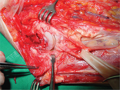
Figure 1a. Thirteen-year-old male patient. Incarceration of the median nerve was observed during exploration.
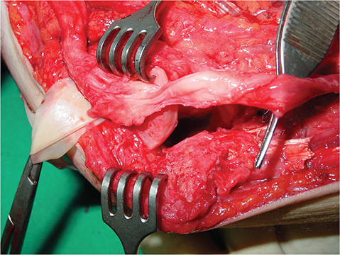
Figure 1b. Same patient with nerve extracted out of the joint. Note the axonotmesis type nerve lesion.
Neurolysis was performed in two patients, and for the third patent, after neurolysis, axonal continuity was observed to be disrupted so sural nerve grafting was performed with four cables. Tendon transfers were performed in all patients. The tendon transfers were brachioradialis for flexor pollicis longus (FPL), side to side transfer for the index profundus to the third finger profundus and extensor indicis proprius transfer for the opposition of the thumb.
Anatomic study
In neuropraxia-type nerve injuries, the recovery period of the nerve should be expected according to the level of the lesion. This anticipated time interval depends on the level of the lesion. Knowing that the healing time of a nerve lesion is 1 mm per day [8], the location of the lesion can be estimated in the case of elbow fracture dislocation. The exact time interval to exploration could be determined by measuring the length of the first motor innervation distal to the lesion of the affected nerve in a cadaver study. The most proximal motor innervation of the median nerve below elbow joint is the pronator teres muscle, but we thought that examination of its function is not easy for assessing nerve recovery. Instead, due to its proximal innervation and ease of assessment, even in a child, by thumb flexion, the muscle innervation of the FPL was planned to be evaluated.
A total number of 20 upper extremities of 10 cadavers were dissected. The mean age of the cadavers was 57.7 years (36–76). The cadaveric specimens had no known history of trauma. After median nerve entrapment in the joint, the nerve should start to heal from the level of the joint line similar to the level of medial epicondyle, so the distance from the medial epicondyle is calculated in the cadaver study. The origin and number of motor branches, and the distance of each motor branch to FPL muscle were measured. For clinical use, the distance between medial epicondyle and each motor branches entrance to the FPL muscle was measured in the extension position of the elbow.
Results
The FPL muscle is generally innervated by the branches from the anterior interosseous nerve, but in four extremities, additional direct innervations from the median nerve were observed. In 15 extremities, the FPL was found to be innervated by two motor branches, in four extremities, by three motor branches, and in one extremity, by only one motor branch (Table 1). The mean length of the motor nerve after branching from the major nerve was measured as 50.27 millimeters (mm) for branches of the median nerve and 35.98 mm for branches of the anterior interosseous nerve (Figure 2). The mean length from the medial epicondyle to the motor innervation of FPL was calculated in each specimen and found to be 101.99 mm (range: 87.5–134.2 mm). The mean longest innervation of FPL was 110.83 mm from (range 87.5–148.1 mm) the medial epicondyle calculated by including each specimen. There was a wide variation in distances between medial epicondyle and entrance of the nerve branch into the muscle, both between individual specimens and between specimens of one individual (Table 1).
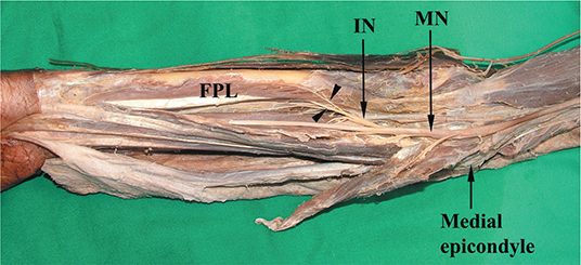
Figure 2. Cadaveric specimen showing median nerve, interosseous nerve, and the motor branches to flexor pollicis longus (marked with arrows).
The mean longest innervation of FPL was 110.83 mm from the medial epicondyle calculated by including each specimen. It was assumed that after injury, the nerve should heal approximately 1 mm a day [8]. It has been reported in the literature that end motor plate regeneration after nerve injuries is irreversible after roughly 18 months [6]. In our cadaveric study, we calculated that the recovery of FPL would take approximately 4 months. We thought that median nerve exploration in elbow fracture dislocation should be performed as soon as possible according to clinical and instrumental data. In the event of waiting decision, if there is no nerve healing at 4 months, no more time needed to be expected for surgical exploration. Tendon transfer surgeries should be performed in addition to nerve exploration if there is no nerve healing in these cases for more than 12–14 months.
Discussion
The most relevant feature of this study was that it is one of the few anatomical studies showing the expected time for exploration in rare median nerve injuries after elbow fracture dislocations. Our cadaveric study emphasizes the time when innervation is expected, and this could be a useful tool in such injuries that cause catastrophic results. The most proximal motor innervation of the median nerve below elbow joint is the pronator teres muscle, but we thought that examination of its function is not easy for assessing nerve recovery. FPL innervation was chosen in our cadaver study because thumb flexion can be easily evaluated in children.
Although median nerve entrapment following elbow dislocation is quite rare, it has been previously classified by Fourrier et al into three types, with the addition of a 4th type by Al-Qattan [9, 10]. This phenomenon is well known and commonly published as case reports in the literature. The type of nerve injury and the delay between that injury and the operation are two particularly important factors in the outcome after nerve repair [1, 5]. Therefore, the median nerve entrapped in the joint is a lesion that will not heal spontaneously and should be treated without delay for functional outcomes. Our two cases were Fourrier type 2. The patient we had grafted with the sural nerve was Fourrier type 4.
Delay in the diagnosis of median nerve entrapment is frequently mentioned in case series reported in the literature [5, 11–13]. There are several reasons for this situation. Since most nerve injuries in closed joint fracture dislocations are in the form of neuropraxia, these injuries are usually underestimated. Symptoms tend to be mild in these cases; moreover, clinical examination of children is also difficult. Sensory examination cannot be evaluated in most children. Two-point discrimination is likely to be impaired or lost, but it is very difficult to distinguish in children. In these cases, the pain may not be severe. For these reasons, the time between these injuries and treatment can be long. Repeated and detailed examinations and a comprehensive neurological examination are of great importance in such cases.
In the literature, there are some findings suggesting median nerve entrapment. In 1976, Matev described a radiographic sign consisting of a focal cortical depression on the ulnar side of the distal humeral metaphysis with interruption of the periosteal reaction in chronic cases [14]. Packer and Lennox have described the “C” sign—an incomplete central circular deficit within the medial epicondyle fracture callus that corresponds to the diameter of the median nerve. They concluded that this sign appears earlier than Matev’s sign, by around 6 weeks after injury, and may help with earlier diagnosis [15]. Starting proximal, a distally migrating Tinel’s sign may indicate progressive reinnervation but loss of functional recovery in expected time rules out its significance. Additionally, this important clinical sign is difficult to evaluate in a young child, and advancing Tinel’s sign is not consistent with a good prognosis for the nerve because this sign can be evoked by percussion over a relatively small number of regenerating axons [16]. EMG is likely to reveal findings consistent with axonotmesis proximal to the branch innervating the pronator teres, so it is not useful preoperatively for deciding whether to perform an exploration [17]. Magnetic resonance imaging (MRI) and ultrasonography (USG) are the gold standard in the evaluation of the median nerve. MRI is effective both in showing the course of the median nerve and in demonstrating muscle atrophy and fatty degeneration. In recent years, the importance of USG has been better understood because it is a dynamic imaging and can be compared with the contralateral extremity [18, 19].
Dolderer et al. examined 19 upper extremities in their cadaver study and found the mean length of the motor nerve innervating the FPL muscle (medial epicondyle to FPL muscle) to be 106.2 ± 16.2 mm [20]. In another anatomical study, it was shown that the FPL is innervated by more than one motor nerve [21]. In our cadaveric study in which we examined 20 upper extremities, we also saw the multiple innervations of the FPL and calculated the mean of the longest motor branches. We found that the mean motor length is 101.99 mm, and the mean longest innervation of FPL is 110.83 mm from medial epicondyle. When nerve healing is expected to be 1 mm a day in axonotmesis type injury, these results could be interpreted, to show that after median nerve palsy following elbow dislocation, thumb flexion should be achieved in the following 4 months.
It is known that irreversible damage occurs at the neuromuscular junction at the end of approximately 18 months [6]. As a result of irreversible fibrosis of a nerve that has been entrapped for a long time, its structural integrity is also impaired. Therefore, delay in treatment in nerve injuries causes poor results. Montanari et al. and Brendan et al. evaluated the publications of median nerve injury after elbow dislocation and showed that there is a significant relationship between nerve healing and surgical timing [1]. Montanari et al. reported that the mean treatment delay in patients with complete recovery was 1.5 months, and the mean treatment delay in patients with poor clinical outcomes was 12.4 months [1]. Brendan et al. reported that the recovery of mixed motor nerves degrades dramatically over time, as repairs delayed more than 1 month. The authors pointed out that the worst results occur beyond 3 months [7]. In our cadaver study, we found the mean longest nerve length from the medial epicondyle to the FPL as 110.8 mm. In other words, if the nerve integrity is preserved in the injury area and has not been entrapped, we think that it should heal within a maximum of 4 months. If radiology also supports entrapment, we should immediately consider exploration with no other time elapsing in surgical decision. Likewise, in late cases (after 12–14 months), we should consider tendon transfers.
There were some limitations in our cadaver-based study. Median nerve injuries after elbow fracture dislocations are frequently seen in children. However, the cadavers in this study were adult cadavers. Healing occurs earlier in children because the distance between the injury site and the end organ is shorter than in adults. Since the nerve regeneration capacity is better in children, recovery is better than adults after nerve surgery in late cases.
Conclusion
In conclusion, for median nerve paralysis after elbow dislocation or fracture dislocation, the surgeon should be aware that the nerve could be entrapped in the joint and should explore mandatorily. The major issue in prognosis for this type of nerve injury is the chronicity of the lesion as supported by the literature that makes the time interval to exploration important. This cadaver-based study objectively defined how long to wait for innervation of the FPL in median nerve injuries in elbow fracture dislocations.
Acknowledgment
We did not receive any contributions from anyone.
Informed consent
Informed consent was obtained from all individual participants included in this study.
ORCID
Yener Yoğun  https://orcid.org/0000-0002-3070-5509
Uğur Bezirgan https://orcid.org/0000-0001-9053-8637
Tülin Şen Esmer https://orcid.org/0000-0002-1879-1790
Sırrı Sinan Bilgin https://orcid.org/0000-0001-6341-8162
Mehmet Armangil https://orcid.org/0000-0003-0433-0253
References
[1] Montanari S, Sartore R, Spina V, et al. Post-traumatic entrapment of the median nerve in the ulno-humeral joint: diagnosis, treatment and literature review. J Orthop Sci. 2022 May 1;27(3): 627–634. https://doi.org/10.1016/j.jos.2021.02.006
[2] Rao SB, Crawford AH. Median nerve entrapment after dislocation of the elbow in children. A report of 2 cases and review of literature. Clin Orthop Relat Res [Internet]. 1995 [cited 2023 Apr 1];312(312):232–237. Available from: https://pubmed.ncbi.nlm.nih.gov/7634608/
[3] Kamble P, Prabhakar A, Wankhade AM, et al. A rare case of lateral displacement of median nerve in a dislocated elbow and its management. J Orthop Case Rep [Internet]. 2021 [cited 2023 Apr 1];11(12):80–83. Available from: https://pubmed.ncbi.nlm.nih.gov/35415152/
[4] Cengiz Ö. Median nerve entrapment in an adolescent medial epicondyle fracture of humerus: A case report. Turk J Trauma Emrg Surgery. 2022 Aug;28(8):1200-1203. doi: 10.14744/tjtes.2020.45742
[5] Simon D, Masquijo JJ, Duncan MJ, et al. Intra-articular median nerve incarceration after spontaneous reduction of a pediatric elbow dislocation: case report and review of the literature. J Pediatr Orthop. 2010 Mar;30(2):125–129. https://doi.org/10.1097/BPO.0b013e3181cf3bfd
[6] Robinson LR. Traumatic injury to peripheral nerves. Muscle Nerve. 2022 Dec 7;66(6):661–670. https://doi.org/10.1002/mus.27706
[7] MacKay BJ, Cox CT, Valerio IL, et al. Evidence-based approach to timing of nerve surgery: a review. Ann Plast Surg. 2021;87:e1–e21.
[8] Lopes B, Sousa P, Alvites R, et al. Peripheral nerve injury treatments and advances: one health perspective. Int J Mol Sci. 2022 Jan 14;23(2):918. https://doi.org/10.3390/ijms23020918
[9] Fourrier P, Levai JP, Collin JP. [Median nerve entrapment in elbow dislocation]. Rev Chir Orthop Reparatrice Appar Mot. 1977;63(1):13–16.
[10] Al-Qattan MM, Zuker RM, Weinberg MJ. Type 4 median nerve entrapment after elbow dislocation. J Hand Surg. 1994 Oct 29;19(5):613–615. https://doi.org/10.1016/0266-7681(94)90127-9
[11] Aggarwal A, Jana M, Kumar V, et al. MR neurography in intraosseous median nerve entrapment. World J Radiol. 2017 Oct 28;9(10):400–404. https://doi.org/10.4329/wjr.v9.i10.400
[12] Korus L, Morhart M, Jarman A, et al. Median nerve reconstruction after entrapment in the elbow. Can J Plast Surg. 2009 Dec 1;17(4):130–132. https://doi.org/10.1177/229255030901700408
[13] Akansel G, Dalbayrak S, Yilmaz M, et al. MRI demonstration of intra-articular median nerve entrapment after elbow dislocation. Skeletal Radiol. 2003 Sep 1;32(9):537–541. https://doi.org/10.1007/s00256-003-0667-4
[14] Matev I. A radiological sign of entrapment of the median nerve in the elbow joint after posterior dislocation. A report of two cases. J Bone Joint Surg Br. 1976 Aug;58-B(3):353–355. https://doi.org/10.1302/0301-620X.58B3.956256
[15] Packer GJ, Lennox CM. Median nerve entrapment after elbow dislocation: a new radiological sign. Br J Clin Pract. 1995;49(4):221. https://doi.org/10.1111/j.1742-1241.1995.tb09958.x
[16] Russell T. Surgical disorders of the peripheral nerves. R. Birch, G. Bonney, C. B. Wynn Parry. 283 × 223 mm. Pp. 539. Illustrated. 1998. Edinburgh: Churchill Livingstone. £95. Br J Surg. 2003 Jan 2;86(3):429. https://doi.org/10.1046/j.1365-2168.1999.1056c.x
[17] Shergill G, Bonney G, Munshi P, et al. The radial and posterior interosseous nerves. J Bone Joint Surg. 2001 Jul 1;83(5):646–649. https://doi.org/10.1302/0301-620X.83B5.0830646
[18] O’Brien AC, Teh Z, Rinaldi M, et al. Intraosseous Type 2 Median Nerve Entrapment After Posterior Elbow Dislocation Diagnosed on Ultrasound With MRI and Surgical Correlation. Cureus. 2021 Oct 8;13(10):e18606. doi: 10.7759/cureus.18606
[19] O’Callaghan PK, Freeman K, Davis LC, et al. A rare case of type 2 entrapment of the median nerve after posterior elbow dislocation with MRI and ultrasound correlation. Skeletal Radiol. 2019 Oct 13;48(10):1629–1636. https://doi.org/10.1007/s00256-019-03201-4
[20] Dolderer JH, Prandl EC, Kehrer A, et al. Solitary paralysis of the flexor pollicis longus muscle after minimally invasive elbow procedures: anatomical and clinical study of the anterior interosseous nerve. Plast Reconstr Surg. 2011 Mar;127(3):1229–1236. https://doi.org/10.1097/PRS.0b013e3182043ac0
[21] Liu J, Pho RWH, Pereira BP, et al. Distribution of primary motor nerve branches and terminal nerve entry points to the forearm muscles. Anat Rec. 1997 Jul;248(3):456–463. https://doi.org/10.1002/(SICI)1097-0185(199707)248:3<456::AID-AR19>3.0.CO;2-O