ORIGINAL RESEARCH ARTICLE
Effect of high-density fat combined with adipose stem cell glue on the success rate of facial filling and its clinical value
aHefei BOE Hospital, Hefei City, Anhui Province, China; bThe First Affiliated Hospital of Anhui Medical University, Hefei City, Anhui Province, China
ABSTRACT
Facial fat grafting is a popular cosmetic procedure, and experts are increasingly endorsing the use of high-density fat with adipose stem cell glue for better results. This study aims to explore the effect of high-density fat combined with adipose stem cell glue on the success rate of facial filling and its clinical value. We conducted a randomized trial with 100 patients who underwent facial fat transplantation between August 2020 and August 2022. They were divided into two groups: a control group receiving traditional Coleman fat transplantation and an observation group receiving high-density fat with adipose stem cells. In the observation and control groups, the excellent and good rate was 98.00 and 80.00%. After 3 months of treatment, the thickness of frontal subcutaneous fat and temporal subcutaneous fat in the observation group was higher (P < 0.05). Observation group retention of fat transplantation was noticeably higher 3 months after treatment (P < 0.05). Three months after treatment, the VISIA (facial imaging system) scores of facial color spots, facial pores and facial wrinkles in the observation group were lower (P < 0.05). After treatment, both groups indicated noticeable improvements in physiological functions, health status, social function, mental health, and somatic diseases compared to before treatment. Notably, the observation group had higher scores (P < 0.05). The observation group had a lower complication rate (4.00% vs. 22.00%) and higher satisfaction rate (98.00% vs. 86.00%) than the control group. Using high-density fat combined with adipose stem cell glue for facial fat grafting yields superior results, reduces complications, and boosts patient satisfaction compared to traditional methods. We have complied with all relevant ethical regulations with regard to the use of stem cells.
KEYWORDS: High density fat; adipose stem cell glue; success rate of facial filling; life quality; complications
Citation: Journal of Plastic Surgery and Hand Surgery 2024; 59: 32–39. DOI: https://doi.org/10.2340/jphs.v59.18683.
Copyright: © 2024 The Author(s). Published by MJS Publishing on behalf of Acta Chirurgica Scandinavica. This is an Open Access article distributed under the terms of the Creative Commons Attribution 4.0 International License (http://creativecommons.org/licenses/by/4.0/), allowing third parties to copy and redistribute the material in any medium or format and to remix, transform, and build upon the material, with the condition of proper attribution to the original work.
Received: 11 September 2023; Revised: 21 November 2023; Accepted: 8 January 2024; Published: 13 March 2024.
CONTACT Yu Zhao Zhaoyu202304@163.com The First Affiliated Hospital of Anhui Medical University, 218 Jixi Road, Shushan District, Hefei City, Anhui Province, China
Supplemental data for this article can be accessed online at https://doi.org/10.2340/jphs.v59.18683
Competing interests and funding: The authors declared no potential conflicts of interest with respect to the research, authorship, or publication of this article.
Introduction
Human facial contour is a system composed of skeleton structure, soft tissue structure and skin (1). Ageing is an inevitable process in the human body. Aging and other abnormal changes will attract people’s attention and cause some psychological pressure (2, 3). For the problem of facial aging, the traditional treatment is rhytidectomy, which can improve the relaxation of facial skin by tightening the skin and removing excess tissue. Rhytidectomy alone cannot solve the problems such as dull skin and poor elasticity. The therapeutic effect is limited (4). With the continuous development of medical technology, facial filling technology has sprung up and become the mainstream way to improve facial aging (5).
Collagen and hyaluronic acid, though effective, are costly and short-lived solutions for age-related facial issues. Autologous fat transplantation, developed by Illouz in 1986, offers a safer alternative since it uses the patient’s own fat tissue, avoiding rejection and allergies (6, 7). However, this method has drawbacks, including strict recipient site conditions, poor infection resistance, easy liquefaction, and limited one-time transplantation amounts. Fat retention rates range from 30 to 60%, with some fat cells failing to absorb nutrients, limiting its broader use.
Yoshimura et al. (8) achieved better results using vascular matrix components (SVF) cells to assist fat transplantation. However, obtaining SVF cells through enzymatic digestion poses safety risks and faces regulatory limitations in most countries (9, 10). Some researchers have obtained SVF-rich adipose stem cell gel (SVF-GEL) through a purely physical process, improving fat retention compared to conventional methods (11, 12). However, SVF-GEL requires significantly more fat extraction. However, it is worth noting that it is necessary to obtain as much pure fat as possible without increasing the liposuction site of the patient, and the upper and middle fat after centrifugation should be used to prepare SVF-GEL for filling fine areas such as around the eye and lacrimal sulcus. The lower layer of high-density fat is used for volume filling in larger areas. Although many studies on the application value of high-density fat-bound adipose stem cell glue in facial fat transplantation have been carried out in the past, there are great differences in experimental design, observation index and object of previous research. As a result, the conclusions are lack of persuasion and consistency. It is necessary to carry out further research. Based on this, 100 patients who received facial fat transplantation from August 2020 to August 2022 were investigated in this study. This study aims to provide a theoretical framework supporting the clinical application of high-density fat combining with fat stem cell glue for facial filling therapy.
Materials and methods
General information
We conducted a randomized controlled trial including 100 patients who underwent facial fat transplantation at our hospital between August 2020 and August 2022. The patients were classified into two groups, with 50 cases in each group. In the control group, patients accepted traditional Coleman fat transplantation, while in the observation group, patients accepted high-density fat combined with adipose stem cell glue. There were eight men and 42 women in the observation group, aged from 35 to 51 years old. The body mass index (BMI) was 17.83–37.29 kg/m2. There were five men and 45 women in the control group. Age 34~50 years old. BMI ranged from 18.05 to 36.61 kg/m2. No noticeable difference was found in baseline related data (P > 0.05), which was comparable, as shown in Table 1.
The inclusion criteria included the following: 1) patients had the relevant conditions for facial granule autologous fat transplantation and can adapt to this study; 2) patient tolerance of this surgical and pharmacological treatment; 3) patients had no other serious primary diseases; 4) patients had normal mental state and normal communication; 5) patients had no other major diseases, no history of allergy or heredity; 6) the clinical data of the patients were complete, and the relevant consent forms were signed.
The exclusion criteria included the following: 1) those who were uncomfortable with the indications related to facial granule autologous fat transplantation; 2) those with serious primary diseases such as cardio-cerebrovascular, liver, kidney and hematopoietic system; 3) patients with abnormal behavior or mental disorders, people with allergic constitution; 4) those with communication disorder and unable to communicate normally; 5) those with organ dysfunction; 6) those who suffered from other major diseases and were allergic to the drugs adopted in the study; 7) those who had a history of allergy or genetic history; 8) those who did not take drugs in accordance with the regulations and were unable to judge the efficacy or safety of the drugs.
Calculation formula of sample size was as follows:

The bilateral α is taken as 0.05 and β as 0.20, the therapeutic effect (excellent and good rate) is taken as the effect index, and set the parameters as: P1 = 0.94, P2 = 0.76. The sample size for each group was calculated to be 45 cases, and based on a 10% shedding rate, there were approximately 50 patients in each group for a total of 100 patients.
Research technology roadmap
Figure 1 shows the research technology roadmap of this experiment.
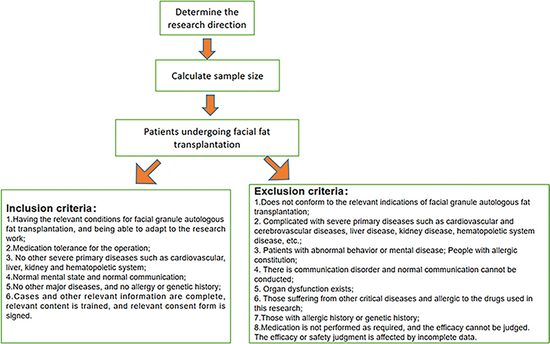
Figure 1. Research Technology Roadmap.
Treatment methods
General anesthesia was used. Swelling infiltration anesthesia was performed in the liposuction area with saline containing 0.001% epinephrine, and adipose tissue was extracted with a liposuction needle with an internal diameter of 2.5 mm. The control group received traditional Coleman fat transplantation. The fat obtained was processed using the Coleman technique standard, which involved centrifugation to separate the fat layer (known as Coleman fat) for transplantation. After operation, the donor area was routinely dressed in body-shaping clothes, and antibiotics and non-steroidal anti-inflammatory drugs were taken orally for 3 days.
A combination of high-density fat and fat stem cell gel was used in the observation group. Swelling anesthesia was administered on the inner thigh under general anesthesia and the swelling solution was configured (500 mL of normal saline + 15 mL of 2% lidocaine + 0.3 mg of epinephrine). After the swelling anesthesia was fully effective, the 20 mL syringe was used to connect the 2.5 mm inner diameter porous liposuction needle for manual liposuction with low negative pressure. A mount of 200 mLpure fat was extracted from each case and then centrifuged for 3 min at 1,200 g. (Xi’an Yizhikang YK-1001 low speed centrifuge).
Preparation of SVF-GEL: After the first centrifugation, the fat was removed from the lower liquid layer, and the lower layer was high density fat. The second time was used to prepare SVF-GEL, where the upper fat component needed to be retained for the preparation of SVF-GEL. The syringe was injected back and forth through a syringe attached to a bidirectional helical conversion head with an internal diameter of 1.4 mm, at a rate of 10 mL/s and a total injection time of 1 min. The emulsified fat was filtered with a 500 μm diameter filter to remove the residual fibrous tissue, then centrifuged 3 min under the action of 2,000 g centrifugal force, and the lowest viscous whitish substance was SVF-GEL.
High-density fat was used for volume filling, such as forehead, question, and cheek filling. SVF-GEL was used for fine filling, such as upper eyelid, lacrimal groove, nasolabial groove filling. An 18G blunt needle connected to a 1 mL syringe was used to fill the high-density fat and a 23G blunt needle connected to a 1 mL syringe was used to fill the SVF-GEL. The injected layers included the periosteal layer, the superficial temporal deep fascia and the subcutaneous fat layer. The harvesting approach employed in this study enables “homologous use” by utilizing mechanically isolated, non-cultured adipose stem cells exclusively derived from subcutaneous tissue and introducing them into the same anatomical or histological environment (13).
After the operation, the liposuction area was fixed with plastic pants to accelerate the recovery of the liposuction area, and the filling area was fixed with 3M adhesive tape and oral antibiotics and non-steroidal anti-inflammatory drugs for 3 days.
Observation index
The main results are as follows:
- Evaluation criteria of therapeutic effect. The therapeutic effect of the patients was evaluated 3 months after treatment, and the evaluation criteria were established according to the clinical experience of doctors after referring to the relevant literature (14). In this study, the therapeutic effect was classified into three levels, including excellent, good, and poor. Excellent means that the facial filler is natural and smooth, the ageing facial profile is noticeably altered and no complications occur. Good means that the facial filler is flat and wrinkles and depressions are improved with no complications or easily recoverable complications; however, the facial profile is improved to some extent but the results are not satisfactory and there are some complications. Poor means that the patient’s facial condition is largely unimproved and there are more complications. Excellent and good rate = (excellent + good + fair) / total number of cases × 100%.
- Before and 3 months after treatment, the facial subcutaneous fat thickness was measured by Japan Fujiwara fujiwara portable subcutaneous fat thickness meter (FMH- 1), and the frontal subcutaneous fat thickness and temporal subcutaneous fat thickness were compared between the two groups. The probe was placed on the patient’s forehead and temporal area, and the instrument could automatically read and display the thickness of subcutaneous fat. Three places on the forehead and temporal area was measured and the results were averaged.
- The retention rate of fat transplantation. At 3 months after treatment, the fat transplantation retention rates of the two groups were compared (Fat graft retention is the volume retention rate 3 months after fat grafting, which is the proportion of stable surviving fat to the total grafted fat. Fat graft retention rate = fat retention/fat injection × 100% 3 months post treatment).
- The improvement of facial skin quality, fine lines, color spots and pores before and 3 months after treatment were assessed by Canfield VISIA system spot residual score (15). Better improvement is measured by a lower score.
- Life quality score. The health status questionnaire (SF-36) (16) was adopted to assess the improvement of patients’ life quality, which involved 8 dimensions. In this study, only five dimensions were selected, including social functioning, physical functioning, health status, mental health, and physical illness. The patients were evaluated before and 3 months after treatment.
- The incidence of postoperative complications. Statistics were made on the number of all kinds of complications occurred within 3 months after operation (common complications included fat liquefaction, infection, nodules, necrosis, hematoma, etc.), and the total incidence of complications was calculated. The total incidence of complications = the sum of the number of complications/the total number of cases × 100%. Fat liquefaction is the rupture of fat cells at the incision after surgery, where fat particles from the ruptured fat cells spill out and break down to form a liquid oil-like substance. An infection is a result of the invasion of pathogens, such as bacteria and viruses, into the body, causing a localized tissue and systemic inflammatory response. Nodule is a palpable, round, ring-shaped, or oval-shaped localized substantial injury, mostly in the dermis or subcutaneous tissue, >1 cm in diameter. Necrosis is the death of localized tissues and cells in vivo in response to injury factors. Haematoma is the rupture of a blood vessel under external forces, with the spilled blood separating from the surrounding tissue and forming a blood-filled cavity.
- Therapeutic satisfaction. Three months after surgery, patients’ treatment satisfaction was surveyed using our own treatment satisfaction questionnaire, which focuses on overall satisfaction, skin improvement and post-filler fullness. The evaluation results were classified into four levels, including very pleased, pleased, general and displeased. Patients can score against the appropriate entries and rate their satisfaction with this treatment accordingly. Total satisfaction = (very pleased + pleased + average) / total number of cases × 100%. The overall Cronbach α coefficient of the scale was 0.92, the Cronbach α coefficient of each factor was 0.82–0.88, and the CVI value was 0.9016. The reliability and validity of the scale were satisfactory.
Statistical analysis
The data were analyzed and processed by SPSS21.0 statistical software. A (x ± s) symbol is used to indicate measurements with a normal distribution or approximate normal distribution. Comparison of the two groups was done using paired t-tests, while comparing the two groups separately using independent sample t-tests. The n (%) was adopted to represent the counting data, and χ2 test was adopted. P < 0.05 was considered statistically remarkable.
Ethics
This study was performed in accordance with the Declaration of Helsinki. Informed consent forms were signed by all patients for this study, which was approved by our hospital’s Medical Ethics Council. See Figure 2 in the inclusion process.
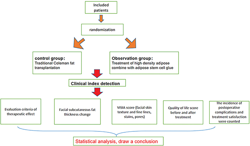
Figure 2. Schematic diagram of screening into groups.
Results
The general data of patients
No statistical difference was found in gender, age, BMI, donor area and years of education (P > 0.05, Table 1).
Comparison of therapeutic effects
In the observation group, the curative effect was excellent in 25 people, good in 16 people, fair in eight people and poor in one person, and the excellent and good rate was 98.00%. In the control group, the curative effect was excellent in 18 people, good in 19 people, fair in three people, and poor in 10 people, and the excellent and good rate was 80.00% (P < 0.05, Table 2).
The average thickness of frontal and temporal subcutaneous fat before and after treatment
Before treatment, no noticeable difference was found in forehead subcutaneous fat thickness and temporal subcutaneous fat thickness (P > 0.05). Three months after treatment, the thickness of frontal subcutaneous fat and temporal subcutaneous fat increased noticeably in both groups, and the frontal subcutaneous fat thickness and temporal subcutaneous fat thickness in the observation group were noticeably higher (P < 0.05, Table 3).
| Group | Thickness of subcutaneous fat in forehead(mm) | Thickness of temporal subcutaneous fat(mm) | ||
| Before treatment | 3 months after treatment | Before treatment | 3 months after treatment | |
| Observation group | 1.08 ± 0.16 | 1.93 ± 0.44a | 1.06 ± 0.14 | 1.95 ± 0.42a |
| Control group | 1.05 ± 0.49 | 1.27 ± 0.52b | 1.12 ± 0.39 | 1.41 ± 0.55b |
| t | 0.412 | 6.851 | 1.024 | 5.518 |
| P | > 0.05 | < 0.05 | > 0.05 | < 0.05 |
| Note: compared with the observation group before treatment, aP < 0.05; compared with the control group before treatment, bP < 0.05. | ||||
The fat transplantation retention rate after treatment
The fat transplantation retention rate of the observation group was noticeably higher 3 months after treatment (P < 0.05, Figure 3).
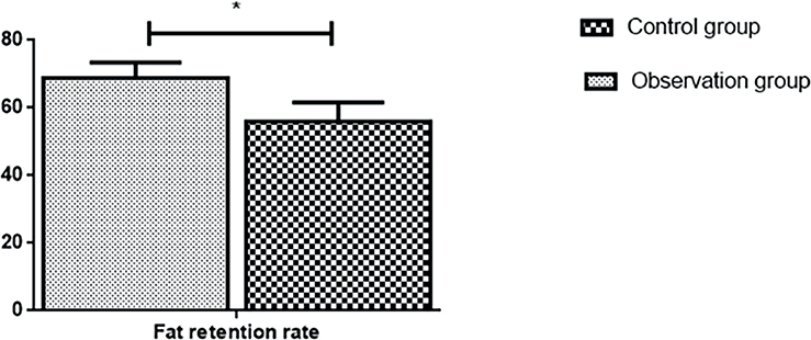
Figure 3. The fat transplantation retention rate after treatment.
VISIA scores of facial color spots, facial pores and facial wrinkles before and after treatment
Before treatment, no noticeable difference was found in VISIA scores of facial color spots, facial pores and facial wrinkles (P > 0.05). After 3 months of treatment, both groups indicated a significant reduction in VISIA scores for facial spots, pores, and wrinkles. However, the reduction in VISIA scores for facial spots, pores, and wrinkles was even greater in the observation group (P < 0.05, Table 4).
| Group | VISIA score of facial spots | Facial pore VISIA score | VISIA score of facial wrinkles | |||
| Before treatment | 3 months after treatment | Before treatment | 3 months after treatment | Before treatment | 3 months after treatment | |
| Observation group | 116.48 ± 25.06 | 66.83 ± 20.28a | 22.39 ± 1.26 | 12.46 ± 2.05a | 9.73 ± 0.71 | 4.26 ± 0.36a |
| Control group | 117.05 ± 27.63 | 85.26 ± 21.05b | 23.01 ± 2.05 | 16.25 ± 2.18b | 9.58 ± 0.66 | 5.83 ± 0.52b |
| t | 0.108 | 4.458 | 1.822 | 8.956 | 1.094 | 17.553 |
| P | > 0.05 | < 0.05 | > 0.05 | < 0.05 | > 0.05 | < 0.05 |
| Note: compared with the observation group before treatment, aP < 0.05; compared with the control group before treatment, bP < 0.05. | ||||||
The life quality scores
No noticeable difference was found in the scores of life quality before treatment (P > 0.05). The scores of physiological functions, health status, social function, mental health, and somatic diseases in both groups significantly improved after treatment compared to before treatment. Furthermore, the scores in the observation group were noticeably higher (P < 0.05, Table 5).
| Group | Physiological function | Health condition | Social function | |||
| Before treatment | After treatment | Before treatment | After treatment | Before treatment | After treatment | |
| Observation group | 41.55 ± 4.13 | 50.71 ± 4.34a | 41.92 ± 4.21 | 45.24 ± 4.07a | 37.93 ± 4.51 | 46.63 ± 4.51a |
| Control group | 42.08 ± 4.18 | 45.83 ± 4.02b | 42.01 ± 4.33 | 51.93 ± 5.12b | 37.82 ± 4.94 | 41.03 ± 4.39b |
| t | 0.638 | 5.833 | 0.105 | 7.232 | 0.116 | 6.292 |
| P | > 0.05 | < 0.05 | > 0.05 | < 0.05 | > 0.05 | < 0.05 |
| Note: compared with the observation group before treatment, aP < 0.05; compared with the control group before treatment, bP < 0.05. | ||||||
| Group | Mental health | Somatic disease | ||
| Before treatment | After treatment | Before treatment | After treatment | |
| Observation group | 48.23 ± 4.81 | 55.41 ± 5.22a | 47.83 ± 5.42 | 56.92 ± 4.12a |
| Control group | 49.03 ± 4.88 | 51.03 ± 5.14b | 48.01 ± 5.39 | 51.49 ± 5.18b |
| t | 0.826 | 4.228 | 0.167 | 5.801 |
| P | > 0.05 | < 0.05 | > 0.05 | < 0.05 |
| Note: compared with the observation group before treatment, aP < 0.05; compared with the control group before treatment, bP < 0.05. | ||||
The postoperative complications
In the observation group, one person had infection and one person had hematoma, and the incidence of complications was 4.00%. In the control group, there were three people of fat liquefaction, four people of infection, one person of nodule and three people of hematoma, and the incidence of complications was 22.00% (P < 0.05, Table 6).
Therapeutic satisfaction between the two groups
In the observation group, 30 people were very pleased, 10 people were pleased, nine people were general, and one person was not pleased; the satisfaction rate was 98.00%. In the control group, 18 people were very pleased, 20 people were pleased, five people were average, seven people were not pleased, and the satisfaction rate was 86.00% (P < 0.05, Table 7).
Discussion
Facial filling is one of the common medical plastic surgeries, which can solve the phenomena of tissue atrophy, deepening wrinkles and sagging skin caused by aging or other reasons (17, 18). Depending on the specific location of the patient’s hollows, appropriate fat fillers can address the ageing problem to a certain extent and rejuvenate the face as a whole.
Finding suitable filling materials has always been one of the important contents of cosmetic plastic surgery research. Autologous fat filler is a surgical procedure in which the patient’s own fat tissue is extracted, transplanted and injected into the desired area to refine the contours, improve local lines and rejuvenate the face. At present, it has been widely used in augmentation mammoplasty, breast reconstruction, sunken scar treatment, facial soft tissue filling, facial contour adjustment, hip filling, and penile thickening (19–21). The concept of high-density fat was first proposed by Allen et al. (22) in 2013, while other scholars confirmed that this part of fat has certain advantages in clinical transplantation (23). After centrifugation of the aspirated adipose tissue at 1,200 g for 3 min, the adipose tissue was separated into upper, middle, and lower layers (24). The high-density fat was located in the lowest layer. Due to its high concentration of adipose stem cells and growth factors associated with the vascular system, high-density fat is more likely to live longer. The main component of SVF-GEL for fine facial fillers is an extracellular matrix rich in fat stem cells, and the main volume component of the filling process is extracellular matrix such as collagen and elastin, so there is no problem of graft survival. The SVF-GEL cell group contains a large number of fat stem cells and endothelial cells (25). These cells can improve the local blood microenvironment after transplantation and participate in the later process of differentiation of adipogenesis. In this study, it was found that in the observation group, 25 people were excellent, 16 people were good, eight people were fair, one people was poor, and the rate of excellent and good treatment was 98.00%. In the control group, 18 people were excellent, 19 people were good, three people were fair, 10 people were poor, and the excellent and good rate was 80.00%. The thickness of subcutaneous fat in the forehead and the thickness of subcutaneous fat in the temporal region in the observation group were noticeably higher 3 months after treatment, and the VISIA scores of facial spots, facial pores and facial wrinkles were lower. Compared to traditional Coleman fat grafting, high density fat combined with adipose stem cell gel provides better facial filler results, noticeably increasing the patient’s subcutaneous fat thickness after treatment, reducing facial blemishes, facial pores and VISIA scores of facial wrinkles, and achieving better aesthetic results. In the early stage of fat transplantation, the transplanted fat cells are exposed to a period of ischemia and hypoxia; those that receive sufficient nutrients survive, while those that do not will apoptosis (26, 27). Because adipocytes are high energy-consuming cells, they are very sensitive to the supply of nutrients, resulting in a survival area of only about 300 μm (28). Fat cells larger than 300 μm will not survive conventional fat grafting, whereas fat stem cells, which are tolerant of ischemic and hypoxic conditions, will survive and participate in the process of fat cell regeneration after revascularization.
Previous studies have indicated that during the process of fat transplantation, the lipid components carried in by the transplant or released by the apoptosis of cells can induce inflammatory reactions and postoperative swelling (29, 30). There may be complications such as calcification and oil sac formation if too much fat is transplanted or if there is a large amount of fat cell apoptosis. The results of this study indicated that the life quality scores of the observation group after treatment were higher, and the incidence of postoperative complications was noticeably lower in the observation group at 4.00% compared to 22.00% in the control group, indicating that adipose-derived stem cell-enriched lipograft transplantation can noticeably enhance the life quality of patients undergoing fat transplantation, with a lower risk of postoperative complications. During conventional fat grafting, the fat cells are stratified during centrifugation due to differences in particle size, with fat cells rich in lipid droplets being less dense and located at the top after centrifugation, and fat cells with lower lipid droplets being relatively dense and located at the bottom after centrifugation. The lipid content carried by the high-density fat selected for transplantation in our study was low, and the amount of lipid released after cell apoptosis was also small. In the preparation of SVF-GEL, shear forces are used to break down the fat cells in the lower layer of low-density fat and a second centrifugation process releases the oil droplets completely, resulting in a mixture consisting mainly of SVF cells and extracellular matrix components. As a result, patients who received high-density fat combined with SVF-GEL grafts indicated lower swelling and faster recovery, with a lower incidence of complications (31). The treatment satisfaction of the patients in the observation group in this study was also noticeably higher, which may be related to the fewer postoperative complications and noticeable improvement in the life quality of patients in the observation group.
Following-up data were not available in this study because the sample size was small. It is recommended that future studies include multi-center, large sample studies to obtain more precise evidence to better serve clinical practice.
SVF-GEL facial fillers can achieve good clinically satisfactory results, better improve patients’ life quality and reduce the risk of post-operative complications. SVF-GEL facial fillers require a relatively large amount of fat extraction and cannot be widely used in people with low BMI. Therefore, we believe that combining high-density fat with SVF-GEL for facial fillers has the following advantages: 1) better clinical results can be achieved; 2) SVF-GEL is effective in filling fine areas; and 3) the amount of fat extracted is relatively small compared to pure SVF-GEL facial grafts.
Acknowledgements
Not applicable.
ORCID
Junsheng Xu:  https://orcid.org/0009-0000-0575-2306
References
[1] Tonnard P, Verpaele A, Carvas M. Fat grafting for facial rejuvenation with nanofat grafts. Clin Plast Surg. 2020; 47(1): 53–62. https://doi.org/10.1016/j.cps.2019.08.006
[2] Hicks KE, Thomas JR. The changing face of beauty: a global assessment of facial beauty. Otolaryngol Clin North Am. 2020; 53(2): 185–194. https://doi.org/10.1016/j.otc.2019.12.005
[3] Azoury SC, Shakir S, Bucky LP, Percec I. Modern fat grafting techniques to the face and neck. Plast Reconstr Surg. 2021; 148(4): 620e–633e. https://doi.org/10.1097/PRS.0000000000008405
[4] Chouikh F, Dierks EJ. The buccal fat pad flap. Oral Maxillofac Surg Clin North Am. 2021; 33(2): 177–184. https://doi.org/10.1016/j.coms.2020.12.005
[5] Felix Bravo B, Bezerra de Menezes Penedo L, de Melo Carvalho R, et al. Improvement of facial skin laxity by a combined technique with hyaluronic acid and calcium hydroxylapatite fillers: a clinical and ultrasonography analysis. J Drugs Dermatol. 2022; 21(1): 102–106. https://doi.org/10.36849/JDD.2022.6333
[6] Dhooghe NS, Maes S, Depypere B, et al. Fat embolism after autologous facial fat grafting. Aesthet Surg J. 2022; 42(3): 231–238. https://doi.org/10.1093/asj/sjab252
[7] Ohashi M. Fat grafting for facial rejuvenation with cryopreserved fat grafts. Clin Plast Surg. 2020; 47(1): 63–71. https://doi.org/10.1016/j.cps.2019.08.007
[8] Lana J, Lana A, da Fonseca LF, et al. Stromal vascular fraction for knee osteoarthritis – an update. J Stem Cells Regen Med. 2022; 18(1): 11–20. https://doi.org/10.46582/jsrm.1801003
[9] Pu LLQ. Fat grafting for facial rejuvenation: my preferred approach. Clin Plast Surg. 2020; 47(1): 19–29. https://doi.org/10.1016/j.cps.2019.08.002
[10] Ude CC, Shah S, Ogueri KS, et al. Stromal vascular fraction for osteoarthritis of the knee regenerative engineering. Regen Eng Transl Med. 2022; 8(2): 210–224. https://doi.org/10.1007/s40883-021-00226-x
[11] Vargel I, Tuncel A, Baysal N, et al. Autologous adipose-derived tissue stromal vascular fraction (AD-tSVF) for knee osteoarthritis. Int J Mol Sci. 2022; 23: 13517. https://doi.org/10.3390/ijms232113517
[12] Sisti A, Boczar D, Forte AJ. Fat injection in facial artery. JAMA Facial Plast Surg. 2019; 21(2): 171. https://doi.org/10.1001/jamafacial.2018.1320
[13] Raposio E, Ciliberti R. Clinical use of adipose-derived stem cells: European legislative issues. Ann Med Surg. 2017; 24: 61–64. https://doi.org/10.1016/j.amsu.2017.11.002
[14] Yin T, Zhao X. Efficacy and safety of nano-fat combined with high-density fat transplantation in facial cosmetic surgery. Cosmet Med China. 2020; 29(12): 75–78.
[15] Holcomb JD. Helium plasma dermal resurfacing: VISIA CR assessment of facial spots, pores, and wrinkles-Preliminary findings. J Cosmet Dermatol. 2021; 20(6): 1668–1678. https://doi.org/10.1111/jocd.14106
[16] Hersnaes PN, Gromov K, Otte KS, et al. Harris Hip Score and SF-36 following metal-on-metal total hip arthroplasty and hip resurfacing – a randomized controlled trial with 5-years follow up including 75 patients. BMC Musculoskelet Disord. 2021; 22(1): 781. https://doi.org/10.1186/s12891-021-04671-1
[17] Prescher H, Hanson SE. Commentary on: supplementation of facial fat grafting to increase volume retention: a systematic review. Aesthet Surg J. 2022; 42(12): NP728–9. https://doi.org/10.1093/asj/sjac184
[18] Paulucci BP. PMMA safety for facial filling: review of rates of granuloma occurrence and treatment methods. Aesthetic Plast Surg. 2020; 44(1): 148–59. https://doi.org/10.1007/s00266-019-01522-2
[19] Brooker JE, Rubin JP, Marra KG. The future of facial fat grafting. J Craniofac Surg. 2019; 30(3): 644–651. https://doi.org/10.1097/SCS.0000000000005274
[20] Ueberreiter CS, Ueberreiter K, Mohrmann C, et al. [Long-term evaluation after autologous fat transplantation for breast augmentation]. Handchir Mikrochir Plast Chir. 2021; 53(2): 149–158. https://doi.org/10.1055/a-1183-4338
[21] Delay E. Commentary on: autologous fat transplantation for aesthetic breast augmentation: a systematic review and meta-analysis. Aesthet Surg J. 2021; 41(6): NP430–2. https://doi.org/10.1093/asj/sjaa419
[22] Allen RJ, Jr, Canizares O, Jr, Scharf C, et al. Grading lipoaspirate: is there an optimal density for fat grafting? Plast Reconstr Surg. 2013; 131(1): 38–45. https://doi.org/10.1097/PRS.0b013e3182729cc6
[23] Tuin AJ, Schepers RH, Spijkervet FKL, et al. Volumetric effect and patient satisfaction after facial fat grafting. Plast Reconstr Surg. 2022; 150(2): 307e–318e. https://doi.org/10.1097/PRS.0000000000009337
[24] Gontijo-de-Amorim NF, Charles-de-Sa L, Rigotti G. Fat grafting for facial contouring using mechanically stromal vascular fraction-enriched lipotransfer. Clin Plast Surg. 2020; 47(1): 99–109. https://doi.org/10.1016/j.cps.2019.08.012
[25] Stuzin JM, Rohrich RJ, Dayan E. The facial fat compartments revisited: clinical relevance to subcutaneous dissection and facial deflation in face lifting. Plast Reconstr Surg. 2019; 144(5): 1070–1078. https://doi.org/10.1097/PRS.0000000000006181
[26] Cohen SR, Womack H, Ghanem A. Fat grafting for facial rejuvenation through injectable tissue replacement and regeneration: a differential, standardized, anatomic approach. Clin Plast Surg. 2020; 47(1): 31–41. https://doi.org/10.1016/j.cps.2019.08.005
[27] Schipper JAM, Vriend L, Tuin AJ, et al. Supplementation of facial fat grafting to increase volume retention: a systematic review. Aesthet Surg J. 2022; 42(12): NP711–27. https://doi.org/10.1093/asj/sjac122
[28] Egro FM, Coleman SR. Facial fat grafting: the past, present, and future. Clin Plast Surg. 2020; 47(1): 1–6. https://doi.org/10.1016/j.cps.2019.08.004
[29] Heller L, Menashe S, Plonski L, et al. 1470-nm radial fiber-assisted liposuction for body contouring and facial fat grafting. J Cosmet Dermatol. 2022; 21(4): 1514–1522. https://doi.org/10.1111/jocd.14767
[30] Denadai R, Buzzo CL, Raposo-Amaral CA, et al. Facial contour symmetry outcomes after site-specific facial fat compartment augmentation with fat grafting in facial deformities. Plast Reconstr Surg. 2019; 143(2): 544–556. https://doi.org/10.1097/PRS.0000000000005220
[31] Cohen SR, Wesson J, Willens S, et al. Standardized anatomic and regenerative facial fat grafting: objective photometric evaluation 1 to 19 months after injectable tissue replacement and regeneration. Aesthet Surg J. 2022; 42(4): 327–339. https://doi.org/10.1093/asj/sjab379