ORIGINAL RESEARCH ARTICLE
Optimizing localization accuracy in peroneal artery perforator sequential flap transplantation with digital subtraction angiography and high-frequency ultrasound
Yong-Pei Chena#, Hai-Yan Zhonga#, Rui Yangb#, Ming-Li Zoua, Qian Wanga, Yong Chena, Min Wanga and Si-Ming Yuana,c
aDepartment of Plastic Surgery, Jinling Hospital, Affiliated Hospital of Medical School, Nanjing University, Nanjing, Jiangsu, China; bDepartment of Ultrasound, Jinling Hospital, Affiliated Hospital of Medical School, Nanjing University, Nanjing, Jiangsu, China; cDepartment of Plastic Surgery, Jinling Hospital, The First School of Clinical Medicine, Southern Medical University, Nanjing, Jiangsu, China
ABSTRACT
The peroneal artery perforator flap is widely used to repair deep defects in the distal lower leg and ankle. However, the success of flap transplantation depends on the accurate location of the peroneal artery perforators, which can be a challenge due to potential vascular damage and anatomical variations. This study utilizes digital subtraction angiography and high-frequency ultrasound to clarify the anatomical features of the peroneal artery and its perforators and accurately locate these perforators, thereby improving preoperative design and clinical outcomes. Peroneal artery perforator sequential flaps were employed to repair the wounds and donor sites, with the second donor site sutured directly. A total of 36 peroneal artery perforators were identified in seven patients, with an average of 5.14 perforators per patient. The majority of these perforators (47.22%) were concentrated in the middle segment of the lower leg. All flaps underwent tension-free primary closure and survived successfully, presenting a smooth appearance, a fine texture, and a color similar to that of the surrounding skin. Only a linear scar was left in the secondary donor site, which did not affect the overall appearance of the limb. This technique can accurately localize peroneal artery perforators, optimize the design of peroneal artery perforator sequential flaps, and facilitate the success of the surgery and postoperative esthetic recovery.
KEYWORDS: Peroneal artery perforator sequential flap; Digital subtraction angiography; High-frequency ultrasound; distal lower leg; Wound repair
Citation: Journal of Plastic Surgery and Hand Surgery 2025; 60: 46–50. DOI: https://doi.org/10.2340/jphs.v60.42954.
Copyright: © 2025 The Author(s). Published by MJS Publishing on behalf of Acta Chirurgica Scandinavica. This is an Open Access article distributed under the terms of the Creative Commons Attribution 4.0 International License (https://creativecommons.org/licenses/by/4.0/), allowing third parties to copy and redistribute the material in any medium or format and to remix, transform, and build upon the material, with the condition of proper attribution to the original work.
Received: 11 September 2024; Accepted: 10 January 2025; Published: 20 February 2025
CONTACT: Si-Ming Yuan yuansm@163.com Department of Plastic Surgery, Jinling Hospital, Affiliated Hospital of Medical School, Nanjing University, Nanjing, Jiangsu 210002, China
#Yong-Pei Chen, Hai-Yan Zhong, and Rui Yang are co-first authors.
Competing interests and funding: The authors declare no competing interests.
Introduction
In modern plastic and trauma surgery, flap transplantation is a crucial technique for repairing skin and soft tissue defects, especially in areas such as the distal lower leg and ankle, where the blood supply is limited and trauma is common. Choosing the appropriate flap is crucial for ensuring surgical success and improving final outcomes. The peroneal artery perforator flap (PAPF) [1], due to its unique blood supply characteristics, has become an ideal option for addressing defects in these regions. However, the success of the surgery heavily depends on accurate preoperative evaluation and localization of the flap vessels, which is essential for enhancing the success rates of transplantation and minimizing postoperative complications. Traditional preoperative evaluation methods often rely on clinical experience and anatomical landmarks. Though these methods can be efficacious, they have constraints, particularly in patients with substantial anatomical variations or concurrent vascular injuries.
In recent years, the rapid advancement of medical imaging technology has led to an increasing application of techniques such as digital subtraction angiography (DSA) and high-frequency ultrasound in clinical practice, providing more precise anatomical information for surgical procedures. DSA [2], a traditional vascular imaging technique, offers high-resolution and real-time imaging capabilities, playing a significant role in diagnosing and treating vascular diseases. High-frequency ultrasound [3], known for its non-invasive nature, convenience, low cost, and ability to provide real-time dynamic information about blood vessels and tissues, has emerged as another vital tool for vascular evaluation. The combined use of DSA and high-frequency ultrasound in PAPF transplantation offers novel prospects for accurate preoperative localization and evaluation. By integrating these imaging modalities, this study aims to optimize preoperative planning and improve clinical outcomes in peroneal artery perforator sequential flap transplantation.
Materials and methods
This study received approval from the Ethics Committee of Jinling Hospital, affiliated to the Medical School of Nanjing University and complied with the Helsinki Declaration. All patients provided informed consent to the study and publication. From January 2021 to June 2024, this study included seven patients who underwent peroneal artery perforator sequential flap repair for soft tissue defects in the distal lower leg and ankle. The cohort consisted of five males and two females, with an average age of 44.7 years (range: 22–67 years). The injuries were caused by traffic accidents (n = 5) and fall accident from high place (n = 2). All the defects were accompanied with bone/steel plate exposure, the size of which ranging from 3 × 5 cm² to 12 × 20 cm² (Table 1).
Preoperative examination
The DSA examination was performed by the same radiologist using the modified Seldinger technique in a flat-panel detector system (AlluraClarity FD 20/15; Philips Healthcare, Best, The Netherlands) in this study. All patients underwent DSA examination to assess potential vascular damage and anatomical variations in the lower limbs as well as to visualize the course and distribution of the peroneal artery branches. Then, the high-frequency ultrasound was used to trace these branches, accurately identifying the locations where they entered the deep fascia for precise localization of the perforators. The high-frequency ultrasonography was all performed by the same sonographer in a LOGIQ E9 color Doppler diagnostic system from GE in Germany, equipped with an L6 probe, with the frequency adjusted to 6–15 MHz, and the peripheral artery condition was selected. Branches that enter the deep fascia were considered as perforators and marked on the skin surface. The distance between the tip of the lateral malleolus and the head of the fibula was divided into three segments to analyze the distribution of the peroneal artery perforators.
Surgical method
Preoperative localization of the peroneal artery perforators was achieved using DSA and high-frequency ultrasound. The peroneal artery perforator sequential flap was designed along the line from the lateral malleolus to the head of the fibula. Based on the wound conditions, the large perforator closest to the wound was selected as the rotation point for the first flap, while a large perforator near the edge of the first donor site was chosen as the rotation point for the second flap. The skin and subcutaneous tissue were incised along the marked lines down to the deep fascia, exposing the peroneal artery perforators. The flap was carefully dissected under the deep fascia, ensuring the protection of the perforators and their surrounding fat and fascial pedicles. The second flap was harvested based on the size of the first donor site. After confirming adequate blood supply to the flaps, they were respectively transferred to repair the wounds, and the second donor site was directly sutured.
Postoperative management
Postoperatively, we closely monitored the blood flow in the flaps and elevated the affected limb to reduce swelling. The wound was kept clean and dry, with regular dressing changes. Once the sutures were removed, the patients started functional exercises gradually. Follow-up assessments focused on the healing of the flaps, the donor site appearance, and the functionality of the affected limb.
Statistical methods
Data analysis was performed using SPSS 22.0 software, with the proportion of perforators in each section expressed as n (%).
Results
A total of 36 peroneal artery perforators were identified in seven patients using DSA combined with high-frequency ultrasound, with an average of 5.14 perforators per patient. The majority of these perforators (47.22%) were located in the middle segment of the lower leg. Regarding the consistency between DSA and high-frequency ultrasound, 33 out of the 36 peroneal artery perforators (91.67%) showed consistency. However, there were still three cases where the perforators were shown by DSA but not recognized by high-frequency ultrasound. Postoperative follow-up assessments (3–12 months) indicated that all flaps survived successfully. The flaps had a smooth appearance, a fine texture, and a color similar to that of the surrounding skin. The second donor site showed only linear scars, which did not affect the overall appearance of the limb.
Case report
The exemplary case is shown in Figure 1.
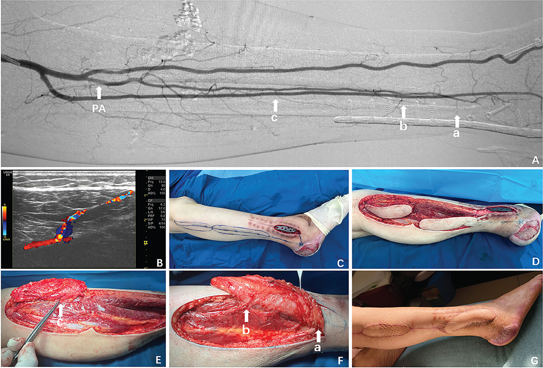
Figure 1. The patient developed a non-healing wound with exposed steel plate after surgery for a right ankle joint fracture. (A) Preoperative DSA revealed the peroneal artery (PA) and its branches a, b, and c (indicated by white arrows). (B) High-frequency ultrasound was used to track and precisely localize the perforators. (C) The wound was evaluated after debridement, and the flap was designed. (D) The flap was harvested intraoperatively. (E, F) Intraoperative exploration confirmed the presence of perforators a, b, and c (indicated by white arrows). (G) The postoperative assessment indicated that the flap survival was satisfactory.
This case showed a 66-year-old female patient who sustained a fracture of the right ankle joint due to trauma, resulting in a non-healing wound with an exposed steel plate after internal fixation surgery. Preoperative DSA and high-frequency ultrasound were used to map the course of the peroneal artery and the distribution of its perforators, allowing for precise localization. Based on the wound condition, perforators a and c were selected as pivotal points for flap rotation, and a peroneal artery perforator sequential flap was designed. The previously identified peroneal artery perforators were carefully explored, revealing perforators a, b, and c. The two flaps were then rotated respectively to cover the wound, and the secondary donor site was directly sutured. Follow-up assessments showed favorable flap esthetics and successful functional recovery of the affected limb (Figure 1).
Discussion
The skin and soft tissue in the distal lower leg and ankle are thin and have a relatively poor blood supply, resulting in limited resistance to infection and impaired healing. Injuries in this region can easily implicate bones and tendons, resulting in osteoarticular infections that complicate the treatment of these complex wounds. Traditional repair methods include pedicled flaps, neurocutaneous flaps, and free flaps. However, pedicled flaps [4] have restricted mobility due to their pedicle, making them unsuitable for covering distant wounds. Neurocutaneous flaps [5–7], such as the sural, superficial peroneal, and saphenous flaps, were born with the risk of damaging nearby nerves, potentially leading to partial sensory loss. Free flaps [8] require microsurgical techniques, which result in longer operation times, increased technical difficulty, and higher risks. PAPFs [9] have become a preferred method for repairing wounds in the distal lower leg and ankle, as they provide a reliable blood supply, primely match the texture of the surrounding skin, and dispense with the need for vascular anastomosis. However, the thin, inelastic skin and limited local tissue volume often hinder direct suturing of the donor site after harvesting, necessitating skin grafting. Unfortunately, skin grafting has its own drawbacks, including poor graft survival, scar contracture, and reduced durability postoperatively, all of which can significantly affect function and appearance. The peroneal artery perforator sequential flap [10] is a technique that utilizes a primary flap to cover the original wound and a secondary flap to repair the defect left by the primary flap’s transplantation. This method allows for direct suturing of the secondary donor site. By using the proximal leg flap for the secondary repair, tension is transferred from the primary donor site to the larger-circumference, less tense area of the proximal leg. This approach allows for direct suturing of the donor site while addressing wounds in the distal lower leg and ankle region. It also improves the appearance and function of the primary donor site, avoids the drawbacks of skin grafting, and reduces complications at the donor site. In this study, all patients showed smooth, well-textured postoperative flap appearance with color similar to that of the surrounding skin. Only linear scars were left on the secondary donor site, which did not damage the overall appearance of the limb.
The peroneal artery is the main artery supplying the lateral skin of the leg. It branches from the posterior tibial artery, descends between the flexor hallucis longus muscle and the medial side of the fibula, and terminates at the lateral malleolus. Studies [11] have shown that the distribution of perforators from the peroneal artery is generally consistent, and harvesting flaps based on clinical experience can yield favorable outcomes. However, this experience-based approach has limitations, particularly in cases with anatomical variations. Additionally, leg and ankle trauma, often resulting from violence, frequently involves vascular injuries. Absolute dependence on clinical experience without preoperative imaging evaluation may increase the risk of surgical failure. With the rapid advancement of medical imaging technology, DSA and high-frequency ultrasound are increasingly used in clinical practice, providing more precise anatomical information for surgical procedures. DSA [12, 13] is an angiographic technique that utilizes electronic computers to enable dynamic observation of the peroneal artery’s course and the distribution of its branches. High-frequency ultrasound [14, 15], known for its non-invasive, convenient, and cost-effective characteristics, provides real-time dynamic information about vessels and tissues, making it a valuable tool for vascular evaluation. It visually depicts the relationship between the branches of the peroneal artery and the surrounding tissues, allowing for precise localization of perforation points through the deep fascia. The combination of DSA and high-frequency ultrasound provides a clear and comprehensive view of the peroneal artery and its branches, facilitating accurate identification of the perforators. This enhances the visualization, precision, and customization of flap design and transplantation. In the case series, the preoperative use of DSA combined with high-frequency ultrasound guided the design and transplantation of flaps, leading to successful flap survival in all patients postoperatively, as well as satisfactory esthetic outcomes and recovery of limb function.
Some studies [16, 17] have shown that the distribution of peroneal artery perforators is in the proximal and distal thirds of the leg. Our findings showed that the peroneal artery perforators were mainly distributed in the middle segment of the fibula. The main reasons are as follows: First, the anatomical structures of the human body exhibit certain variabilities, and the distribution of peroneal artery perforators may differ among individuals. Our study sample might have included some individuals with specific anatomical characteristics, resulting in a relatively dense distribution of perforators in the middle segment. Second, the differences in research methods and samples might contribute to the varying results. The specific detection techniques and equipment (DSA combined with high-frequency ultrasound) used in our study might have been more sensitive in detecting the perforators, enabling the discovery of distribution patterns that were not fully noticed in previous studies. Regarding the consistency between DSA and high-frequency ultrasound, our findings showed a high degree of concordance between the two modalities. However, there were still three cases where the perforators were shown by DSA but not recognized by high-frequency ultrasound. We believe this discrepancy may be attributed to the higher sensitivity of DSA in detecting smaller perforators, which might not be as easily visualized using high-frequency ultrasound.
The surgical considerations for peroneal artery perforator sequential flaps are as follows: 1) It is essential to protect the sural and deep peroneal nerves during surgery to prevent sensory and motor dysfunction in the leg and foot. 2) The vascular pedicle can be placed through either an open or subcutaneous tunnel. It is necessary to ensure that the tunnel is unobstructed and loose to avoid compression, kinking, or twisting of the pedicle. 3) Moderate dissection of the perforator pedicle is advisable, with 0.5–1.0 cm of deep fascia spared around the perforator to prevent accidental damage to both the perforator and the venous vessel. 4) The skin possesses inherent elasticity and mobility, allowing for the reduction of the donor site defect width by gently pulling the defect edges. Research [18] indicates that when the width of the secondary flap is within 6 cm, proximal defects of the lower leg can be directly closed. In the flap design process, the width of the secondary flap can be planned to be 1–2 cm narrower than that of the primary donor site defect. This is followed by performing a ‘pinch test’ to ensure primary closure of the secondary donor site.
While this study achieved good clinical outcomes, it has several limitations. First, the sample size is small, comprising only seven patients. Future studies should involve larger sample sizes to improve the generalizability of the findings. Second, this study focused solely on the application of peroneal artery perforator sequential flaps. Future research could explore the utility of DSA and high-frequency ultrasound in other types of flap transplants. Additionally, as imaging technology advances, more sophisticated techniques such as 3D reconstruction and dynamic contrast enhancement could be employed in perforator flap transplants, potentially enhancing surgical precision and success rates.
In conclusion, the combination of DSA and high-frequency ultrasound in peroneal artery perforator sequential flap transplantation significantly enhances the accuracy of preoperative perforator localization, optimizes surgical design, and improves both the success rate and postoperative esthetic outcomes of flap transplantation. As imaging technology continues to evolve, this approach is expected to be increasingly utilized in various flap transplantations, providing patients with more precise and efficient treatment options.
Acknowledgments
This study was supported by the Foundation for Key Medical Specialty of Nanjing (SZDZK202001, awarded to Dr. Si-Ming Yuan), the Natural Science Foundation of Jiangsu Province, China (No. BK20231504, awarded to Dr. Si-Ming Yuan), and the Medical Project of Jinling Hospital (No. 22LCZLXJS4 awarded to Dr. Si-Ming Yuan, No. 22JCYYYB37 awarded to Qian Wang, and No. 22JCYYYB28 awarded to Ming-li Zou).
ORCID
Si-Ming Yuan  https://orcid.org/0000-0003-1380-5425
References
[1] Altinkaya A, Yazar S, Bengur FB. Reconstruction of soft tissue defects around the Achilles region with distally based extended peroneal artery perforator flap. Injury. 2021;52(7):1985–1992. https://doi.org/10.1016/j.injury.2021.04.015
[2] Uricchio M, Gupta S, Jakowenko N, Levito M, Vu N, Doucette J, et al. Computed tomography angiography versus digital subtraction angiography for postclipping aneurysm obliteration detection. Stroke. 2019;50(2):381–388. https://doi.org/10.1161/STROKEAHA.118.023614
[3] Aziz MU, Eisenbrey JR, Deganello A, Zahid M, Sharbidre K, Sidhu P, et al. Microvascular flow imaging: a state-of-the-art review of clinical use and promise. Radiology. 2022;305(2):250–264. https://doi.org/10.1148/radiol.213303
[4] Ducic I, Attinger CE. Foot and ankle reconstruction: pedicled muscle flaps versus free flaps and the role of diabetes. Plast Reconstr Surg. 2011;128(1):173–180. https://doi.org/10.1097/PRS.0b013e3182173d3a
[5] Huang Q, Wang Q, Xu Y, Ren C, Lin H, Zhang C, et al. Dual-perforator flap with wide pedicle versus sural neurocutaneous flap with peroneal artery perforator in the treatment of soft tissue defects of foot and ankle. J Foot Ankle Surg. 2023;62(1):150–155. https://doi.org/10.1053/j.jfas.2022.06.003
[6] Wang C, Yang W, Zhang F, Lineaweaver WC, Wen G, Chai Y. Superficial peroneal neurocutaneous flap for coverage of donor site defect after the combined transfer of toe and dorsal foot flap. Ann Plast Surg. 2021;86(4):440–443. https://doi.org/10.1097/SAP.0000000000002520
[7] Zhong W, Lu S, Chai Y. Distally based saphenous neurocutaneous perforator flap: a versatile donor site for reconstruction of soft tissue defects of the medial malleolar region. J Foot Ankle Surg. 2016;55(2):391–396. https://doi.org/10.1053/j.jfas.2015.02.002
[8] Ciudad P, Kaciulyte J, Torto FL, Vargas MI, Bustamante A, Chen HC, et al. The profunda artery perforator free flap for lower extremity reconstruction. Microsurgery. 2022;42(1):13–21. https://doi.org/10.1002/micr.30745
[9] Shih YJ, Wang YC, Lin CT, Chang SC, Tzeng YS, Dai NT, et al. Clinical application of the peroneal artery perforator flap for reconstruction of lateral malleolus defects. Int J Low Extrem Wounds. 2023;22(2):378–384. https://doi.org/10.1177/15347346211015828
[10] Franchi A, Fritsche E, Scaglioni MF. Sequential propeller flaps in the treatment of post-traumatic soft tissue defects of the lower limb – a case series. Injury. 2020;51(12):2922–2929. https://doi.org/10.1016/j.injury.2020.02.031
[11] Schaverien M, Saint-Cyr M. Perforators of the lower leg: analysis of perforator locations and clinical application for pedicled perforator flaps. Plast Reconstr Surg. 2008;122(1):161–170. https://doi.org/10.1097/PRS.0b013e3181774386
[12] Cang ZQ, Xu Y, Wang M, Xu MN, Yuan SM. Application of digital subtraction angiography in reconstruction of plantar forefoot with reverse medial plantar flap. Ann Plast Surg. 2021;86(5):573–576. https://doi.org/10.1097/SAP.0000000000002521
[13] Cang ZQ, Xu Y, Wang M, Xu MN, Yuan SM. Anterior tibial artery injury is not the contraindication of medial plantar flap: digital subtraction angiography evidence and clinical application. J Plast Reconstr Aesthet Surg. 2021;74(10):2512–2518. https://doi.org/10.1016/j.bjps.2021.03.054
[14] Gong J, Jia YP, Luo WD, Li CJ. Preoperative high-frequency color Doppler ultrasound assessment of the blood vessels of the fibular myocutaneous flap. J Plast Reconstr Aesthet Surg. 2022;75(11):3964–3969. https://doi.org/10.1016/j.bjps.2022.08.045
[15] Haribhakti VV, Thorat TS, Powle VR, Virarkar MK. Prospective study to analyse the efficiency of Acoustic Doppler v/s Colour Duplex Ultrasound in locating perforators for antero-lateral thigh flap in reconstructive head and neck surgery. J Plast Reconstr Aesthet Surg. 2021;74(9):2026–2033. https://doi.org/10.1016/j.bjps.2020.12.088
[16] Yoshimura M, Shimada T, Hosokawa M. The vasculature of the peroneal tissue transfer. Plast Reconstr Surg. 1990;85(6):917–921. https://doi.org/10.1097/00006534-199006000-00012
[17] Lykoudis EG, Koutsouris M, Lykissas MG. Vascular anatomy of the integument of the lateral lower leg: an anatomical study focused on cutaneous perforators and their clinical importance. Plast Reconstr Surg. 2011;128(1):188–198. https://doi.org/10.1097/PRS.0b013e3182174303
[18] Sharma M, Balasubramanian D, Thankappan K, et al. Propeller flaps in the closure of free fibula flap donor site skin defects. Ann Plast Surg. 2013;71(1):76–79. https://doi.org/10.1097/SAP.0b013e318248b813