ORIGINAL RESEARCH ARTICLE
The efficacy of immediate lymphatic reconstruction in preventing breast cancer-related lymphedema: long-term follow-up study
Ki-Jae Kima, Sun-Hyeok Kima, Seung-Pil Jungb, Eul-Sik Yoona and Jae-Ho Chunga,c
aDepartment of Plastic and Reconstructive Surgery, Korea University Hospital, Seoul, Republic of Korea; bDivision of Breast and Endocrine Surgery, Korea University Hospital, Seoul, Republic of Korea; cInstitute of Advanced Regeneration and Reconstruction, College of Medicine, Korea University, Seoul, Republic of Korea
ABSTRACT
Background: Immediate lymphatic reconstruction (ILR) has emerged as an effective intervention to reduce breast cancer-related lymphedema, which affects 2–30% of patients who undergo axillary lymph node dissection (ALND). Our previous research validated ILR’s effectiveness over 14 months, but the duration was short and warranted further study. This study provides long-term evidence of its benefits in a well-defined patient cohort.
Method: This retrospective cohort study included unilateral breast cancer patients who underwent ALND between November 2019 and February 2021 with ≥ 24 months of follow-up. Patients were grouped by whether ILR was attempted intraoperatively. Exclusion criteria were recurrence, prophylactic mastectomy, and pre-existing lymphedema. The primary outcome was lymphedema incidence at ≥ 24 months; the secondary was lymphedema-free survival. Outcomes were evaluated using Cox regression models.
Results: Among 73 patients, we analyzed ALND patients divided into a control group (n = 57, defined as no-try or failure) and an ILR group (n = 16). The pooled median follow-up was 37 months. (range, 26~47 months). Among the ILR group, 13 underwent end-to-end anastomosis and 3 end-to-side. Postoperative lymphedema was higher in the control group (24.6%) compared to the ILR group (6.3%). Multivariate Cox regression analysis showed a significantly lower hazard ratio for the ILR group (HR: 0.117, 95% CI: 0.014–0.965), emphasizing ILR’s effectiveness in reducing lymphedema risk post-ALND. Additionally, survival plots illustrating lymphedema-free survival showed a significant difference.
Conclusion: Our study emphasizes ILR’s efficacy over extended follow-up. The ILR group exhibited a lower rate of postoperative lymphedema, supporting ILR as an effective preventive measure against Breast Cancer-Related Lymphedema (BCRL) following ALND.
Level of Evidence: III
KEYWORDS: Lymphedema; axillary reverse mapping; lymphovenous anastomosis
Citation: Journal of Plastic Surgery and Hand Surgery 2025; 60: 138–143. DOI: https://doi.org/10.2340/jphs.v60.43738.
Copyright: © 2025 The Author(s). Published by MJS Publishing on behalf of Acta Chirurgica Scandinavica. This is an Open Access article distributed under the terms of the Creative Commons Attribution 4.0 International License (http://creativecommons.org/licenses/by/4.0/), allowing third parties to copy and redistribute the material in any medium or format and to remix, transform, and build upon the material, with the condition of proper attribution to the original work.
Received: 14 November 2024; Accepted: 22 April 2025; Published: 25 June 2025
CONTACT: Jae-Ho Chung cjh665@korea.ac.kr Department of Plastic and Reconstructive Surgery, Korea University Anam Hospital, 73, Korea-university-ro, Seongbuk-gu, Seoul 02841, Republic of Korea
Supplemental data for this article can be accessed online at https://doi.org/10.2340/jphs.v60.43738
Competing interests and funding: We have no potential conflicts of interest relevant to this article to report.
None
Introduction
Breast Cancer-Related Lymphedema (BCRL) affects 2–30% of patients post-surgery, particularly those undergoing lymphadenectomy. To mitigate this risk, simultaneous immediate lymphaticovenular bypass, introduced by Boccardo and Campisi in 2009 [1], has gained attention. This technique, commonly known as the Lymphatic Microsurgical Preventing Healing Approach (LYMPHA) or immediate lymphatic reconstruction (ILR), connects arm lymphatics to a collateral branch of the axillary vein. While Boccardo’s group originally introduced LYMPHA, ILR has been more widely adopted in the US since 2020, particularly by Singhal and colleagues at Harvard. ILR is now commonly used to distinguish this approach from delayed lymphatic reconstruction, which has recently emerged at some institutions, including ours.
In 2011, Boccardo published a pioneering prospective study on LYMPHA, demonstrating a marked reduction in BCRL incidence (4.34%) among treated patients compared to the untreated group (30.43%) [2]. Subsequently, a 2014 follow-up study examined 78 patients who underwent LYMPHA following ALND over 4 years. The results showed a significantly lower lymphedema rate of 4% in the 74 successfully treated patients, in stark contrast to lymphedema rates of 13–65% observed in untreated cohorts in other studies [3]. The efficacy of LYMPHA for the lower extremities was also reported in 2016. Recent meta-analyses, based on a comprehensive analysis of various studies, have confirmed ILR’s effectiveness in preventing lymphedema, demonstrating a relative risk reduction of 0.22 [4].
However, a significant 2023 study spanning 57 months, the longest follow-up to date, challenged these findings by suggesting that LYMPHA may not provide continuous long-term protection against lymphedema in ALND patients, thus igniting ongoing scientific discourse [5, 6]. Conversely, our research team published results in 2023 supporting the efficacy of ILR over a 14-month follow-up period with 26 patients. Unlike previous studies with conflicting conclusions, our research establishes a foundation for an exhaustive subsequent review of ILR procedures. Building on these initial findings, our ongoing study aims to broaden and meticulously review ILR procedures across a larger patient cohort and extended timeframe. This investigation seeks to further elucidate the sustained advantages and optimal protocols of ILR in managing BCRL.
In our previous study, patients who had underwent ALND exhibited a significantly higher hazard ratio (HR 6.582; 95% CI, 1.654–26.198, p < 0.05) as analyzed by a multivariable Cox’s hazard regression model [7]. ALND is a recognized risk factor for lymphedema, presenting an incidence rate nearly fourfold that of sentinel lymph node biopsy [8, 9]. Consequently, this study aims to conduct a detailed long-term follow-up to assess the impact of ILR on patients post-ALND over an extensive period.
Materials and method
Patients
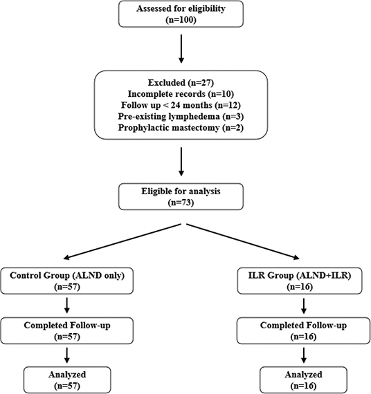
This retrospective analysis was approved by our institutional review board (protocol number 2024AN0465). Medical records, including operation details, follow-up records, and images, were thoroughly reviewed. The study included patients who underwent mastectomy with ALND for node-positive unilateral breast cancer between November 2019 and February 2021. The intervention group comprised patients who underwent ILR during ALND, while the control group included those who underwent ALND without ILR or who failed procedure during the same period. Patients were included if they had a minimum follow-up duration of 24 months. Patients were excluded if they had incomplete medical records or lacked clinical photographs, follow-up durations of less than 24 months, a history of recurrent breast cancer, prophylactic mastectomy, or pre-existing lymphedema symptoms before mastectomy. Ultimately, 16 patients who met the inclusion criteria were included in the ILR group (Figure 1).
Surgical technique
At our center, ILR is available to patients slated for ALND who consent to the procedure. Three oncologic surgeons decided the type of axillary intervention during surgery. Following axillary surgery, all ILRs after nodal extirpation were performed by the corresponding author.
Before the axillary procedure began, axillary reverse mapping (ARLM) was carried out 6 cm distal to the axilla following Thompson’s technique [10]. A total of 3cc of isosulfan blue dye was injected subdermally at 5–6 sites, with subsequent massage to aid dye migration to the axilla. After completing ALND, the arm lymphatics and an axillary vein branch were examined under a microscope. Blue lymphatics or nodes were usually identified; if not, further exploration proceeded along the expected lymphatic course. Lymphatic vessels unsuitable for anastomosis were clipped. An appropriate axillary vein branch was selected, and ILR was executed using 10-0 nylon sutures (Ethicon, Somerville, N.J.) in an end-to-end manner. Employing Boccardo’s technique, a U stitch inserted the lymphatic vessel into the vein, with perilymphatic tissues and venous walls were joined using interrupted sutures [3]. For larger veins, an end-to-side anastomosis was utilized. And the patency was verified microscopically. After anastomosis, the clamp on the lymphatic vessel was released while checking ICG flow to confirm patency. (Supplementary Figure 1, 2 , Supplementary Video).
Surveillance
Breast cancer patients at our center are routinely followed every 6 months with mammography and ultrasonography for 5 years, then annually, based on pathology. If lymphedema was suspected, patients were referred to the certified rehabilitation physician and plastic surgeon through our institutional co-management system. ILR patients are monitored every 3 months until 2 years post-surgery, then every 6 months up to 4 years if there are no abnormal signs.
Limb circumference was measured at six points: the superior margin of the upper arm, mid-upper arm, both superior and inferior elbow margins, mid-forearm, and wrist. A positive arm circumference measurement was defined as a difference of ≥2 cm at two or more locations between the affected and contralateral arms. Patients were also monitored for symptoms such as swelling and heaviness.
Postoperative lymphedema assessment additionally included bioimpedance analysis (BIA) by InBody S10 (InBody, Seoul, South Korea) at each visit and annual lymphoscintigraphy. BIA was performed in patients who underwent ILR and in selective patients with suspected lymphedema in the control group. Imaging primarily referred to lymphoscintigraphy, while ICG lymphography was selectively performed when needed. Suspected cases were referred to a single rehabilitation specialist who conducted all evaluations, while two nuclear medicine specialists interpreted lymphoscintigraphy findings.
Study design
The primary endpoint of this study was the diagnosis of lymphedema. Cases in which swelling occurred during adjuvant treatment (excluding hormone or immunotherapy) or within 6 months after the last oncologic therapy were classified as transient lymphedema, following the definition by Singhal et al. [11] If no events occurred, the observation was censored at the last follow-up visit.
The primary aim of this study was to compare the risk of lymphedema between the ILR and control groups in patients who underwent axillary lymph node dissection (ALND) for unilateral breast cancer. To assess the impact of ILR on the risk of upper extremity lymphedema, outcomes were compared between the ILR success group (n = 16) and the no-try or failure group (n = 57) over the same period. The secondary aim was to evaluate the long-term effect of ILR on lymphedema-free survival.
Failure of ILR was defined as cases where the intended anastomosis or lymphatic reconstruction did not achieve functional outcomes. This included instances where no suitable lymphatic vessels (n = 3) or adjacent veins (n = 2) were identified for anastomosis, or where lymphatic flow was insufficient despite a seemingly successful anastomosis (n = 4), as confirmed intraoperatively by ICG lymphography. Failure was determined by the absence of flow enhancement, indicating unsuccessful lymphatic drainage reconstruction due to anatomical variations, inadequate vessel caliber, or back-flow phenomena.
Statistical analyses
We conducted statistical analyses to determine the effect of ILR on the incidence of postmastectomy lymphedema, adjusting for clinicopathologic variables. Patient demographics and baseline characteristics were compared using suitable statistical tests. Continuous variables, such as the follow-up period, age, and body mass index (BMI), were analyzed with either two independent t-tests or Mann-Whitney tests, depending on the data distribution. Categorical variables, such as comorbidities, tumor grade, and type of mastectomy, were evaluated using Chi-squared tests or Fisher’s exact tests.
Postoperative outcomes following ILR, such as operative methods and lymphedema occurrences, were analyzed using Fisher’s exact tests for categorical variables. Multivariate Cox’s proportional hazards regression models were utilized to identify factors influencing lymphedema. Survival plots, based on time-to-event data, illustrated the beneficial effects of ILR. All analyses were performed using IBM SPSS Statistics and SAS, with a significance threshold set at p < 0.05.
Result
Patient demographics and baseline characteristics
Patient demographics and baseline characteristics were compared between the control group (ALND with no ILR) and the intervention group (ILR following ALND). Mean follow-up duration was 31.1 ± 13.3 months in the control group and 30.1 ± 7.2 months in the ILR group (p = 0.38). There were no significant differences in age (53.5 ± 12.0 years vs. 53.6 ± 11.2 years, p = 0.32), BMI (24.0 ± 3.7 vs. 23.9 ± 2.7, p = 0.81), or most comorbidities between the groups. A significant difference, however, was observed in tumor grade distribution (p = 0.03), with varying proportions across different grades between the groups. Additionally, the types of mastectomies differed between the two groups (p < 0.01). The ILR group had more patients who underwent breast-conserving surgery (BCS), whereas the control group had more patients who underwent mastectomy (Table 1).
| Variables | Control (ALND and no ILR) (%) | Intervention (ILR following ALND) (%) | p |
| No. of patients | 57 | 16 | |
| Follow-up period, months | 31.1 ± 13.3 | 30.1 ± 7.2 | 0.38 |
| Mean age, year | 53.5 ± 12.0 | 53.6 ± 11.2 | 0.32 |
| Mean BMI | 24.0 ± 3.7 | 23.9 ± 2.7 | 0.81 |
| Hypertension | 9 (15.8) | 5 (31.3) | 0.11b |
| Diabetes mellitus | 4 (7.0) | 0 (0) | 0.57b |
| Smoking history | 2 (3.5) | 1 (6.3) | 0.53b |
| Tumor grade | 0.03b* | ||
| 0 | 3 (5.3) | 2 (12.5) | |
| I | 7 (12.3) | 1 (6.3) | |
| II | 39 (68.4) | 6 (37.5) | |
| III | 8 (14.0) | 7 (43.7) | |
| Mastectomy type | < 0.01b* | ||
| MRM | 37 (64.9) | 10 (62.4) | |
| SSM | 8 (14.0) | 1 (6.3) | |
| NSM | 12 (21.1) | 1 (6.3) | |
| BCS | 0 (0) | 4 (25.0) | |
| Reconstruction | 21 (36.8) | 4 (25.0) | 0.38a |
| Autologous flap | 11 (19.3) | 1 (6.3) | |
| DIEP | 9 (15.8) | 1 (6.3) | |
| LD | 2 (3.5) | 0 | |
| Implant-based | 11 (19.3) | 3 (18.7) | |
| Chemotherapy | 53 (93.0) | 14 (87.5) | 0.61b |
| Neoadjuvant | 35 (61.4) | 10 (62.4) | |
| Adjuvant | 25 (43.9) | 10 (62.4) | |
| Radiotherapy | 27 (47.4) | 10 (62.4) | 0.28a |
| Hormonal therapy | 25 (43.9) | 3 (18.7) | 0.07a |
| LR: lymphatic reconstruction; BMI: body mass index; MRM: modified radical mastectomy; SSM: skin sparing mastectomy; NSM: nipple sparing mastectomy; BCS: breast conserving surgery; DIEP: deep inferior epigastric perforator flap; LD: latissimus dorsi; ILR: immediate lymphatic reconstruction. Patients underwent implant-enhanced LD flap were classified to LD flap. aChi-square analysis; bFisher’s exact test; Above p-value was calculated by chi-square test or Fischer exact test or t-test or Mann-Whitney test for the difference between ‘immediate lymphatic reconstruction’ group and ‘delayed lymphatic reconstruction’ group. *Statistical significance. |
|||
Postoperative outcomes of immediate lymphatic reconstruction
In the intervention group, ILR predominantly utilized end-to-end anastomosis (81.3%). Transient lymphedema was minimal in both groups, with two cases (3.5%) observed in the control group and one case (6.3%) in the ILR group, respectively. Postoperative lymphedema was less common in the ILR group (6.3%) compared to the control group (24.6%), although the difference was not statistically significant (p = 0.17) (Table 2). Table 3 displays individual patient characteristics of 15 cases with clinically diagnosed postoperative lymphedema.
| Variables | Control (ALND and no ILR) (%) | Intervention (ILR following ALND) (%) | p |
| No. of patients | 57 | 16 | |
| Operative method | N/A | < 0.01a* | |
| End-to-end anastomosis | 13 (81.3) | ||
| End-to-side anastomosis | 3 (18.7) | ||
| Transient lymphedema | 2 (3.5) | 1 (6.3) | 0.22a |
| Postoperative lymphedema | 14 (24.6) | 1 (6.3) | 0.17b |
| LR: lymphatic reconstruction; ILR: immediate lymphatic reconstruction. bFisher’s exact test; *Statistical significance. |
|||
Risk factors for postoperative lymphedema
Multivariate Cox’s proportional hazards regression analysis was used to assess the risk of postmastectomy lymphedema. Variables such as age, BMI, comorbidities (including hypertension and diabetes), smoking history, tumor grade, type of mastectomy, reconstructive surgery and adjuvant therapies (chemotherapy, radiotherapy) were incorporated into the model. In this multivariate analysis, ILR was associated with a significantly reduced risk of developing postmastectomy lymphedema (HR 0.117, p = 0.046). The final model, derived using stepwise variable selection, identified ILR as a protective factor (p = 0.046) and radiotherapy as a significant risk factor (p = 0.004) for postoperative lymphedema (Table 4). Survival plots depicted the time-to-event data for lymphedema occurrence, showing a lower cumulative incidence in the ILR group compared to the control group, with the discrepancy increasing over time (Figure 2).
| Variables | Hazard ratio | p | 95% Hazard ratio confidence | |
| Limits | ||||
| Hypertension (+) versus Hypertension (-) | 3.664 | 0.097a | 0.792 | 16.955 |
| Radiotherapy (+) versus Radiotherapy (-) | 11.835 | 0.004* | 1.967 | 31.84 |
| ILR versus No intervention | 0.117 | 0.046* | 0.014 | 0.965 |
| ALND: axillary lymph node dissection; SLND: sentinel lymph node dissection; ILR: immediate lymphatic reconstruction. aBorderline significance; *Statistical significance. We used multivariable cox’s hazard survival model for statistical analysis. Variables including age, BMI, comorbidity (hypertension, diabetes), smoking, tumor grade, mastectomy type, lymph node dissection type, reconstructive type, chemotherapy, radiotherapy, hormonal therapy, and ILR success were fitted to this model, and stepwise selection was performed. |
||||
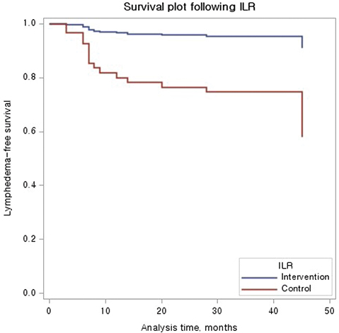
Figure 2. Survival plot following immediate lymphatic reconstruction. Survival plots demonstrates a lower comulative incidence of lymphedea in the ILR group, compared to the control group.
Discussion
Lymphedema remains a significant complication following breast cancer treatment, particularly after axillary dissection and radiation therapy [12]. It is challenging to treat, requiring continuous monitoring and follow-up for both physicians and patients. ILR aims to prevent lymphedema by establishing a lymphovenous anastomosis immediately after axillary surgery. Prior studies, including Boccardo’s 2014 follow-up, demonstrated significantly lower lymphedema rates among ILR-treated patients. While multiple institutions have validated ILR’s preventive efficacy, its long-term effectiveness remains debated due to limited comprehensive studies.
Recent studies, including those by Boccardo, Singhal, and Rhode, have examined ILR efficacy with follow-up periods ranging from 17 to 57 months. Boccardo reported a lymphedema incidence of 4.05% in the ILR group, and Singhal reported 9% in their ILR cohort. In contrast, Rhode’s study, although it included a control group, found no statistically significant difference in lymphedema incidence between the ILR and control groups [3, 6, 13]. Our study, with an extended follow-up, observed a lymphedema rate of 6.3% in the ILR group, aligning with Boccardo and Singhal’s results, reinforcing the preventive role of ILR. The control group exhibited an incidence of 24.6%, similar to recent literature (14.1–29.4%) [3, 6, 13–15].
A critical issue when evaluating the preventive impact of ILR is the distinction between transient and persistent lymphedema. Several studies have proposed definitions for a distinct category of transient lymphedema [16–18]. In this study, we adopted the definition by Singhal et al., which classifies swelling occurring during adjuvant treatment (excluding hormone or immunotherapy) or within 6 months of completing oncologic therapy as transient lymphedema [11]. This classification is essential to avoid overestimating lymphedema incidence and to better isolate the true long-term effects of ILR. For instance, Feldman et al. noted that 8.3% of their patients exhibited persistent lymphedema, a figure that rose to 12.5% with the inclusion of cases classified as transient lymphedema [16]. In our study, we observed one instance of transient lymphedema in the ILR group, which spontaneously resolved and did not progress to persistent lymphedema. Consequently, our final analysis included only one case of persistent lymphedema in the ILR group.
In this study, the diagnosis of lymphedema was based on objective criteria, including imaging findings and limb circumference differences, and was confirmed by a certified rehabilitation physician. This strategy minimized reliance on subjective symptoms such as heaviness or numbness, which can be nonspecific and variable. Given the inherent challenges in defining and diagnosing lymphedema, it is essential to control for variables associated with temporary symptoms and apply rigorous statistical methods to accurately evaluate the preventive efficacy of ILR. This methodological rigor ensures a more accurate assessment of ILR’s effect on reducing lymphedema incidence in patients undergoing ALND.
Key insights from the patient demographic data in our study demand attention. A systematic review by Mohamed A. identified several demographic risk factors, such as BMI, tumor size, breast size, type of axillary surgery, and adjuvant therapy, as potential influencers on the development of lymphedema [17]. In our study, although most patient demographics showed no significant differences, notable variations were observed in tumor grade and mastectomy type between the ILR and control group. The control group had a higher proportion of patients with tumor grade II, while the ILR group included a larger proportion of patients with the more severe grade III tumors. Prior research has indicated that higher tumor grades and larger tumor sizes may elevate the risk of lymphedema. Despite these factors, our results showed that postoperative lymphedema was less frequent in the ILR group, suggesting that ILR status significantly influences the incidence of lymphedema more than tumor grade.
Regarding the type of mastectomy, both groups exhibited similar rates of modified radical mastectomy (MRM). However, BCS was significantly more prevalent in the ILR group (25%) compared to the control group (0%). Previous research by Inger-Lise Nesvold indicated that, for the same tumor stage, the BCS group experienced significantly lower lymphedema rates compared to the mastectomy group (24% vs. 7%, p < 0.01). Conversely, a 2010 study by Renata Freitas-Silva reported no significant difference in lymphedema incidence between BCS and various mastectomy types (MRM 12%, BCS 18%, p = 0.66), [19] illustrating the continued debate in previous research findings. While we matched groups based on the presence of ALND, it is pertinent to note that BCS is generally considered less invasive than mastectomy, and despite lacking a consensus yet, it is thought to cause less lymphedema. In our study, statistical analysis did not identify a significant association between surgical type and lymphedema incidence. However, we acknowledge that the higher rate of BCS in the ILR group may serve as a potential confounder. Future research is needed to focus more on this relationship, particularly in relation to radiotherapy status.
Our surgical approach initially utilized the lymphaticovenous Implantation (LVI) technique; however, we have since transitioned to supermicrosurgical lymphovenous anastomosis (LVA) as our preferred method. Since this study analyzes data from 2019 to 2021, it reflects our early surgical experience when LVI was predominantly used. Regardless of the technique, we ensured patency assessment through intraoperative ICG lymphography, which likely contributed to higher patency rates compared to cases performed without ICG confirmation [20–22].
This study acknowledges several potential limitations that warrant attention. First, the sample size is relatively small, and the diversity in terms of gender and racial representation is limited, which affects the generalizability of the findings [14, 23, 24, 25]. Second, differences in lymphedema diagnostic criteria compared to other studies may pose challenges in comparing and interpreting results, thereby hindering standardization and comparability of outcomes. Most studies identify lymphedema by the presence of symptoms together with either in (1) an increase of L-Dex by 10 units or (2) a volume change of more than 10%. However, the availability of these diagnostic tools (perometry, L-dex) is limited, as they are not accessible in the author’s country. Third, variations in the measurement location can lead to inconsistencies in identifying and categorizing lymphedema cases. Addressing these limitations could improve the reliability and applicability of future research in this field. Another limitation of this study is the potential for selection bias, as the decision to perform ILR was made intraoperatively by a small group of oncologic surgeons. This may have introduced variability in patient selection and led to differences in baseline characteristics between groups, potentially influencing the observed lymphedema outcomes. Additionally, as ILR was only performed in patients who had provided preoperative consent, the study population was inherently preselected. These factors should be considered when interpreting the results.
Finally, breast reconstruction was not initially categorized as a distinct factor in our demographics analysis. Upon reviewing the data, we found that all autologous reconstructions were immediate, while implant-based reconstructions were exclusively immediate direct-to-implant (DTI) procedures. Due to the small sample size, further subgroup analysis (e.g. reconstruction type or timing) was not feasible. However, given previous studies suggesting a potential protective effect of breast reconstruction on lymphedema, future research should incorporate a more detailed analysis of reconstruction type and timing to better assess its impact on lymphedema risk.
Conclusion
Based on our study and the existing literature, ILR appears to be effective in reducing secondary lymphedema following axillary surgery for breast cancer. Our results indicate a significant reduction in lymphedema incidence with ILR compared to conventional treatments. However, challenges persist in standardizing diagnostic criteria and ensuring consistent interpretation of results across studies. Future research should focus on larger, multicenter studies with extended follow-up periods to enhance statistical reliability and generalizability.
Ethical approval
Not required
References
[1] Boccardo F, Campisi C, De Cian F, et al. Lymphedema microsurgical preventive healing approach: a new technique for primary prevention of arm lymphedema after mastectomy. Ann Surg Oncol. 2009; 16: 703–708. https://doi.org/10.1245/s10434-008-0270-y
[2] Boccardo F, Campisi C, Friedman D, et al. Surgical prevention of arm lymphedema after breast cancer treatment. Ann Surg Oncol. 2011; 18: 2500–2505. https://doi.org/10.1245/s10434-011-1624-4
[3] Boccardo F, Campsisi C, De Cian F, et al. Lymphatic microsurgical preventing healing approach (LYMPHA) for primary surgical prevention of breast cancer-related lymphedema: over 4 years follow-up. Microsurgery. 2014; 34: 421–424. https://doi.org/10.1002/micr.22254
[4] Temple-Oberle C, Fraser WK, Platt A, et al. Immediate lymphatic reconstruction during axillary node dissection for breast cancer: a systematic review and meta-analysis. Plast Reconstr Surg Glob Open. 2022; 10: e4291. https://doi.org/10.1097/GOX.0000000000004291
[5] Levy AS, Murphy AI, Ishtihar S, et al. Lymphatic microsurgical preventive healing approach for the primary prevention of lymphedema: a 4-year follow-up. Plast Reconstr Surg. 2023; 151(2): 413–420. https://doi.org/10.1097/PRS.0000000000009857
[6] Melisa D, Singhal D, Shillue K, et al. A 4-year institutional experience of immediate lymphatic reconstruction. Plast Reconstr Surg. 2023; 152(5): 773–778. https://doi.org/10.1097/PRS.0000000000010381
[7] Chung JH, Kwon S-H, Jung S-P, et al. Assessing the preventive effect of immediate lymphatic reconstruction on the upper extremity lymphedema. Gland Surg. 2023; 12(3): 334–343. https://doi.org/10.21037/gs-22-554
[8] Disipio T, Rye S, Newman B, et al. Incidence of unilateral arm lymphoedema after breast cancer: a systematic review and meta-analysis. Lancet Oncol. 2013; 14(6): 500–501. https://doi.org/10.1016/S1470-2045(13)70076-7
[9] McLaughlin SA, Wright MJ, Morris KT, et al. Prevalence of lymphedema in women with breast cancer 5 years after sentinel lymph node biopsy or axillary dissection: objective measurements. J Clin Oncol. 2008; 26(32): 5213–9. https://doi.org/10.1200/JCO.2008.16.3725
[10] Thompson M, Korourian S, Henry-Tilman R, et al. Axillary reverse mapping (ARM): a new concept to identify and enhance lymphatic preservation. Ann Sug Oncol. 2007; 14(6): 1890–5. https://doi.org/10.1245/s10434-007-9412-x
[11] Johnson AR, Fleishman A, Granoff SD, et al. Evaluating the impact of immediate lymphatic reconstruction for the surgical prevention of lymphedema. Plast Reconstr Surg. 2021; 147(3): 373–381. https://doi.org/10.1097/PRS.0000000000007636
[12] Pressman PI. Surgical treatment and lymphedema. Cancer. 1998; 83(12 Suppl American): 2782–2787. https://doi.org/10.1002/(SICI)1097-0142(19981215)83:12B+%3C2782::AID-CNCR26%3E3.0.CO;2-2
[13] Levy AS, Rhode CH, et al. Lymphatic microsurgical preventive healing approach for primary prevention of lymphedema : a 4-year follow up. Plast Reconstr Surg. 2023; 151(2): 413–420. https://doi.org/10.1097/PRS.0000000000009857
[14] Weinstein B, Le NK, Robertson E, et al. Reverse lymphatic mapping and immediate microsurgical lymphatic reconstruction reduced early risk of breast cancer-related lymphedema. Plast Reconstr Surg. 2022; 149(5): 1061–1069. https://doi.org/10.1097/PRS.0000000000008986
[15] Magnus J, Saeg F, Meade A, et al. Immediate lymphatic reconstruction for prevention of secondary lymphedema: a meta-analysis. J Plast Reconstr Aesthet Surg. 2022; 75(3): 1130–1141. https://doi.org/10.1016/j.bjps.2021.11.094
[16] Feldman S, Bansil H, Ascherman J, et al. Single institution experience with lymphatic microsurgical preventive healing approach (LYMPHA) for the primary prevention of lymphedema. Ann Surg Oncol. 2015; 22(10): 3296–3301. https://doi.org/10.1245/s10434-015-4721-y
[17] Mohamed A, Mohamed E, Sherif M, et al. Breast lymphedema after conservative breast surgery: an up-to-date systematic review. Clin Breast Cancer. 2020; 21(3): 156–161. https://doi.org/10.1016/j.clbc.2020.11.017
[18] Inger-Lise N, Dahl AA, Løkkevik E, et al. Arm and Shoulder morbidity in breast cancer patients after breast-conserving therapy versus mastectomy. Acta Oncol. 2008;47;835–42. https://doi.org/10.1080/02841860801961257
[19] Freitas-Silva R, Conde DM, De Freitas-Júnior R, et al. Comparison of quality of life, satisfaction with surgery and shoulder-arm morbidity in breast cancer survivors submitted to breast-conserving therpay or mastectomy followed by immediate breast reconstruction. Clinical Science. 2010; 65(8): 781–787. https://doi.org/10.1590/S1807-59322010000800009
[20] Pafitanis G, Narushima M, Yamamoto T, et al. Insights to establish early learning curve in clinical supermicrosurgery. Microsurgery. 2019; 39(1): 100–101. https://doi.org/10.1002/micr.30401
[21] Ishiura R, Yamamoto T, Saito T, et al. Comparison of lymphovenous shunt methods in a rat model: supermicrosurgical lymphaticovenular anastomosis versus microsurgical lymphaticovenous implantation. Plast Reconstr Surg. 2017; 139(6): 1407–1413. https://doi.org/10.1097/PRS.0000000000003354
[22] Brahma B, Yamamoto T, Panigoro SS, et al. Supermicrosurgery lymphaticovenous and lymphaticolymphatic anastomosis: technical detail and short-term follow-up for immediate lymphatic reconstruction in breast cancer treatment-related lymphedema prevention. J Vasc Surg Venous Lymphat Disord. 2024; 12(4): 101863. https://doi.org/10.1016/j.jvsv.2024.101863
[23] Nicole K, Weinstein B, Parikh J, et al. Immediate lymphatic reconstruction in 77 consecutive breast cancer patients: 2-year follow up. J Reconstr Microsrug. 2024; 40(04); 262–267. https://doi.org/10.1055/a-2153-2203
[24] Corridi M, Mehrara B, Dayan J, Bloomfield E, et al. Efficacy of immediate lymphatic reconstruction to decrease incidence of breast cancer-related lymphedema. Ann Surg. 2023; 278(4): 630–637. https://doi.org/10.1097/SLA.0000000000005952
[25] Hahamoff M, Gupta N, Munoz D, et al. A lymphedema surveillance program for breast cancer patients reveals the promise of surgical prevention. J Surg Res. 2019; 244; 604–611. https://doi.org/10.1016/j.jss.2017.10.008