ORIGINAL RESEARCH ARTICLE
Results of finger extension reconstruction in hemiplegic patients after stroke by transferring the flexor carpi radialis branch to the posterior interosseous nerve
Sihan Hu, MDa#, Lingkang Zhu, MDb#, Xuanyu Zhao, MDb#, Wanling Zheng, MDa, Guobao Wang, MD, PhDc, Yundong Shen, MD, PhDa,b, Yan-Qun Qiu, MDa,b* and Wendong Xu, MD, PhDa,b,d,e
aNational Clinical Research Center for Aging and Medicine, Department of Hand Surgery, Huashan Hospital, Fudan University, Shanghai, China; bDepartment of Hand and Upper Extremity Surgery, Jing’an District Central Hospital, Fudan University, Shanghai, China; cDepartment of Plastic and Cosmetic Surgery, Tongji Hospital, Tongji University School of Medicine, Shanghai, China; dInstitute of Brain Science, State Key Laboratory of Medical Neurobiology and Collaborative Innovation Center for Brain Science, Fudan University, Shanghai, China; eResearch Unit of Synergistic Reconstruction of Upper and Lower Limbs after Brain Injury, Chinese Academy of Medical Sciences, Shanghai, China
ABSTRACT
Purpose: Contralateral C7 nerve transfer (CC7) surgery has demonstrated success in restoring upper limb motor function after central nervous system injuries. However, deficits in finger extension limit patient independence. This study evaluates a nerve transfer that transfers the motor branch of the flexor carpi radialis (FCR) to the posterior interosseous nerve (PIN) to improve finger extension.
Methods: Two patients with chronic brain injuries, who showed minimal finger extension recovery for over 1 year after CC7 surgery, were enrolled. Both patients then underwent the nerve transfer procedure. Upper extremity motor function was measured using the Fugl-Meyer upper extremity scale (UEFM), and changes in muscle tone were quantified with the Modified Ashworth Scale (MAS). Regular follow-up evaluations were conducted over an 18-month postoperative period to monitor motor recovery and spasticity.
Results: Within the first postoperative month, both patients exhibited significant improvements in spasticity. Although a minor rebound occurred in the second month, spasticity levels stabilized in subsequent evaluations. By 18 months after surgery, one patient regained functional finger extension, underscoring the potential efficacy of the procedure. Importantly, wrist flexion, governed by the flexor carpi radialis, remained unaffected throughout recovery.
Conclusion: FCR-to-PIN nerve transfer alleviates spasticity and partially restores finger extension in patients with limited recovery following contralateral C7 nerve transfer. Although full motor recovery was not achieved, these findings offer promising clinical implications. Overall, the results support the procedure’s value in clinical practice. Further studies with larger cohorts are needed to confirm these results and elucidate underlying mechanisms.
KEYWORDS: Hemiplegia; nerve transfer; reinnervation; paralysis; stroke
Citation: Journal of Plastic Surgery and Hand Surgery 2025; 60: 146–152. DOI: https://doi.org/10.2340/jphs.v60.44128.
Copyright: © 2025 The Author(s). Published by MJS Publishing on behalf of Acta Chirurgica Scandinavica. This is an Open Access article distributed under the terms of the Creative Commons Attribution 4.0 International License (http://creativecommons.org/licenses/by/4.0/), allowing third parties to copy and redistribute the material in any medium or format and to remix, transform, and build upon the material, with the condition of proper attribution to the original work.
Received: 20 February 2025; Accepted: 9 June 2025; Published: 11 August 2025
CONTACT: Yan-Qun Qiu yanqunqiu@fudan.edu.cn Department of Hand and Upper Extremity Surgery, Jing’an District Central Hospital, Fudan University, Shanghai, China
#Sihan Hu, Lingkang Zhu and Xuanyu Zhao contributed equally to this work.
Competing interests and funding: The authors declare no conflicts of interest/competing interests.
Introduction
Stroke is the second leading cause of death worldwide, accounting for 11% of all global deaths (approximately 55.4 million annually) [1]. Among stroke survivors, roughly one-third experience persistent motor dysfunction, which significantly diminish their quality of life [2]. Various interventions, such as botulinum toxin type A injections, muscle lengthening procedures, and tendon transfer surgeries, have been developed to address post-stroke sequelae [3–5]. These approaches primarily aim to reduce spasticity, alleviate contractures, and restore muscle balance to improve motor function. However, these muscle-targeted treatments often fall short of achieving long-term functional recovery, likely due to their inability to effectively address the neurological damage underlying abnormal spasticity [6–8].
To tackle neurological impairments, selective dorsal rhizotomy and highly selective muscle branch neurotomy have been increasingly employed in clinical practice. The distal ulnar intrinsic release procedure reduces intrinsic tightness; however, its benefits in enhancing function are limited [9]. Although these surgeries effectively reduce spasticity, their impact on motor function improvement is limited because central motor control remains compromised poststroke [10, 11]. To address this challenge, Xu et al. introduced a novel surgical approach involving contralateral C7 (CC7) nerve transfer from the unaffected side to the affected C7 nerve, aiming to improve upper limb motor function [12]. CC7 surgery significantly enhances motor recovery in poststroke hemiplegic patients by redirecting the central control of the spastic limb to the healthy hemisphere [12–14].
Nevertheless, clinical observations reveal that some patients continue to experience finger extension impairments following CC7 transfer. This phenomenon is primarily attributed to an imbalance between spastic flexor muscles and weakened extensor muscles. Similar finger extension deficits are commonly observed in brachial plexus injuries due to denervation of the extensor muscles. In cases of traditional brachial plexus injuries, Mackinnon et al. applied the principle of muscle synergy to perform localized nerve transfers, effectively improving upper limb dysfunction caused by radial and median nerve damage. Recent nerve transfer studies provide key anatomical insights supporting selective transfers. Kang et al. demonstrated that dual nerve transfers—spinal accessory nerve to suprascapular nerve combined with a partial median or ulnar nerve transfer to the axillary nerve—can significantly improve shoulder function in C5-7 avulsion injuries even when conventional donor nerves are compromised [15]. Similarly, Kaiser et al. confirmed that direct dorsal transfer of the spinal accessory nerve through a bony canal under the scapular spine to the infraspinatus branch is anatomically feasible [16]. These studies highlight the importance of precise anatomical matching and minimal reinnervation distance. To restore finger extension after radial nerve injury, Mackinnon et al. proposed alternative strategies based on the principle of muscle synergy, which contributes to subsequent cortical integration [3, 17, 18]. The muscular branch of the flexor digitorum superficialis (FDS) was transferred to the extensor carpi radialis brevis (ECRB), while the muscular branch of the flexor carpi FCR or palmaris longus (PL) was transferred to the PIN [3, 18], resulting in excellent results in the treatment of severe lesions of the radial nerve [19]. Additionally, in some patients with spinal cord injuries, it has also been reported that the supinator branch of the radial nerve was transferred to the PIN for finger extension reconstruction [20, 21].
Based on this, we innovatively propose local nerve transfer to relieve spasm in patients after CC7 surgery by allowing the C7 nerve from the healthy side to assume control of key muscles, and autonomous movement can be achieved. This technique involves the transfer of functional muscle branches to the PIN to facilitate functional recovery and assist cortical integration. Such procedures have shown promising results in treating finger extension impairments caused by severe radial nerve injuries and spinal cord injuries.
Anatomical and electrophysiological studies indicate that the radial FCR is primarily innervated by the C7 nerve [22, 23]. For stroke patients with persistent finger extension impairments after CC7 transfer, transferring the FCR muscle branch to the PIN may serve as an effective adjunctive surgical option. This procedure theoretically reduces the tension of the spastic flexors while activating the PIN through neural reconstruction, thereby enhancing the independent control of the extensors. Moreover, transferring the FCR branch does not significantly impair the wrist flexion function.
In this study, we report two cases of stroke patients who exhibited persistent finger extension impairments after CC7 transfer and were treated with FCR-to-PIN nerve transfer. The patients were followed for 12 months to evaluate the functional recovery and assess the feasibility and efficacy of this novel adjunctive surgery in improving finger extension function poststroke.
Materials and methods
This study was a retrospective case analysis involving two patients with sequelae of brain hemorrhage. One patient suffered from a right brain hemorrhage, and the other had a ruptured left arteriovenous malformation, both resulting in hemiplegia. Each patient underwent contralateral C7 to C7 cross-nerve transfer (CC7) surgery 1 year prior. While CC7 transfer significantly improved some upper limb functions, both patients exhibited persistent finger extension impairments that severely impacted their daily living activities. The time from injury to the current surgical intervention was 3 and 9 years, respectively, reflecting the typical characteristics of patients with chronic brain hemorrhage sequelae.
Preoperative and postoperative clinical evaluations
Systematic clinical evaluations were performed for all patients both preoperatively and postoperatively. The assessments included the Fugl-Meyer Upper Extremity Scale (UEFM) to evaluate the recovery of motor function in the affected upper limb. The UEFM, a widely recognized clinical tool, was used to accurately quantify motor function levels. In addition to upper limb function assessment, muscle tone evaluations were conducted using the Modified Ashworth Scale (MAS). The MAS was employed to measure muscle tone and spasticity in the affected upper limb, with a specific focus on changes in flexor and extensor muscle tension.
Follow-up and postoperative monitoring
Patients were followed for 18 months postoperatively to dynamically assess functional improvement. Follow-up evaluations were conducted at 1, 2, 4, 12, and 18 months after surgery. These evaluations included repeated UEFM and MAS scoring to monitor clinical functional recovery. Both patients underwent pre- and postoperative nerve conduction studies to assess the effectiveness of nerve reconstruction. Additionally, one patient underwent pre- and postoperative transcranial magnetic stimulation (TMS) studies to evaluate central nervous system reorganization.
Data analysis
Clinical scores and neurophysiological indicators were compared between the preoperative and postoperative follow-up periods to evaluate the efficacy of the surgical intervention in restoring finger extension function. Results were presented in tabular format (see Table 1).
Case 1
The first patient was a 48-year-old man who presented to our center for limb function reconstruction due to sequelae of right-sided brain hemorrhage. Two years prior, the patient had undergone emergency treatment for a sudden right-sided cerebral hemorrhage. He underwent contralateral C7 to C7 cross-nerve transfer (CC7) surgery to improve upper limb function 1 year ago. Despite 12 months of routine postoperative rehabilitation, significant improvement was observed in shoulder joint function, but no further progress was noted in finger extension, suggesting a plateau in functional recovery.
The patient exhibited severe spasticity in the left wrist, metacarpophalangeal joints, and interphalangeal joints. His MAS scores were 4/4 for the elbow, 4/4 for the wrist, 3/4 for the thumb, and 4/4 for the second to fifth digits, indicating pronounced spasticity in the left upper limb. The Fugl-Meyer UEFM score was 4/66, reflecting a severe limitation in overall upper limb motor function. Despite the marked spasticity in the wrist, the patient was unable to perform active wrist flexion, indicating a significant imbalance between extensor and flexor muscle functions.
Neurophysiological evaluations revealed that stimulation of the contralateral C7 nerve elicited motor action potentials in the paralyzed radial FCR but failed to generate any detectable motor action potentials in the finger extensor muscles. This finding indicated a complete lack of neural innervation in the extensor muscle group. Furthermore, TMS assessments demonstrated that stimulation of the contralateral (healthy) hemisphere failed to evoke motor action potentials in the paralyzed finger extensors, suggesting that central motor control had not established an effective functional connection.
Based on the clinical presentation and neurophysiological findings, the patient’s finger extension impairment was attributed to severe muscular imbalance and a lack of neural innervation. These observations indicated that the patient might benefit from additional nerve transfer surgery to address these deficits.
Case 2
The second patient was a 19-year-old male who developed right-sided hemiplegia following a left-sided brain hemorrhage 7 years prior. After the hemorrhage, the patient underwent systematic rehabilitation therapy. However, by 1 year poststroke, recovery of right-sided limb function was limited, particularly hand function, which severely impacted his independence in daily activities.
At the time of assessment, the patient exhibited mild to moderate spasticity in the right wrist, metacarpophalangeal joints, and interphalangeal joints. The MAS scores were 2/4 for the elbow, 2/4 for the wrist, 2/4 for the thumb, and 1+/4 for the second to fifth fingers, indicating significantly lower spasticity compared with the first case. Functional assessment with the Fugl-Meyer UEFM yielded a score of 58/66, suggesting relatively good recovery of upper limb motor function. Notably, the patient retained the ability to actively flex and extend the wrist, consistent with preserved muscle strength and control in the wrist flexors and extensors.
Neurophysiological examination revealed that stimulation of the contralateral C7 nerve elicited motor action potentials in the paralyzed radial FCR, indicating relatively intact neural innervation of this muscle group. However, no motor action potentials were detected in the finger extensor muscles on the affected side, indicating a significant loss of neural innervation in these muscles.
Compared with the first case, this patient exhibited less severe spasticity and retained partial wrist function. However, significant impairment in finger extension persisted. This suggests that while central and peripheral neural recovery was partially achieved, the extensor muscle group remained insufficiently reinnervated. Targeted interventions, such as nerve transfer surgery, could potentially enable more precise recovery of hand motor function in this patient.
Surgical procedures
All surgical procedures were performed under general anesthesia with the use of a pneumatic tourniquet to ensure a clear operative field. An S-shaped incision was made anterior to the elbow joint, and the soft tissue on the medial side of the biceps tendon was dissected to expose the deeper structures of the median nerve. The branches of the median nerve, including those innervating the pronator teres, the radial FCR, and PL, were identified and marked with visible dyes to protect the main trunk of the nerve (Figure 1).
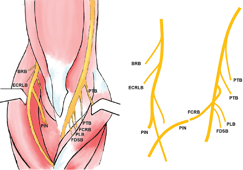
Figure 1. Schematic representation illustrates our technique.
AIN: anterior interosseous nerve; BRB: brachioradialis branch; ECRLB: extensor carpi radialis longus branch; FCRB: flexor carpi radialis brevis; FDSB: flexor digitorum superficialis branch; PTB: pronator teres branch; PLB: palmar longus branch.
The brachioradialis muscle was retracted radially to expose the intermuscular space between the brachioradialis and the FCR. The muscle branch of the FCR was carefully isolated up to its entry point. Functional branches were confirmed intraoperatively using electromyography to ensure the presence of robust compound muscle action potentials (CMAP).
Next, the radial nerve was exposed by dissecting the intermuscular plane between the biceps and brachioradialis muscles. The supinator tunnel was identified, and the PIN was traced to its three distal branches. The PIN was sharply transected at the distal end of the supinator tunnel, with local application of lidocaine to minimize postoperative pain. The length of the nerves was measured to determine the optimal transfer route from the FCR branch to the PIN. The FCR branch was transected at its entry point, providing a length of approximately 6 cm, which could be extended by an additional 4.5 cm with the inclusion of the PL branch (Figure 2).
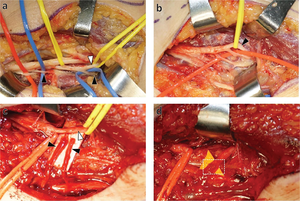
Figure 2. Intraoperative photograph illustration. (a) Identification of the median nerve within the internal bicipital canal. The first branch is directed toward the pronator teres muscle (red rubber bands), followed by two branches to the flexor carpi radialis (FCR) muscles (black arrowhead). One branch fully enters the flexor carpi radialis muscles, while the other branch has a sub-branch that includes a portion of the palmaris longus muscle (PL) (white arrowhead). (b) Identification of the radial nerve and its branches around the supinator muscle canal. In this case, there are two branches of the supinator muscle (red rubber bands) appearing before the posterior interosseous nerve (black arrowhead). (c) Identification of the FCR branches and PIN. It shows two branches to the flexor carpi radialis (FCR) muscles (black arrowhead) and the posterior interosseous nerve (white arrowhead). (d) Intraoperative picture shows the coaptation of the FCR motor branch transferred to the PIN (white-dotted box).
Under an operating microscope, the distal end of the FCR branch was sutured to the PIN in a tension-free, end-to-end fashion, ensuring a secure and precise anastomosis.
Postoperatively, the patient’s elbow was immobilized in a flexed position for 4 weeks to avoid tension or compression in the nerve transfer region. Early rehabilitation was initiated to support nerve regeneration and functional recovery. The entire surgical process emphasized microsurgical precision and intraoperative protection of the nerves and surrounding structures to achieve optimal surgical outcomes.
Ethics
The study was conducted in accordance with the Declaration of Helsinki and approved by the Institutional Review Board of Jing’an District Central Hospital. Informed consent was obtained from all subjects involved in the study.
Results
Both surgeries were successfully completed with an average duration of 2 h. Intraoperative monitoring indicated stable vital signs, including blood pressure, heart rate, respiration, and body temperature, without any abnormal fluctuations. Postoperatively, the patients experienced only mild pain, with no evidence of infection, bleeding, or other complications. All discomfort resolved within 3 months. Wrist flexion functionality remained unaffected by the surgery, and no long-term complications were observed during the 18-month follow-up, confirming the safety and feasibility of the procedure.
Spasticity and functional improvements
Postoperative follow-up revealed a reduction in upper limb spasticity. By 2 weeks after surgery, the first patient showed improvements in MAS scores, with reductions from 4/4 to 3/4 in the elbow, from 4/4 to 2/4 in the wrist, and from 4/4 to 3/4 in the thumb and fingers. The UEFM score increased from 4/66 to 7/66. Similarly, the second patient exhibited a decrease in MAS scores, with the elbow improving from 2/4 to 0/4, the wrist from 2/4 to 1/4, the thumb from 2/4 to 1/4, and the fingers from 1+/4 to 0/4. The UEFM score for this patient increased from 58/66 to 60/66 (Table 2).
At the 1-month follow-up, there was a temporary recurrence of spasticity. The first patient’s MAS scores returned to preoperative levels (4/4 for the elbow, 2/4 for the wrist, and 4/4 for the thumb and fingers), with the UEFM score decreasing to 4/66. The second patient showed partial retention of improvements, though MAS scores increased slightly (elbow: 1/4, wrist: 2/4, thumb: 2/4, fingers: 4/4), with the UEFM score reverting to 58/66 (Table 2).
From the 2-month follow-up onward, both patients exhibited reductions in spasticity, which remained stable throughout the follow-up period. By 18 months postoperatively, the first patient showed further MAS score improvements, with reductions to 1/4 in the elbow, wrist, thumb, and fingers. The UEFM score increased from 4/66 to 7/66. While this patient could actively extend the fingers in a relaxed state, the extension remained difficult under tension. The second patient demonstrated comprehensive MAS score improvements at 18 months (elbow: 0/4, wrist: 1/4, thumb: 1/4, fingers: 1/4), with the UEFM score increasing from 58/66 to 63/66. Notably, this patient achieved active extension of the second through fifth fingers under stress conditions, a function not attained within the first postoperative year (Table 2).
Neural innervation and electrophysiological changes
At the 18-month follow-up, neurophysiological assessments showed no motor action potentials in the paralyzed FCR when the contralateral C7 nerve was stimulated. However, clear motor action potentials were recorded in the paralyzed finger extensor muscles, indicating the successful establishment of functional connections through nerve transfer. Furthermore, TMS in the first patient demonstrated motor responses in the paralyzed finger extensors when stimulating the healthy hemisphere, suggesting partial central integration of function.
Functional preservation
Wrist flexion functionality was unaffected by the surgery, with both patients retaining full flexion capabilities of the wrist (Table 2). The transfer of the FCR branch effectively achieved neural reinnervation of the finger extensors, enabling functional recovery without compromising other essential functions.
Discussion
In this study, we conducted nerve transfer from FCR to PIN in two stroke patients who still experienced finger extension impairment after CC7 transfer. A 18-month longitudinal follow-up demonstrated that both patients regained finger extension in a state of extreme relaxation. Following the transfer of the FCR branch, neither flexion nor extension of the wrist was impaired, and no serious complications or disabling sequelae were observed. These two patients experienced significant relief of wrist and finger spasticity, not limited to muscles controlled by the severed nerve. This aligns with previous research, where CC7 transfer could not only improve upper limb function in spastic hemiplegic patients but also diminished spasticity in the lower limb [24].
The occurrence of upper limb spasticity is closely associated with damage to or dysfunction of the motor cortex, particularly the corticospinal tract. The motor cortex plays a critical role in generating commands to regulate muscle movement and control. When the motor cortex is damaged, as seen in conditions such as cerebral palsy, stroke, or brain injury, its regulation of muscle activity becomes abnormal, leading to an imbalance between excessive activation and inadequate inhibition of muscles, ultimately resulting in spasticity [25, 26].
The efficacy of neurectomy in alleviating spasms has been proved; however, its long-term efficacy and impact on motor function improvement are not entirely satisfactory. Nerve transfer after neurectomy, which uses functional nerves to reinnervate dysfunctional muscles, can effectively enhance motor function.
Imaging data indicate that in brain-injured patients who underwent CC7 transfer, pre-existing activation in the ipsilateral hemisphere is reduced at the 12-month mark compared to baseline, accompanied by the appearance of activation in the contralateral hemisphere starting at the 8th month and continuing to increase until the 12th month [12]. A similar process has been observed in rodent models of brain injury with CC7 transfer [13]. Recently published studies in mice also demonstrate that cortical reshaping occurs in the contralateral hemisphere after CC7 surgery, separating the functional areas of the healthy forelimb from the impaired forelimb [14].
Furthermore, previous research suggests that ipsilateral sensory input after nerve transfer, as a trigger, is sufficient to stimulate neural plasticity for the recovery of skilled motor control after adult brain injury [14]. According to Rotterman et al. after regeneration of peripheral sensory axons, the regenerated sensory axons do not have a direct impact on the motor neurons in the ventral horn [27]. However, they are able to efficiently transmit sensory information from the impaired forelimb to the intact hemisphere [28]. If sensory input to the cerebral cortex is lacking, the reduced sensory input will directly decrease spinal reflexes. However, if a sensory pathway to the healthy cortex is established, abundant sensory input will improve the motor function of the affected limb. Unlike the C7 nerve, the motor nerves lack sensory innervation. The lack of sensory input might be one of the reasons for the long-term adverse outcomes of this procedure.
Moreover, precise nerve matching and shortening reinnervation distances play a critical role in restoring motor function. For example, the first (Targeted Muscle Reinnervation, TMR) study demonstrated that selecting donor nerves with matching diameters and minimal reinnervation distances significantly improved functional outcomes in upper limb reconstruction [29]. These findings reinforce our approach of transferring the FCR branch to the PIN for the nerve matching, as precise anatomical matching is essential to relieve spastic flexor tension and promote effective reinnervation of the weakened extensor muscles, ultimately supporting favorable cortical integration and improved finger extension.
One of the key clinical implications of this surgery lies in its ability to complement and enhance existing treatments. As an adjunct to CC7 transfer, it offers an additional therapeutic option for patients who have not achieved complete recovery through conventional approaches. By addressing persistent motor deficits, the procedure fills a crucial gap in poststroke rehabilitation.
Additionally, this nerve transfer promotes dynamic CNS remodeling through neural reinnervation. By reestablishing functional connections in the peripheral nervous system, the surgery generates new inputs that trigger adaptive changes in central motor networks. This continuous interplay between the periphery and the CNS provides a sustained impetus for functional recovery.
Moreover, the FCR-to-PIN transfer enhances the efficiency and breadth of motor recovery. It not only alleviates local spasticity but also significantly improves overall motor control, extending its impact beyond the targeted muscles. These findings underscore the procedure’s potential to address both localized and systemic motor dysfunctions, establishing it as a promising strategy in the management of complex poststroke motor impairments.
Although this study demonstrates that radial FCR branch-to-PIN transfer surgery significantly alleviates spasticity and improves finger extension function, several limitations remain. These limitations require further investigation and refinement in future studies.
This study included only two patients, limiting its ability to fully evaluate the generalizability and effectiveness of the procedure. Individual variations may have had a substantial impact on the outcomes, emphasizing the need for larger, multicenter, randomized controlled trials to validate the procedure’s efficacy and safety. Such trials should particularly assess its applicability in patients with varying etiologies and severities of poststroke finger dysfunction.
The follow-up period was restricted to 18 months, during which significant short-term functional improvements were observed. However, the lack of long-term data precludes a comprehensive assessment of the procedure’s sustained impact on motor function. Future studies should extend follow-up durations to evaluate the long-term stability of nerve regeneration and the ultimate outcomes of central nervous system remodeling. Additionally, prolonged follow-up could uncover potential late-onset complications, such as nerve scarring, functional deterioration, or new motor control impairments.
The study did not include a control group, making it difficult to delineate the specific contributions of nerve transfer surgery to functional improvement. Future research should include comparator groups undergoing alternative interventions, such as highly selective neurotomy or conventional rehabilitation therapy. This comparative approach would help differentiate the roles of nerve excision and nerve transfer in reducing spasticity and improving motor function, thereby validating the unique advantages of nerve transfer surgery.
For some patients who reach a rehabilitation plateau after CC7 surgery, additional procedures, such as transferring the FCR to the PIN, can further reconstruct extensor function and enhance cortical reorganization. This combined approach aims to optimize functional recovery and strengthen brain plasticity.
In a word, this study provides preliminary clinical evidence supporting the efficacy of FCR branch-to-PIN nerve transfer surgery. However, its conclusions require validation through larger-scale, long-term follow-up studies with robust control designs. Future research should aim to address limitations related to sample size, follow-up duration, and comparative frameworks while exploring the procedure’s mechanisms and broader applicability. Refining study designs and surgical techniques has the potential to enhance the clinical utility of this approach across a range of spastic disorders. FCR branch-to-PIN nerve transfer is an innovative and effective surgical strategy for restoring finger function in patients with poststroke spastic hemiplegia. It holds promise as both a standalone treatment and an adjunctive procedure to CC7 transfer or other therapeutic interventions. This technique offers new possibilities for the management of spastic disorders and upper limb functional restoration, demonstrating substantial clinical potential and research value.
Acknowledgments
This work was supported by CAMS Innovation Fund for Medical Sciences (2019-I2M-5-007) and National Natural Science Foundation of China [81972157, 81830063, 82021002]. The funding bodies had no influence on the study design, conduct, or analyses.
References
[1] WHO. Global health estimates 2019: life expectancy, 2000–2019. 2020. Available from:https://www.who.int/data/gho/data/themes/mortality-and-global-health-estimates/. Cited 18 February 2025.
[2] Dimyan MA, Cohen LG. Neuroplasticity in the context of motor rehabilitation after stroke. Nat Rev Neurol. 2011;7(2):76–85. https://doi.org/10.1038/nrneurol.2010.200
[3] Brown JM, Mackinnon SE. Nerve transfers in the forearm and hand. Hand Clin. 2008;24(4):319–340. https://doi.org/10.1016/j.hcl.2008.08.002
[4] Waljee JF, Chung KC. Surgical management of spasticity of the thumb and fingers. Hand Clin. 2018;34(4):473–485. https://doi.org/10.1016/j.hcl.2018.06.005
[5] Genet F, Denormandie P, Keenan MA. Orthopaedic surgery for patients with central nervous system lesions: concepts and techniques. Ann Phys Rehabil Med. 2019;62(4):225–233. https://doi.org/10.1016/j.rehab.2018.09.004
[6] Lin DJ, Finklestein SP, Cramer SC. New directions in treatments targeting stroke recovery. Stroke. 2018;49(12):3107–3114. https://doi.org/10.1161/STROKEAHA.118.021359
[7] Zhang ZG, Buller B, Chopp M. Exosomes – beyond stem cells for restorative therapy in stroke and neurological injury. Nat Rev Neurol. 2019;15(4):193–203. https://doi.org/10.1038/s41582-018-0126-4
[8] Cook DJ, Teves L, Tymianski M. Treatment of stroke with a PSD-95 inhibitor in the gyrencephalic primate brain. Nature. 2012;483(7388):213–217. https://doi.org/10.1038/nature10841
[9] Reinholdt C, Fridén J. Selective release of the digital extensor hood to reduce intrinsic tightness in tetraplegia. J Plast Surg Hand Surg. 2011;45(2):83–89. https://doi.org/10.3109/2000656X.2011.558729
[10] Reynolds RM, Morton RP, Walker ML, et al. Role of dorsal rhizotomy in spinal cord injury-induced spasticity. J Neurosurg Pediatr. 2014;14(3):266–270. https://doi.org/10.3171/2014.5.PEDS13459
[11] Leclercq C. Selective neurectomy for the spastic upper extremity. Hand Clin. 2018;34(4):537–545. https://doi.org/10.1016/j.hcl.2018.06.010
[12] Zheng MX, Hua XY, Feng JT, et al. Trial of contralateral seventh cervical nerve transfer for spastic arm paralysis. N Engl J Med. 2018;378(1):22–34. https://doi.org/10.1056/NEJMoa1615208
[13] Hua XY, Qiu YQ, Wang M, et al. Enhancement of contralesional motor control promotes locomotor recovery after unilateral brain lesion. Sci Rep. 2016;6:18784. https://doi.org/10.1038/srep18784
[14] Gao Z, Pang Z, Lei G, et al. Crossing nerve transfer drives sensory input-dependent plasticity for motor recovery after brain injury. Sci Adv. 2022;8(35):eabn5899. https://doi.org/10.1126/sciadv.abn5899
[15] Kang GH, Yong FC. Shoulder abduction reconstruction for C5-7 avulsion brachial plexus injury by dual nerve transfers: spinal accessory to suprascapular nerve and partial median or ulnar to axillary nerve. J Plastic Surg Hand Surg. 2022;56(2):87–92. https://doi.org/10.1080/2000656X.2021.1934842
[16] Kaiser R, Krajcová A, Makel M, et al. Anatomical aspects of the selective infraspinatus muscle neurotization by spinal accessory nerve. J Plastic Surg Hand Surg. 2021;55(4):220–225. https://doi.org/10.1080/2000656X.2020.1856680
[17] Lowe JB, 3rd, Tung TR, Mackinnon SE. New surgical option for radial nerve paralysis. Plast Reconstr Surg. 2002;110(3): 836–843. https://doi.org/10.1097/00006534-200209010-00019
[18] Brown JM, Tung TH, Mackinnon SE. Median to radial nerve transfer to restore wrist and finger extension: technical nuances. Neurosurgery. 2010;66(3 Suppl Operative):75–83; discussion. https://doi.org/10.1227/01.NEU.0000354364.13224.C8
[19] Garcia-Lopez A, Navarro R, Martinez F, et al. Nerve transfers from branches to the flexor carpi radialis and pronator teres to reconstruct the radial nerve. J Hand Surg Am. 2014;39(1):50–56. https://doi.org/10.1016/j.jhsa.2013.10.011
[20] Javeed S, Dibble CF, Greenberg JK, et al. Upper limb nerve transfer surgery in patients with tetraplegia. JAMA Netw Open. 2022;5(11):e2243890. https://doi.org/10.1001/jamanetworkopen.2022.43890
[21] Bertelli JA, Ghizoni MF. Nerve transfers for elbow and finger extension reconstruction in midcervical spinal cord injuries. J Neurosurg. 2015;122(1):121–127. https://doi.org/10.3171/2014.8.JNS14277
[22] Bell SW, Brown MJ, Hems TJ. Refinement of myotome values in the upper limb: evidence from brachial plexus injuries. Surgeon. 2017;15(1):1–6. https://doi.org/10.1016/j.surge.2015.08.004
[23] Wang GB, Yu AP, Ng CY, et al. Contralateral C7 to C7 nerve root transfer in reconstruction for treatment of total brachial plexus palsy: anatomical basis and preliminary clinical results. J Neurosurg Spine. 2018;29(5):491–499. https://doi.org/10.3171/2018.3.SPINE171251
[24] Yu BF, Chen LW, Qiu YQ, et al. Contralateral seventh cervical nerve transfer can affect the pennation angle of the lower limb in spastic hemiplegia patients: an observational case series study. Brain Behav. 2019;9(12):e01460. https://doi.org/10.1002/brb3.1460
[25] Burke D, Wissel J, Donnan GA. Pathophysiology of spasticity in stroke. Neurology. 2013;80(3 Suppl 2):S20–S26. https://doi.org/10.1212/WNL.0b013e31827624a7
[26] Ward NS. Restoring brain function after stroke – bridging the gap between animals and humans. Nat Rev Neurol. 2017;13(4):244–255. https://doi.org/10.1038/nrneurol.2017.34
[27] Rotterman TM, Akhter ET, Lane AR, et al. Spinal motor circuit synaptic plasticity after peripheral nerve injury depends on microglia activation and a CCR2 mechanism. J Neurosci. 2019;39(18):3412–3433. https://doi.org/10.1523/JNEUROSCI.2945-17.2019
[28] Yamashita H, Chen S, Komagata S, et al. Restoration of contralateral representation in the mouse somatosensory cortex after crossing nerve transfer. PLoS One. 2012;7(4):e35676. https://doi.org/10.1371/journal.pone.0035676
[29] Kenji K, Hasegawa H, Omokawa S, et al. Anatomical study of vulnerable sensory and expendable motor nerves for targeted muscle reinnervation in the upper extremity. J Plast Surg Hand Surg. 2025;60:35–39. https://doi.org/10.2340/jphs.v60.42655