ORIGINAL RESEARCH ARTICLE
Risk factors for free flap failure in the reconstruction of diabetic foot ulcers
Tomoya Satoa  , Norihiko Ohurab, Shien Seikec, Masahide Furukawad and Shigeru Ichiokaa
aDepartment of Plastic and Reconstructive Surgery, Saitama Medical University, Saitama, Japan; bDepartment of Plastic and Reconstructive Surgery, Kyorin University School of Medicine, Tokyo, Japan; cDepartment of Plastic Surgery, Osaka University Graduate School of Medicine, Osaka, Japan; dDepartment of Plastic Surgery, Oita Oka Hospital, Oita, Japan
ABSTRACT
Background: The aim of study was to investigate the risk factors for free flap failure in the reconstruction of diabetic foot ulcers.
Methods: We retrospectively analyzed patient records collected from three teaching hospitals in Japan from April 2007 to March 2019. The exclusion criteria were potentially fatal severe systemic diseases and being unable to ambulate. We evaluated the risk factors associated with free flap failure in the reconstruction of diabetic foot ulcers. Univariate analysis and a multivariate logistic regression were used to identify the independent risk factors for free flap failure.
Results: A total of 86 patients were enrolled. Among all the records reviewed, 13 patients (15.1%) experienced free flap failure. According to the univariate analysis, 84.6% of patients in the failure group and 49.3% of patients in the success group had peripheral artery disease (PAD), and the difference was statistically significant (p = 0.031). The wound location and tissue donor site were not related to the result of surgery. The p values for sex (p = 0.134), white blood cell count (p = 0.183), serum calcium concentration (p = 0.066), and HbA1c level (p = 0.078) were lower than 0.2. Multivariate logistic regression analysis revealed that PAD was the major independent risk factor for free flap failure (OR = 6.076, 95%CI: 1.031–35.821).
Discussion: This study revealed that PAD was a major risk factor for free flap failure in the reconstruction of diabetic foot ulcers.
KEYWORDS: Amputation; diabetes; diabetic foot; free flap; lower extremity wounds; peripheral artery disease
Citation: Journal of Plastic Surgery and Hand Surgery 2025; 60: 204–208. DOI: https://doi.org/10.2340/jphs.v60.44803.
Copyright: © 2025 The Author(s). Published by MJS Publishing on behalf of Acta Chirurgica Scandinavica. This is an Open Access article distributed under the terms of the Creative Commons Attribution 4.0 International License (http://creativecommons.org/licenses/by/4.0/), allowing third parties to copy and redistribute the material in any medium or format and to remix, transform, and build upon the material, with the condition of proper attribution to the original work.
Received: 7 March 2025; Accepted: 25 August 2025; Published: 21 October 2025
CONTACT: Tomoya Sato tomsat@saitama-med.ac.jp Department of Plastic and Reconstructive Surgery, Saitama Medical University, 38 Morohongo, Moroyama, Iruma-gun, Saitama, 350-0495, Japan
Competing interests and funding: None of the authors have a conflict of interest to disclose.
Introduction
Diabetes is a substantially burdensome disease worldwide. In 2018, 34.2 million individuals were diagnosed with diabetes in the United States [1]. The total prevalence of diabetes (diagnosed and undiagnosed cases) was 14% in the United States in 2010, a percentage that is predicted to increase to 21–33% by 2050 [1]. Diabetes increases the risk of many serious health problems. A person with diabetes is at high risk of heart disease, stroke, and other serious complications, such as kidney failure, blindness, and amputation of the toe, foot, or leg. However, advances in medicine and healthcare are prolonging the life span. While these advances benefit the general population, older adults and minority groups continue to experience morbidity associated with diabetes [2].
Diabetic foot is a foot disease in patients with diabetes that is as prevalent as vision issues and blindness in this population, not to mention a major public health issue worldwide. The economic burden of diabetic foot disease is also significant. The average annual cost of diabetic foot per patient is $10,604 in the United States, which is similar to the cost of diabetic kidney disease [3].
Severe cases of diabetic foot may require major amputation (below-the-knee or above-the-knee amputation). According to research conducted in the United Kingdom, the 5-year amputation rate (YEAR to YEAR) in patients with new-onset diabetic foot ulcers is 11–29% [4]. Data from the United States [5] and Europe [6] show that approximately 8–14% of patients who undergo major amputation die within 30 days after surgery. Even those who survive may no longer walk independently. At the University of Colorado Health Sciences Center, Nehler et al. studied the physical function of surviving patients 17 months after major amputation and reported that 46% of patients could not walk by themselves, 25% ambulated indoors only, and only 29% ambulated outdoors [7]. Although young, healthy amputees can continue walking with a prosthesis, leg loss can lead to a decrease in or complete loss of walking ability, which is also true for most diabetes patients. Therefore, amputation prevention is critical for maintaining the quality of life of diabetes patients.
One of the objectives of Healthy People 2030 is to reduce the rate of lower extremity amputations among individuals with diabetes. The incidence of amputation in 2016 was 4.9 per 1,000 adults aged 18 years or older with diabetes, and one of the objectives of Healthy People 2030 is to decrease the number to 4.3 per 1,000 in 2030 [8]. Various strategies for the prevention of foot ulcers have been studied scientifically. There is robust evidence indicating that long-term blood sugar control and a multidisciplinary approach to managing diabetic foot ulcers can reduce amputation rates in people with diabetes. However, managing large foot ulcers is still challenging. In particular, diabetic foot ulcers reaching the bone, gangrene, and large wounds are known factors associated with amputation [9].
Free flap surgery has been reported as an alternative for amputation. A free flap is especially useful for closing large wounds associated with diabetic foot ulcers and therefore reducing the risk of amputation. Surgery is associated with higher rates of independent ambulation [10], maintained physical activity and 5-year survival [11]. However, the high failure rate is the most significant drawback of free flap surgery for diabetic foot ulcers. Approximately 9–27% of patients experience free flap failure and require reoperation or amputation [12, 13]. The percentage is higher than that at the other reconstruction sites. For example, the failure rate is 2–4% for head and neck reconstruction and 2–5% for breast reconstruction [14–17]. Therefore, to ensure a good outcome, only the most suitable patients should be selected. Moreover, as diabetic foot ulcers are generally understudied, there is a paucity of data on the risk factors for free flap failure in the reconstruction of diabetic foot ulcers.
The aim of this study was to investigate the risk factors for free flap surgery failure in the reconstruction of diabetic foot ulcers by retrospectively reviewing the electronic medical records of selected patients.
Methods
A multicenter retrospective record review was conducted in three teaching hospitals in Japan. We reviewed the electronic medical records of patients who underwent free flap surgery for diabetic foot ulcers between April 2007 and March 2019. Patient characteristics, surgical data, laboratory data, and postoperative complications were collected from electronic medical records, and an Excel database was developed.
Patients were considered eligible for inclusion on the basis of ulcer size (approximately 4 cm in size or larger), ulcer depth (extending to the bone, fascia, or tendon), overall health status, and physical function. Surgery was also appropriate if the patient had an extensive tissue defect on their foot that could not be managed via conservative treatment or other types of surgery except free flap surgery or amputation. Additionally, patients with severe peripheral artery disease (PAD) and wound ischemia should have undergone surgical or endovascular revascularization at least 1 month before free flap surgery. The exclusion criteria were potentially fatal severe systemic diseases (recent myocardial infarction, cerebrovascular accident, ongoing cardiac ischemia, severe valve dysfunction, severe reduction in ejection fraction, shock, and sepsis) and being unable to ambulate (Figure 1).
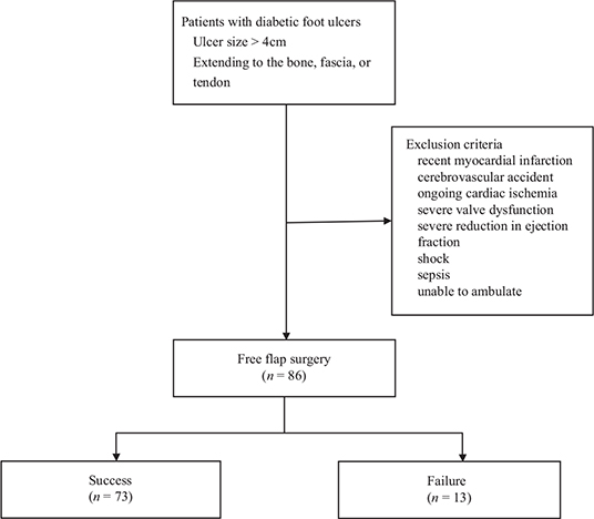
Figure 1. Flow diagram of patient recruitment.
The patients were divided into a success group and a failure group according to the surgical results. The primary study outcome was free flap surgery failure. Representative cases of successful and failed free flaps are shown in Figure 2. The independent factors were age, sex, end-stage renal disease, ischemic heart disease, white blood cell count, C‐reactive protein (CRP) level, hemoglobin level, serum albumin concentration, serum calcium concentration, hemoglobin A1C level, wound location, and donor site. Patients were classified as having PAD if they had ischemic lower extremity wounds due to peripheral arterial disease and had undergone revascularization before free flap surgery. The 13 independent factors mentioned earlier are also risk factors identified in previous studies [14, 18], as well as those deemed high impact by physicians performing these surgeries at the research sites.
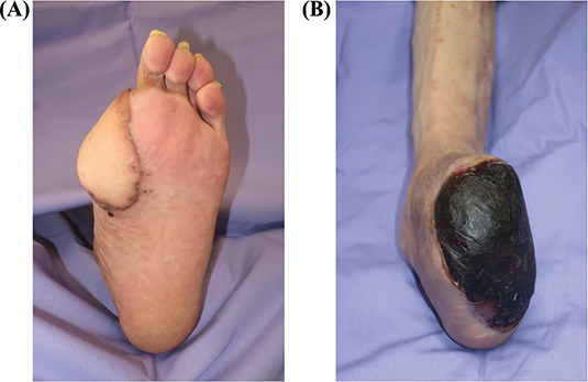
Figure 2. Representative cases of successful (A) and failed (B) free flaps.
Statistical analysis
We assessed the associations of surgery failure with each independent variable via the chi-square test, Fisher’s exact test, and Student’s t test, as appropriate. Variables with p < 0.20 in the univariate analyses were entered into the multivariate logistic regression model. The significance threshold was set at 0.05. All the statistical analyses were performed using SPSS version 24 (IBM Corp., Armonk, NY).
Ethics
This study was approved by the institutional review boards of all participating hospitals. The lead institution was Saitama Medical University Hospital, where the study received IRB approval under reference number 2022-038.
Results
The demographic data are listed in Table 1. Patient characteristics are presented as the means and standard deviations for continuous variables and as frequencies and percentages for categorical variables. A total of 86 patients underwent free flap surgery at one of the three hospitals between April 2007 and March 2019. The mean age of the patients was 58.8 ± 11.8 years; 68 patients were male (79.1%), and 18 were female (20.9%). Thirteen patients experienced free flap failure, resulting in a failure rate of 15.1%. Half of the patients had end-stage renal disease, and 31.4% had ischemic heart disease. Among the 47 patients (54.7%) who had PAD, 29 patients (33.7%) underwent endovascular treatment, and 41 patients (47.7%) underwent bypass surgery prior to free flap surgery.
The wound locations are shown in Figure 3. In all, 46.5% of patients had wounds on the heel of the plantar area, 18.6% were around the metatarsal bone, 18.6% were around the ankle, 10.5% were at a previous Chopart amputation site, and 4.7% were at a previous Lisfranc amputation site. In terms of the donor sites, the latissimus dorsi flap was used in 73.3% of patients, followed by the rectus abdominis musculocutaneous flap in 14.0%, the anterolateral thigh flap in 12.8%, and the groin flap in 1.2%.
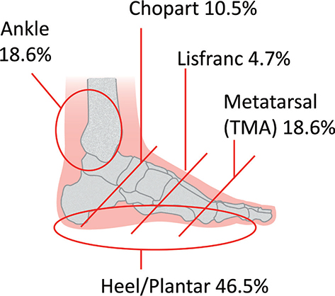
Table 2 shows the results of the univariate analysis. The percentage of patients with end-stage renal disease was higher in the failure group (46.6 vs. 69.2%), but the difference was not statistically significant. The distribution of ischemic heart disease was similar across the two groups. In the failure group, 84.6% of patients had PAD, whereas 49.3% of patients in the success group had PAD, and the difference was statistically significant (p = 0.031). Wound location and donor site were not significantly associated with the result of surgery. The p values of sex (p = 0.134), white blood cell count (p = 0.183), serum calcium concentration (p = 0.066), and HbA1c level (p = 0.078) were lower than 0.2, so these five variables were entered into a logistic regression model.
Multiple regression analysis revealed that sex, white blood cell count, serum calcium concentration, and hemoglobin A1c level were not significantly associated with the surgical result. However, PAD was significantly associated with free flap failure (OR, 6.076; 95% CI, 1.032–35.820; see Table 3).
Discussion
Our study team investigated the risk factors associated with free flap failure in diabetic foot reconstruction. The overall failure rate for the study population was 15.1%. This study highlights two critical issues with respect to surgical treatment options for eligible diabetic patients. First, the prevalence of PAD is high in diabetic foot patients (54.7% of the cases reviewed in this study). In addition, PAD was identified as the only statistically significant independent risk factor for free flap surgery failure in this sample.
In this study, 13 of the 86 patients (15.1%) experienced free flap failure, according to our record review. The failure rate was comparable to that reported in previous studies [12, 13]; however, the rate was still higher than that of other types of recipient sites, such as the breast, head, and neck. Diabetes has been reported as a risk factor for free flap failure and postoperative complications in breast [19], head and neck [20] reconstructions and in posttraumatic extremity reconstruction [14]. Most patients who develop diabetic foot ulcers have a long history of diabetes. Although the use of free flaps has become a popular strategy to prevent amputation resulting from severe diabetic foot ulcers, many surgeons may hesitate to use free flaps because of the high failure rate.
In more than half of the cases (54.7%), PAD was present, which is higher than that reported in the literature. Recent data show that the incidence of PAD in patients with diabetes ranges from 25 to 30% in the United States [21] and China [22]. Our data suggest that the prevalence of PAD in diabetes patients with foot ulcers may be higher than that in diabetes patients without foot ulcers. PAD is an underrecognized complication of diabetes. The main reason for underreporting is the mostly asymptomatic nature of PAD in individuals with diabetes [23]. People with diabetes are recommended to undergo regular PAD evaluations [24]. PAD is also a sign of systemic atherosclerosis; therefore, timely detection of PAD reduces the risks of amputation, cardiovascular disease, and stroke [25].
Our study also revealed that PAD was an independent risk factor for free flap failure. Free flap surgery is technically challenging in patients with calcified vessels due to atherosclerosis. Atherosclerotic changes reduce vascular elasticity and increase the risk of thrombosis in anastomotic vessels [26]. Vascular atherosclerosis is also a known risk factor for flap failure in reconstructive breast surgery, with an adjusted odds ratio as high as 10.61 [27]. The presence of a calcified atherosclerotic lesion, identified via computed tomography, was also associated with failure of the free flap for necrotizing soft tissue defects of the lower extremities [18]. The results of our research are concordant with those of these previous studies. A thorough vascular assessment is crucial for diabetes patients before free flap surgery. Computed tomography for angiographic study is useful for identifying and determining the size of an atherosclerotic lesion and for assessing the degree of stenosis of the patient’s arteries [26].
The percentage of patients who had end-stage renal disease was higher in the failure group (69.2%) than in the success group (46.6%), but the difference was not statistically significant; this may be due to the small sample size of our study. Renal disease, especially associated with diabetes, has been reported to be a strong indicator of possible complications in free flap surgery [18]. Wound healing is impaired in patients with end-stage renal disease, and this impairment has been reported to be one of the significant obstacles to limb salvage [28]. Additionally, the combination of diabetes and renal disease can significantly predispose patients to infection secondary to a suppressed immune system. Postoperative infection is also an independent risk factor for free flap surgery [14]. Aggressive wound debridement and infection control before free flap surgery are crucial for obtaining a high success rate.
To our knowledge, this is the first study to evaluate the risk factors for free flap failure in the reconstruction of diabetic foot ulcers. However, this study is not without its limitations. First, only 86 patient records were reviewed. However, the cases spanned three hospitals across 12 years, providing a robust study sample for examination. Second, this study was a retrospective review of patient data. Therefore, data were not directly collected from the patients, thus some essential data may be missing. Finally, the study was conducted in Japan; therefore, the results may not be generalizable to other populations. However, given that the study involved cases from multiple centers over a 12-year period, we believe that the lessons learned are relevant to clinicians in other communities.
Conclusion
This study revealed that PAD was the most prominent independent risk factor for free flap surgery failure. Patients, especially high-risk diabetic patients, should be screened for PAD before free flap surgery. Patients with a large wound, no ischemia, and no signs of infection are the most suitable candidates for free flap surgery. These assessments are conducive for ensuring good surgical outcomes and potentially preventing amputation, thereby maintaining maximum mobility and preserving quality of life.
References
[1] Boyle JP, Thompson TJ, Gregg EW, et al. Projection of the year 2050 burden of diabetes in the US adult population: dynamic modeling of incidence, mortality, and prediabetes prevalence. Popul Health Metr. 2010; 8: 29. https://doi.org/10.1186/1478-7954-8-29
[2] Deshpande AD, Harris-Hayes M, Schootman M. Epidemiology of diabetes and diabetes-related complications. Phys Ther. 2008; 88(11): 1254–1264. https://doi.org/10.2522/ptj.20080020
[3] Schirr-Bonnans S, Costa N, Derumeaux-Burel H, et al. Cost of diabetic eye, renal and foot complications: a methodological review. Eur J Health Econ HEPAC Health Econ Prev Care. 2017; 18(3): 293–312. https://doi.org/10.1007/s10198-016-0773-6
[4] Moulik PK, Mtonga R, Gill GV. Amputation and mortality in new-onset diabetic foot ulcers stratified by etiology. Diabetes Care. 2003; 26(2): 491–494. https://doi.org/10.2337/diacare.26.2.491
[5] Aulivola B, Hile CN, Hamdan AD, et al. Major lower extremity amputation: outcome of a modern series. Arch Surg Chic Ill 1960. 2004; 139(4): 395–399; discussion 399. https://doi.org/10.1001/archsurg.139.4.395
[6] Fard B, Dijkstra PU, NEDA Study Group, et al. Mortality, reamputation, and preoperative comorbidities in patients undergoing dysvascular lower limb amputation. Ann Vasc Surg. 2020; 64: 228–238. https://doi.org/10.1016/j.avsg.2019.09.010
[7] Nehler MR, Coll JR, Hiatt WR, et al. Functional outcome in a contemporary series of major lower extremity amputations. J Vasc Surg. 2003; 38(1): 7–14. https://doi.org/10.1016/S0741-5214(03)00092-2
[8] Reduce the rate of foot and leg amputations in adults with diabetes – D‑08 – Healthy People 2030 | health.gov [Interest]. [cited 2021 Mar 9]. Available from: https://health.gov/healthypeople/objectives-and-data/browse-objectives/diabetes/reduce-rate-foot-and-leg-amputations-adults-diabetes-d-08
[9] Wang N, Yang BH, Wang G, et al. A meta-analysis of the relationship between foot local characteristics and major lower extremity amputation in diabetic foot patients. J Cell Biochem. 2019; 120(6): 9091–9096. https://doi.org/10.1002/jcb.28183
[10] Sato T, Yana Y, Ichioka S. Free flap reconstruction for diabetic foot limb salvage. J Plast Surg Hand Surg. 2017; 51(6): 399–404. https://doi.org/10.1080/2000656X.2017.1285782
[11] Oh TS, Lee HS, Hong JP. Diabetic foot reconstruction using free flaps increases 5-year-survival rate. J Plast Reconstr Aesthetic Surg JPRAS. 2013; 66(2): 243–250. https://doi.org/10.1016/j.bjps.2012.09.024
[12] Kim HB, Altiparmak M, Pak CJ, et al. Reconstruction using free flaps for diabetic heel defects: outcomes and risk factor analysis. J Reconstr Microsurg. 2020; 36(7): 494–500. https://doi.org/10.1055/s-0040-1709477
[13] Lee ZH, Daar DA, Stranix JT, et al. Free-flap reconstruction for diabetic lower extremity limb salvage. J Surg Res. 2020; 248: 165–170. https://doi.org/10.1016/j.jss.2019.12.008
[14] Las DE, de Jong T, Zuidam JM, et al. Identification of independent risk factors for flap failure: a retrospective analysis of 1530 free flaps for breast, head and neck and extremity reconstruction. J Plast Reconstr Aesthetic Surg JPRAS. 2016; 69(7): 894–906. https://doi.org/10.1016/j.bjps.2016.02.001
[15] Massenburg BB, Sanati-Mehrizy P, Ingargiola MJ, et al. Flap failure and wound complications in autologous breast reconstruction: a national perspective. Aesthetic Plast Surg. 2015; 39(6): 902–909. https://doi.org/10.1007/s00266-015-0575-8
[16] Wang KY, Lin YS, Chen LW, et al. Risk of free flap failure in head and neck reconstruction: analysis of 21,548 cases from a nationwide database. Ann Plast Surg. 2020; 84(1S Suppl 1): S3–S6. https://doi.org/10.1097/SAP.0000000000002180
[17] Zhou W, Zhang WB, Yu Y, et al. Risk factors for free flap failure: a retrospective analysis of 881 free flaps for head and neck defect reconstruction. Int J Oral Maxillofac Surg. 2017; 46(8): 941–945. https://doi.org/10.1016/j.ijom.2017.03.023
[18] Lee YK, Park KY, Koo YT, et al. Analysis of multiple risk factors affecting the result of free flap transfer for necrotising soft tissue defects of the lower extremities in patients with type 2 diabetes mellitus. J Plast Reconstr Aesthet Surg. 2014; 67(5): 624–628. https://doi.org/10.1016/j.bjps.2014.01.047
[19] Chung CU, Wink JD, Nelson JA, et al. Surgical site infections after free flap breast reconstruction: an analysis of 2,899 patients from the ACS-NSQIP datasets. J Reconstr Microsurg. 2015; 31(6): 434–441. https://doi.org/10.1055/s-0035-1548739
[20] Valentini V, Cassoni A, Marianetti TM, et al. Diabetes as main risk factor in head and neck reconstructive surgery with free flaps. J Craniofac Surg. 2008; 19(4): 1080–1084. https://doi.org/10.1097/SCS.0b013e3181763531
[21] Marso SP, Hiatt WR. Peripheral arterial disease in patients with diabetes. J Am Coll Cardiol. 2006; 47(5): 921–929. https://doi.org/10.1016/j.jacc.2005.09.065
[22] Yang S, Zhu L, Han R, et al. Pathophysiology of peripheral arterial disease in diabetes mellitus. J Diabetes. 2017; 9(2): 133–140. https://doi.org/10.1111/1753-0407.12474
[23] Mascarenhas JV, Albayati MA, Shearman CP, et al Peripheral arterial disease. Endocrinol Metab Clin North Am. 2014; 43(1): 149–166. https://doi.org/10.1016/j.ecl.2013.09.003
[24] Balletshofer B, Ito W, Lawall H, et al. Position paper on the diagnosis and treatment of peripheral arterial disease (PAD) in people with diabetes mellitus. Exp Clin Endocrinol Diabetes. 2019; 127(Suppl. 01): S105–S113. https://doi.org/10.1055/a-1018-9250
[25] On behalf of the ADVANCE Collaborative Group, Mohammedi K, Woodward M, et al. Presentations of major peripheral arterial disease and risk of major outcomes in patients with type 2 diabetes: results from the ADVANCE-ON study. Cardiovasc Diabetol. 2016; 15(1): 129. https://doi.org/10.1186/s12933-016-0446-x
[26] Bouaoud J, Honart JF, Bennis Y, et al. How to manage calcified vessels for head and neck microsurgical reconstruction. J Stomatol Oral Maxillofac Surg. 2020; 121(4): 439–441. https://doi.org/10.1016/j.jormas.2020.03.015
[27] Masoomi H, Clark EG, Paydar KZ, et al. Predictive risk factors of free flap thrombosis in breast reconstruction surgery. Microsurgery. 2014; 34(8): 589–594. https://doi.org/10.1002/micr.22250
[28] Edwards JM, Taylor LM, Porter JM. Limb salvage in end-stage renal disease (ESRD). Comparison of modern results in patients with and without ESRD. Arch Surg Chic Ill 1960. 1988; 123(9): 1164–1168. https://doi.org/10.1001/archsurg.1988.01400330144022