ORIGINAL RESEARCH ARTICLE
Partial intracranial volumes in metopic synostosis – pre- and postoperative comparisons with healthy controls
Hanna Lif, PhDa  , Rehan Chakari, MDa , Per Enblad, MD, PhDb and Daniel Nowinski, MD, PhDa
aDepartment of Surgical Sciences, Plastic Surgery, Uppsala University, Uppsala, Sweden; bDepartment of Medical Sciences, Neurosurgery, Uppsala University, Uppsala, Sweden
ABSTRACT
Surgical intervention for metopic synostosis has been much debated but aims to correct trigonocephaly and hypotelorism while ensuring enough space for undisturbed brain growth. The aim of this study was to evaluate changes in partial intracranial volumes and the interfrontal angle following fronto-orbital advancement. Twenty-six non-syndromic metopic synostosis patients treated with fronto-orbital advancement at Uppsala University Hospital between 2012 and 2022 who had undergone computed tomography preoperatively and at age three were included, as well as 40 age- and sex-matched controls who had undergone computed tomography for post-traumatic evaluation. Demographic information, imaging, pre-, peri-, and postoperative data were collected. The frontal-, middle-, and posterior intracranial volume and their relative distribution were calculated in the softwares Craniosyn and ITK-SNAP 3.8.0. Preoperatively, patients had smaller interfrontal angles (p < 0.001), total intracranial volumes (p = 0.03), and frontal volumes (p = 0.02), compared with controls. At age three, the total intracranial volume (p = 0.96) and frontal volume (p = 0.51) did not differ significantly between patients and controls, whereas the interfrontal angle remained smaller in the synostosis group (p < 0.001). The relative intracranial volume distribution between frontal-, middle-, and posterior volumes did not match the distribution in healthy controls pre- or postoperatively, where the middle volume ratio was notably greater, and both the frontal- and posterior volume ratios were smaller in patients. Fronto-orbital advancement and subsequent growth improve the total- and partial intracranial volume in metopic synostosis to match the volumes of healthy controls at age three but does not restore the relative intracranial volume distribution.
KEYWORDS: Metopic synostosis; intracranial volume; partial intracranial volume; frontal volume; interfrontal angle; fronto-orbital advancement
Citation: Journal of Plastic Surgery and Hand Surgery 2025; 60: 219–225. DOI: https://doi.org/10.2340/jphs.v60.44913.
Copyright: © 2025 The Author(s). Published by MJS Publishing on behalf of Acta Chirurgica Scandinavica. This is an Open Access article distributed under the terms of the Creative Commons Attribution 4.0 International License (http://creativecommons.org/licenses/by/4.0/), allowing third parties to copy and redistribute the material in any medium or format and to remix, transform, and build upon the material, with the condition of proper attribution to the original work.
Received: 7 August 2025; Accepted: 3 September 2025; Published: 18 December 2025.
CONTACT: Hanna Lif hanna.lif@uu.se Akademiska sjukhuset ing 85, 9 tr, 751 85 Uppsala, Sweden
Competing interests and funding: Nothing to declare.
This study was funded by ALF grants Region Uppsala. No funders were involved in the study design, data collection, analyses, interpretation of data, writing the manuscript, or in the decision to submit this paper.
Introduction
Metopic synostosis is the second most common single-suture craniosynostosis and is characterized by trigonocephaly, hypotelorism, bitemporal narrowing, biparietal widening, and a midline forehead ridge [1 – 10]. The incidence of craniosynostosis in Sweden is 7.7 cases per 10,000 live births [11], and approximately 1 in 5,000 live births for metopic synostosis with a slight male predominance, and most cases occur spontaneously [1, 9]. Recent literature has shown an increasing incidence for unknown reasons [12, 13].
The metopic suture is the only cranial suture that fuses physiologically during the time-period when the brain grows most rapidly, beginning at an approximate age of 3 months and fusing completely around 9 months of age [14]. A patent metopic suture in adulthood is, however, considered a physiological variant [14]. The consequences of premature metopic suture fusion exhibit a diverse phenotypic presentation, with deformities along a spectrum raging from metopic ridge to severe trigonocephaly [15, 16]. Previous studies have reported an increased risk for cognitive, mental, and behavioral challenges in children with trigonocephaly [3, 9, 17–24]. As of today, the correlation between the severity of metopic synostosis and mental development is not fully understood. Moreover, the incidence of intracranial hypertension in metopic synostosis has been much debated and remains uncertain [3, 9, 18, 25]. Early surgical intervention, often performed between 6 and 12 months of age, mainly aims to create the best possible circumstances for the developing brain by restoring forehead and skull shape [2, 3, 5, 7, 9]. Surgical techniques for treating metopic synostosis can grossly be separated into two main groups: various types of fronto-orbital advancement and endoscopic strip craniectomy, followed by helmet-assisted molding [2, 3, 26, 27]. Metopic ridge is not operated, and mild metopic synostosis cases seldomly require surgical intervention due to the increased evidence on spontaneous correction of skull shape with growth [15, 28, 29].
Severity of metopic synostosis can be measured from radiological imaging or 3D photogrammetry, for example, by determining the interfrontal angle (IFA) [30] or using CranioRate [31]. Calculations of total- and frontal intracranial volume (FIV) have been used as an indirect measure of deformity and to quantify the frontal expansion achieved by surgery and subsequent growth. According to prior studies, the total intracranial volume (TIV) in children with metopic synostosis appears to be within the range observed in the healthy population or slightly smaller [32–35]. Few previous studies have analyzed the distribution of partial intracranial volumes [35, 36], but the frontal volume has been reported to be smaller in patients preoperatively but approaching the volumes of controls following fronto-orbital advancement and subsequent growth [34]. To our knowledge, only one previous study has aimed to measure the posterior cranial volume in this condition, based on 3D photogrammetry, reporting no difference compared to controls preoperatively or 1 year after surgery [34]. The conflicting reports on the relevance and effect of surgical intervention in metopic synostosis are reflected in the diverse management ranging from centers not operating any patients regardless of severity to centers operating even mild cases. There is an ongoing clinical trial in Rotterdam (NCT06069479), evaluating conservative versus surgical treatment for metopic synostosis with the aim to inform future treatment guidelines.
The aim of the present study was to evaluate morphological- and volumetric outcomes following fronto-orbital advancement and subsequent growth in children with non-syndromic-isolated metopic synostosis compared to controls, hypothesizing that the shape and volumes would be normalized at 3 years of age. The novelty of this study lies in improved understanding of the intracranial volume distribution following fronto-orbital advancement, an area that has been poorly investigated. This may contribute with improved insight on cranial growth and compensatory mechanisms in metopic synostosis following surgical treatment, which could guide the development of future treatment strategies.
Material and methods
Patients and controls
This single-center retrospective cohort study included patients with non-syndromic isolated metopic synostosis treated with fronto-orbital advancement between 2012 and 2022 at the Uppsala University Hospital. Inclusion criteria were single-suture non-syndromic metopic synostosis with trigonocephaly, surgical treatment by fronto-orbital advancement, and available computed tomography (CT) of sufficient quality preoperatively and at age three. All patients were examined by a clinical geneticist preoperatively for the evaluation of potential signs of syndromic stigma and genetically tested if there was any suspicion of syndromic status based on clinical examination or family history. The following data were collected from the Uppsala Craniofacial Quality Registry: sex, age at surgery, syndromic status, and length of hospital stay. Data on head circumference and blood loss were obtained from medical records and the system for perioperative care and monitoring. One patient had 22q11 deletion syndrome but was left included as this was considered a co-incidental diagnosis. All patients were confirmed to not have any craniofacial syndrome. Sex- and aged-matched children without craniosynostosis who underwent CT-examination for post-traumatic evaluation were used as controls. Exclusion criteria for controls were insufficient CT quality or any condition or injury that could potentially affect cranial growth or shape.
Surgical technique
Fronto-orbital advancement was performed according to the following main steps (Figure 1): (1) lazy s bicoronal skin incision, (2) subgaleal dissection and raising a separate pericranial flap as well as release of the temporal muscles, (3) bifrontal craniotomy in front of the coronal sutures, (4) transbasal orbital osteotomies for the supraorbital bar with tongue in gooves, (5) frontal bone and supraorbital bar split in midline, (6) subpraorbital bar widened with interpositional parietal bone graft and resorbable plate fixation and angle to tongue in grooves reduced and armoring at the angle with 3/0 PDS sutures, (7) the two piece forehead fixed to supraorbital bar to optimal shape with multiple 3/0 PDS sutures, (8) the whole construct fixed with horizontal resorbable plates at tongue in grooves and 3/0 PDS suture at nasal radix, (9) temporal muscles rotated anteriorly and suture to lateral aspect of supraorbital bar, (10) bone dust glued to remaining defects and periosteum, galea and skin sutured in layers with 3/0 and 4/0 Monocryl suture, and placement of an active surgical drain in the subgaleal plane.
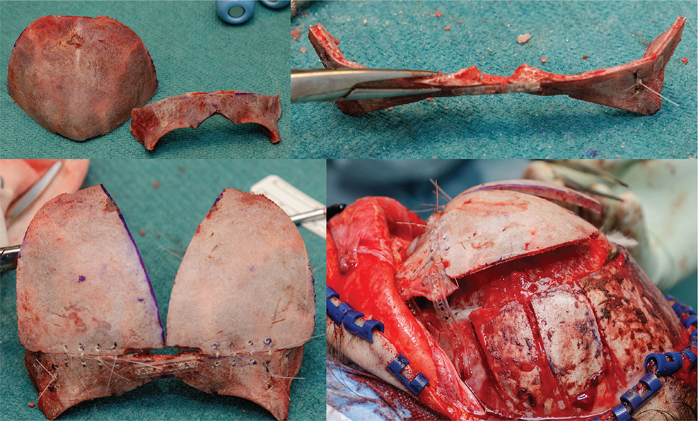
Figure 1. Fronto-orbital advancement as performed to treat metopic synostosis at Uppsala Craniofacial Center.
Total- and partial intracranial volume
The software Craniosyn, developed in collaboration with the Center for Image Analysis at Uppsala University, was used to define and segment total- and partial intracranial volumes, as previously described [37]. Semi-automatic segmentation of the TIV was achieved through automatic identification of bony boundaries based on Hounsfield units. In cases with incomplete bone formation/calvarial defects, manual outlining was required. TIV was automatically calculated from the software. To divide TIV into partial volumes, four landmarks (opisthion, frontosphenoid suture, and the anterior borders of both frontozygomatic sutures) were placed before segmenting to create landmark-based infinitive planes to determine the FIV, middle intracranial volume ( ), and the posterior intracranial volume (PIV) (Figures 2 and 3). ITK-SNAP, a widely used open-source segmentation software, was used to visualize and calculate the three partial volumes. Division into partial volumes allowed for the calculation of relative volumes: the frontal to total volume (FIV%), middle to total volume (MIV%), and posterior to total volume (PIV%).
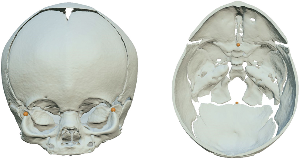
Figure 2. Three predefined landmarks in the skull base: frontozygomatic suture, frontosphenoid suture, and opisthion.
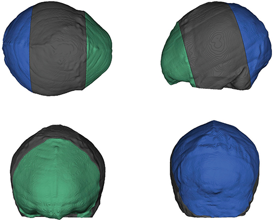
Figure 3. The portioning of the Frontal Intracranial Volume (FIV), Middle Intracranial Volume (MIV), and Posterior Intracranial Volume (PIV).
Determining the IFA
The method employed in this article was the same as developed by Kellog et al. [30, 32]. MITK, an interactive open-source software for viewing and performing measurements on medical images, was used to enable the implementation of this method. In a two-dimensional axial plane on each CT-scan, the IFA was determined between the most anterior point of the cranium and the supraorbital notches (Figure 4).
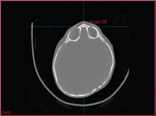
Figure 4. Measurement of the interfrontal angle.
Statistical analysis
Statistical analyses were performed in R (R Foundation for Statistical Computing), and p ≤ 0.05 was considered statistically significant. Mann–Whitney U-tests were employed to analyze potential differences in age between patients and controls at both ages. Additionally, Mann–Whitney U-tests tests were utilized to investigate differences in TIV, FIV, MIV, PIV, FIV%, MIV%, PIV%, and IFA between patients and controls at both ages, and to compare IFA by age-group in controls. Wilcoxon signed rank tests were used to compare FIV%, MIV%, PIV%, and IFA pre- and postoperatively. Linear regression investigated potential correlation between FIV and IFA in pre- and postoperative patients.
Ethics
This study followed the Declaration of Helsinki and was approved by the Ethical Review Board of Uppsala (Dnr 2013-402), and informed consent was obtained from the parents of all participants.
Results
Patients and controls
Twenty-six patients including 20 males and 6 females were included. In the synostosis group, the mean age at CT-scanning was 157 ± 92 (3–323) days pre-operatively and 1,080 ± 158 (511–1,236) days postoperatively. Two patients in the postoperative group had undergone CT-examination at 2 years of age even though the Swedish national guideline for craniosynostosis aims for CT at age three. Forty controls were included: 16 were age-matched with the preoperative metopic group and 24 with the postoperative metopic group. The mean age at CT-scan in the control group matching the preoperative synostosis group was 186 days ± 44 (118–253), p = 0.24 when compared to the age of patients. The mean age at CT-scan in the control group matching the postoperative synostosis group was 1192 ± 68 (1074–1330), p < 0.001 when compared to the age of patients. There were no available controls with CT-scans at both the preoperative age for patients and at age three.
Demographic and perioperative data are summarized in Table 1. Two patients were missing follow-up CT-examination and were excluded from the analyses of postoperative data. The mean age at fronto-orbital advancement was 241 ± 61 days. The weight the day before surgery was 8.5 ± 3.2 kg. The mean operation time was 261 ± 42 min (including pre- and postoperative care in connection to the surgery). Perioperative mean blood loss was 35.5 ± 21 % of total circulating blood volume. Mean hospitalization time was 5 ± 2 days. One patient was reoperated on postoperative day seven due to a fracture of an absorbable plate. Preoperatively, six patients had a head circumference below -2 SD, eight between -2 SD and -1, 11 between -1 and +1 SD, and one above 1 SD.
Preoperative results
The median IFA in the preoperative patient group was 133.25, compared to 157.44 in the age-matched control group (p < 0.001). In the preoperative patient group, the median TIV was 856.6 cm3, while it was 931.9 cm3 in the age-matched control group (p = 0.028). The median FIV in the preoperative patient group and the age-matched control group was 87.32 cm3 and 115.6 cm3, respectively (p = 0.018). The preoperative patient group exhibited a median FIV%, MIV%, and PIV% of 11.29, 65.75, and 19.05%, respectively, compared with the age-matched control group, where the medians were 12.43% (p = 0.56), 59.17% (p < 0.001), and 25.80 (p = 0.02)%, respectively. A summary of preoperative data is presented in Table 2.
Postoperative results
The median TIV at the postoperative CT-scan and in the age-matched control group was 1299.85 cm3 and 1273.30 cm3, respectively (p = 0.96). The median FIV was 166.15 cm3 at the postoperative CT-scan in the patient group and 163.80 cm3 in the age-matched control group (p = 0.51). The median FIV%, MIV%, and PIV% in the postoperative group were 13.85, 61.64, and 24.46%, respectively, compared to the age-matched control group, where these values were 11.73% (p = 0.35), 57.30% (p = 0.005), and 31.26% (p = 0.008), respectively. The median IFA was 143.32 postoperatively, and in the age-matched control group, it was 151.16 (p < 0.001). Table 3 displays a summary of postoperative data. There was no linear relationship between FIV and IFA for the patient group postoperatively (p = 0.48, Adjusted R-squared = 0.02). IFA was smaller in older controls compared to younger controls (p = 0.003), as indicated in Table 4.
Discussion
In this study, we investigated the distribution of partial intracranial volume in patients with non-syndromic metopic synostosis following fronto-orbital advancement. Based on these findings, fronto-orbital advancement and subsequent growth improve the total- and partial intracranial volume in metopic synostosis to match the volumes of healthy age-matched controls at 3 years of age. However, the relative intracranial volume distribution did not match that of healthy controls postoperatively, with significantly larger medial volumes and significantly smaller posterior volumes in patients both pre- and postoperatively. Whether this was a result of the surgical intervention itself or due to compensatory mechanisms remains to be determined. These findings may have implications for developing improved treatment and long-term follow-up protocols and highlight the need for further research on intracranial dynamics in metopic synostosis.
Both the TIV and FIV were significantly smaller preoperatively in patients compared to controls, an expected finding. However, in the postoperative patient group at age three, there was no significant disparity observed for either TIV or FIV compared to controls, indicating successful reconstruction of frontal- and overall volume by fronto-orbital advancement. Future studies including functional outcome in relation to partial volume measurements could elucidate the relevance of this finding further regarding the complex relationship between shape and function in craniosynostosis. In addition, there were no statistically significant differences between FIV% pre- and postoperatively compared with controls, and PIV% was smaller both pre- and postoperatively compared with controls, suggesting a complex interaction between frontal- and posterior skull growth in metopic synostosis. The MIV% was notably greater in both pre- and postoperative patients compared with controls, but still significantly reduced by 3 years of age, indicating a postoperative decrease in biparietal widening. The IFA was significantly reduced in both pre- and postoperative metopic patients when compared with controls and was significantly increased after surgery, suggesting successful morphological improvement.
Previous studies have presented conflicting results with respect to TIV in metopic synostosis compared to controls. The present study confirms the recurrent finding that children with metopic synostosis have significantly reduced TIV when compared with healthy children [32–35, 38]. In addition, the present study found that the absolute frontal volume was initially smaller in children with metopic synostosis compared with controls, but that following fronto-orbital advancement, this difference was no longer present postoperatively at age three, in line with previous studies [4, 34]. Consistent with the present study, both previous studies treated metopic synostosis with fronto-orbital advancement. However, there was a disparity in the follow-up period, with Qing et al. having a 1-year follow-up and Freudlsperger et al. presented immediate results 8-days after surgery. Consequently, this study added an additional perspective of stability in sufficient frontal volume at age three.
Regarding comparable research based on CT-scans with identical ages for CT, two previous publications looked into similar matters at the same ages [39, 40]. In contradiction to the present study, Maltese et al. showed significantly smaller frontal volumes compared to controls both pre-operatively and at 3 years of age. Notably, the study from Maltese et al. found comparable TIV between preoperative patients and controls, as well as reduced TIV in patients at 3 years of age. Conversely, the present study found the opposite: smaller TIV before surgery and normalized TIV at 3 years of age. Future comparative studies looking further into demographic parameters and comparing different variations of fronto-orbital advancement could elucidate this finding further.
In the present study, no differences in frontal volume ratio compared with controls were found either before or after surgery. Both Maltese et al. and Bhatti-Söfteland et al. showed an increase in the frontal volume ratio after surgery although it did not reach to that of controls [35, 39]. This disparity could be due to different methodologies and landmarks for defining the frontal volume. In the current study, the frontosphenoid suture was used as landmark to separate the frontal volume, while previous authors used the coronal suture. The rationale for choosing the frontosphenoid suture was stability over time with growth, where its relative position changes less than that of the coronal sutures. Other potential factors for the conflicting findings could be the range in severity of trigonocephaly or differences in controls.
Patients had a significantly smaller posterior volume ratio compared with controls, which remained at 3-year of age. In contrast, the middle volume ratio was larger in the metopic group both pre-operatively and at age three when compared with controls but showed a significant reduction at the 3-year follow-up. This finding supports the fact that metopic synostosis affects not only the frontal volume but also the entire calvarium, which may explain the compensatory increase in cephalic width observed in the parietal region.
The IFA has been widely used to characterize severity of trigonocephaly, with its main benefits being that it is simple to measure and conceptually easy to comprehend. However, it has its limitations, including that the supraorbital notch may not be as visible on follow-up CT-scans. In the context of postoperative evaluation, previous studies have employed diverse methods for assessing IFA. Nguyen et al., the only study employing the same method as the present study, demonstrated a significant improvement in IFA on two-dimensional axial CT-scan 1 year following surgery with equivalent results to controls [2]. The results of the present study align with these findings, indicating a postoperative increase in IFA, although it did not reach the levels observed in the healthy population. This difference might be attributed to the different time points for follow-up CT or caused by statistical differences between control samples. The same results have also been confirmed by other authors [41, 42]. In theory, IFA should correlate to FIV, but our findings did not align with this expectation, which is most likely explained by the limited sample size.
The largest limitation of this study was that postoperative metopic patients were not ideally aged-matched to the healthy controls, owing to the age spread within the patient group and few performed CT without identified pathology in otherwise healthy children. A common challenge and limitation noted in both the current and prior studies is the small sample size, making it challenging to detect subtle differences. Further studies on larger sample sizes are needed to address this limitation, preferably including collaborations between multiple craniofacial centers. Strengths of the study include a high participation rate, preoperatively matched groups, the access to CT-scans preoperatively, and at a standardized follow-up age, as well as the use of advanced and stable measurements to determine partial volumes and IFA.
In summary, the present study found successful correction of trigonocephaly and improvement of total- and partial intracranial volumes by 3 years of age after fronto-orbital advancement, however, without elucidating how much of this was explained by the surgery itself, spontaneous improvement with growth, or a combination of both. This highlights the current conflicting data and praxis on surgical indication in metopic synostosis within the field. At Uppsala Craniofacial Center, our current protocol is to follow all metopic patients at the clinic and only treat them surgically by fronto-orbital advancement in severe cases, if there is a suspicion of raised intracranial pressure or if that is the informed preference of the parents. All moderate and mild cases are followed until 2 years of age, and so far, we have observed sufficient spontaneous correction with growth in these groups of metopic patients without any signs of functional impact.
Acknowledgments
The authors would like to thank Johan Nysjö for developing Craniosyn and the workflow for standardized measurement of partial intracranial volumes.
Contributors
All authors meet the authorship criteria. No others meeting the criteria were omitted. All authors contributed to the study conception and design, interpretation of analyses, revising, and final approval of the article. HL contributed to intracranial analyses, statistical analyses, tables, figures, and drafting the article. RC contributed to data acquisition, IFA analysis, tables, and drafting the article. PE and DN had senior responsibility for the study.
References
[1] van der Meulen J. Metopic synostosis. Child’s Nerv Syst. 2012; 28(9): 1359–1367. https://doi.org/10.1007/s00381-012-1803-z
[2] Nguyen DC, Patel KB, Skolnick GB, et al. Are endoscopic and open treatments of metopic synostosis equivalent in treating trigonocephaly and hypotelorism? J Craniofac Surg. 2015; 26(1): 129–134. https://doi.org/10.1097/SCS.0000000000001321
[3] Hormozi AK, Shahverdiani R, Mohammadi HR, et al. Surgical treatment of metopic synostosis. J Craniofac Surg. 2011; 22(1): 261–265. https://doi.org/10.1097/SCS.0b013e3181f7b80c
[4] Yan Q, He J, Gao Z, et al. Evaluation of fronto-orbital reconstruction surgery for the treatment of metopic synostosis in Chinese population. Child’s Nerv Syst. 2021; 37(4): 1167–1174. https://doi.org/10.1007/s00381-020-04977-w
[5] Wes AM, Paliga JT, Goldstein JA, et al. An evaluation of complications, revisions, and long-term aesthetic outcomes in nonsyndromic metopic craniosynostosis. Plastic Reconstr Surg (1963). 2014; 133(6): 1453–1464. https://doi.org/10.1097/PRS.0000000000000223
[6] Taşkapılıoğlu MÖ, Ocakoğlu G, Kaya S, et al. Statistical shape analyses of trigonocephaly patients. Child’s Nerv Syst. 2020; 36(2): 379–384. https://doi.org/10.1007/s00381-019-04269-y
[7] Francis CS, Shetty A, Frank R, et al. In situ Fronto-Orbital advancement with medial orbital osteotomies for trigonocephaly-associated hypotelorism. J Craniofac Surg. 2011; 22(1): 281–284. https://doi.org/10.1097/SCS.0b013e3181fb8851
[8] Hicdonmez T. Children with metopic ridge. Turk Neurosurg. 2017; 27(4): 585–589.
[9] Natghian H, Song M, Jayamohan J, et al. Long-term results in isolated metopic synostosis: the Oxford experience over 22 years. Plast Reconstr Surg. 2018; 142(4): 509e–515e. https://doi.org/10.1097/PRS.0000000000004768
[10] Selber J, Reid RR, Gershman B, et al. Evolution of operative techniques for the treatment of single-suture metopic synostosis. Ann Plastic Surg. 2007; 59(1): 6–13. https://doi.org/10.1097/01.sap.0000264836.54760.32
[11] Tarnow P, Kölby L, Maltese G, et al. Incidence of non-syndromic and syndromic craniosynostosis in Sweden. J Craniofac Surg. 2022; 33(5): 1517–1520. https://doi.org/10.1097/SCS.0000000000008457
[12] Kolar JC. An epidemiological study of nonsyndromal craniosynostoses. J Craniofac Surg. 2011; 22(1): 47–49. https://doi.org/10.1097/SCS.0b013e3181f6c2fb
[13] Cornelissen M, Ottelander B, Rizopoulos D, et al. Increase of prevalence of craniosynostosis. J Craniomaxillofac Surg. 2016; 44(9): 1273–1279. https://doi.org/10.1016/j.jcms.2016.07.007
[14] Vinchon M. The metopic suture: natural history. Neuro-chirurgie. 2019; 65(5): 239–245. https://doi.org/10.1016/j.neuchi.2019.09.006
[15] Bloch K, Geoffroy M, Taverne M, et al. New diagnostic criteria for metopic ridges and trigonocephaly: a 3D geometric approach. Orphanet J Rare Dis. 2024; 19(1): 204. https://doi.org/10.1186/s13023-024-03197-8
[16] Geoffroy M, François PM, Khonsari RH, et al. Paediatric skull growth models: a systematic review of applications to normal skulls and craniosynostoses. J Stomatol Oral Maxillofac Surg. 2022; 123(5): e533–e543. https://doi.org/10.1016/j.jormas.2022.01.002
[17] Almeida MN, Alper DP, Parikh N, et al. Comparison of emotional and behavioral regulation between metopic and sagittal synostosis. Childs Nerv Syst. 2024; 40(9): 2789–2799. https://doi.org/10.1007/s00381-024-06387-8
[18] Junn AH, Long AS, Hauc SC, et al. Long-term neurocognitive outcomes in 204 single-suture craniosynostosis patients. Childs Nerv Syst. 2023; 39(7): 1921–1928. https://doi.org/10.1007/s00381-023-05908-1
[19] Long AS, Hauc SC, Almeida MN, et al. Morphologic severity and age at surgery are associated with school-age neurocognitive outcomes in metopic craniosynostosis. Plast Reconstr Surg. 2024; 154(4): 824–835. https://doi.org/10.1097/PRS.0000000000010999
[20] Alperovich M, Tonello C, Mayes LC, Kahle KT. Non-syndromic craniosynostosis. Nat Rev Dis Primers. 2025; 11(1): 24. https://doi.org/10.1038/s41572-025-00607-4
[21] Almeida MN, Alper DP, Barrero C, et al. Radiographic severity is associated with worse executive function in metopic craniosynostosis. Childs Nerv Syst. 2024; 40(12): 3971–3982. https://doi.org/10.1007/s00381-024-06493-7
[22] Gabrick KS, Wu RT, Singh A, et al. Radiographic severity of metopic craniosynostosis correlates with long-term neurocognitive outcomes. Plast Reconstr Surg. 2020; 145(5): 1241–1248. https://doi.org/10.1097/PRS.0000000000006746
[23] Almeida MN, Alper DP, Long AS, et al. Risk of attention-deficit/hyperactivity disorder, autism spectrum disorder, and executive function impairment in metopic craniosynostosis. Plast Reconstr Surg. 2024; 154(5): 979e–992e. https://doi.org/10.1097/PRS.0000000000011249
[24] Cabrejo R, Lacadie C, Chuang C, et al. What is the functional difference between sagittal with metopic and isolated sagittal craniosynotosis? J Craniofac Surg. 2019; 30(4): 968–973. https://doi.org/10.1097/SCS.0000000000005288
[25] Gaillard L, Tjaberinga MC, Dremmen MHG, et al. Brain volume in infants with metopic synostosis: less white matter volume with an accelerated growth pattern in early life. J Anat. 2024; 245(6): 894–902. https://doi.org/10.1111/joa.14028
[26] Safi A-F, Kreppel M, Grandoch A, et al. Clinical evaluation of standardized Fronto-Orbital advancement for correction of isolated trigonocephaly. J Craniofac Surg. 2018;29(1):72–75. https://doi.org/10.1097/SCS.0000000000004058
[27] Di Rocco F, Arnaud E, Marchac D, et al. Anterior fronto-orbital remodeling for trigonocephay. Child’s Nerv Syst. 2012; 28(9): 1369–1373. https://doi.org/10.1007/s00381-012-1841-6
[28] Tio PAE, Abdel Alim T, Roshchupkin G, et al. Forehead shape analysis following surgical and conservative treatment in metopic synostosis: a 3D photogrammetry analysis. Plastic Reconstr Surg (1963). 2024; 256(1): 103e–111e. https://doi.org/10.1097/PRS.0000000000011753
[29] Tio P, van Staalduinen M, Okkerse J, et al. Comparing effectiveness of conservative policy to craniofacial surgery in children with metopic synostosis: protocol for an observational cohort study on clinical outcomes, psychosocial well-being and costs in a Dutch academic hospital. BMJ Open. 2025; 15(5): e094112. https://doi.org/10.1136/bmjopen-2024-094112
[30] Kellogg R, Allori AC, Rogers GF, et al. Interfrontal angle for characterization of trigonocephaly: part 1: development and validation of a tool for diagnosis of metopic synostosis. J Craniofac Surg. 2012; 23(3): 799–804. https://doi.org/10.1097/SCS.0b013e3182518ad2
[31] Beiriger JW, Tao W, Bruce MK, et al. CranioRate: an image-based, deep-phenotyping analysis toolset and online clinician interface for metopic craniosynostosis. Plastic Reconstr Surg (1963). 2024; 153(1): 112e–119e. https://doi.org/10.1097/PRS.0000000000010452
[32] Sisti A, Bassiri Gharb B, Papay F, et al. Anthropometric cranial measurements in metopic craniosynostosis/trigonocephaly: diagnostic criteria, classification of severity and indications for surgery. J Craniofac Surg. 2022; 33(1): 161–167. https://doi.org/10.1097/SCS.0000000000008196
[33] Cronin BJ, Brandel MG, McKee RM, et al. A comparison of intracranial volume growth in normal children and patients with metopic craniosynostosis. J Craniofac Surg. 2020; 31(1): 142–146. https://doi.org/10.1097/SCS.0000000000005946
[34] Freudlsperger C, Steinmacher S, Bächli H, et al. Metopic synostosis: measuring intracranial volume change following fronto-orbital advancement using three-dimensional photogrammetry. J Cranio-maxillo-fac Surg. 2015; 43(5): 593–598. https://doi.org/10.1016/j.jcms.2015.02.017
[35] Bhatti-Söfteland M, Maltese G, Tarnow P, et al. The degree of surgical frontal volume correction in metopic synostosis determines long-term outcomes. J Craniofac Surg. 2017; 28(5): 1161–1163. https://doi.org/10.1097/SCS.0000000000003586
[36] Maltese G, Tarnow P, Wikberg E, et al. Intracranial volume before and after surgical treatment for isolated metopic synostosis. J Craniofac Surg. 2014; 25(1): 262–266. https://doi.org/10.1097/SCS.0000000000000423
[37] Fischer S, Unander-Scharin J, Bhatti-Söfteland M, et al. Springs produce favorable morphologic outcomes relative to H-craniectomy according to a two-center comparison of matched cases. Plast Reconstr Surg. 2024; 154(2): 317e–325e. https://doi.org/10.1097/PRS.0000000000010761
[38] Graf AR, Klement KA, Denny AD. Quantifying craniometric change early after Fronto-Orbital advancement in metopic synostosis. J Craniofac Surg. 2016; 27(7): 1727–1731. https://doi.org/10.1097/SCS.0000000000002977
[39] Maltese G, Tarnow P, Tovetjärn R, et al. Correction of hypotelorism in isolated metopic synostosis. J Plastic Surg Hand Surg. 2014; 48(1): 63–66. https://doi.org/10.3109/2000656X.2013.812967
[40] Tarnow P, Kölby L, Maltese G, et al. Incidence of non-syndromic and syndromic craniosynostosis in Sweden. J Craniofac Surg. 2022; 33(5): 1517–1520. https://doi.org/10.1097/SCS.0000000000008457
[41] Ha AY, Skolnick GB, Chi D, et al. School-aged anthropometric outcomes after endoscopic or open repair of metopic synostosis. Pediatrics (Evanston). 2020; 146(3): 1. https://doi.org/10.1542/peds.2020-0238
[42] Glener AD, Allori AC, Shammas RL, et al. Post-surgical relapse in metopic synostosis and limitations of the interfrontal angle as an outcome measure. J Craniofac Surg. 2017; 28(5): e494–e500. https://doi.org/10.1097/SCS.0000000000003800