ORIGINAL RESEARCH ARTICLE
Vascularized bone grafting using the 1,2 intercompartmental supraretinacular artery for the treatment of Preiser’s disease
Yoshinori Takemura, Narihito Kodama, Kosei Ando, Takafumi Yayama and Shinji Imai
Department of Orthopedic Surgery, Shiga University of Medical Science, Seta Tsukinowa-cho Otsu Shiga Japan
ABSTRACT
Introduction: Preiser’s disease refers to a rare idiopathic avascular necrosis of the scaphoid that often leads to pole collapse and poor clinical outcomes when managed conservatively. Vascularized bone grafting (VBG) using the 1,2 intercompartmental supraretinacular artery (ICSRA) flap has been used to restore perfusion, but its treatment efficacy across different disease stages remains unclear.
Methods: We retrospectively reviewed eight women (mean age 60.6 years) who were treated using 1,2 ICSRA VBG between 2006 and 2022. Disease stage was determined using the Herbert radiographic classification (one stage 1, four stage 2, and three stage 3 cases). Clinical parameters [grip strength, range of motion, Disabilities of the Arm, Shoulder, and Hand (DASH) scores, Mayo Wrist Scores] and radiographic progression were assessed after a mean follow-up of 3.6 years. Contralateral scaphoid morphology was evaluated to explore any potential anatomical predisposition.
Results: On average, there were improvements in grip strength (9.5 to 16.6 kg), dorsal flexion (49° to 65°), DASH score (49.5 to 10.6), and Mayo score in all cases. Radiographically, the stage 1 case did not exhibit any progression, one stage 2 case progressed, and all stage 3 cases progressed despite symptomatic improvement. Three patients exhibited type 2 scaphoid morphology, but no correlation with outcomes was observed.
Conclusion: VBG using the 1,2 ICSRA flap results in favorable clinical/radiographic outcomes in early Preiser’s disease (stages 1–2), effectively preserving function and stability. In stage 3 disease, VBG may provide symptomatic relief but does not prevent progression. This warrants careful patient selection and consideration of alternative reconstructive options.
KEYWORDS: Hand; Necrosis; Vascularized bone graft; intercompartmental supraretinacular artery; Preiser’s disease
Citation: Journal of Plastic Surgery and Hand Surgery 2025; 60: 226–230. DOI: https://doi.org/10.2340/jphs.v60.45011.
Copyright: © 2025 The Author(s). Published by MJS Publishing on behalf of Acta Chirurgica Scandinavica. This is an Open Access article distributed under the terms of the Creative Commons Attribution 4.0 International License (http://creativecommons.org/licenses/by/4.0/), allowing third parties to copy and redistribute the material in any medium or format and to remix, transform, and build upon the material, with the condition of proper attribution to the original work.
Received: 15 September 2025; Accepted: 17 October 2025; Published: 18 December 2025
CONTACT: Yoshinori Takemura takemura@belle.shiga-med.ac.jp Seta Tsukinowa-cho Otsu-shi Shiga Japan 520-2192
Competing interests and funding: The authors declare no potential conflicts of interest with respect to the research, authorship, and/or publication of this article.
Introduction
Preiser’s disease, a rare form of idiopathic avascular necrosis of the scaphoid, can be challenging to treat in clinical practice due to its obscure etiology and progressive nature. First described by Preiser in 1910, the condition was initially identified in patients who exhibited radiographic signs of scaphoid pathology without a history of overt trauma. Although subsequent investigations suggested that some of Preiser’s original cases could have involved undiagnosed fractures, the term ‘Preiser’s disease’ has since evolved to denote non-traumatic osteonecrosis of the scaphoid, which typically affects the entire bone and is distinct from sequelae of pseudarthrosis.
The vascular anatomy of the scaphoid is quite vulnerable, particularly in the proximal pole, which is primarily perfused by dorsal branches of the radial artery with limited collateral circulation. This vascular vulnerability renders the bone highly susceptible to ischemic insults, leading to progressive fragmentation, collapse, and eventual carpal instability. Despite multiple theories, including repetitive microtrauma, systemic disease, and anatomical variation, no definitive cause has been established for Preiser’s disease. At present, the underlying pathophysiological mechanism is believed to involve a compromised vascular supply.
Conservative management of Preiser’s disease, even in its early stages, has limited efficacy, since many patients continue to experience radiographic deterioration and persistent pain. As a result, surgical intervention has gained prominence, especially with regard to revascularization techniques. Among these, vascularized bone grafting (VBG) using the 1,2 intercompartmental supraretinacular artery (1,2 ICSRA) flap [1] has been widely adopted due to its anatomical accessibility and reliable vascular supply. However, the effectiveness of VBG in halting disease progression – especially in more advanced stages – remains a topic of ongoing debate. In addition, the very low prevalence of Preiser’s disease and the lack of coherent reports have made it difficult to establish evidence for treatment options.
This study aims to evaluate the clinical and radiographic outcomes of patients with Preiser’s disease who were treated with VBG, primarily using the 1,2 ICSRA flap technique. By analyzing surgical outcomes across different disease stages, this report seeks to clarify the indications, limitations, and future potential of vascularized grafting techniques in the management of this rare yet debilitating disorder.
Materials and methods
From April 2006 to January 2022, eight patients (all female) with Preiser’s disease underwent VBG at our institution. The average follow-up period after surgery was 3.6 years. The right wrist was affected in seven patients, and the left wrist was affected in one patient. The average age at surgery was 60.6 years. Diagnosis was based on radiographic and MRI findings, including sclerosis, fragmentation, erosion, or collapse of the scaphoid, low signal intensity overall on T1-weighted images, high signal intensity on T2-weighted images, in the absence of any definitive history of trauma.
Using the Herbert radiographic classification system [2], one case was classified as Stage 1, four cases were classified as Stage 2, and three cases were classified as Stage 3. All patients were managed surgically; none received conservative treatment. In all cases, we performed the transplantation of a vascularized bone flap harvested from the distal radius using the 1,2 ICSRA technique as described by Zaidemberg [1]. We evaluated the stage, grip strength, wrist range of motion, Disability of the Arm, Shoulder, and Hand (DASH/Disability and Symptom) score, and Mayo Wrist Score before surgery and at the final follow-up.
To evaluate scaphoid morphology, contralateral wrist radiographs with dedicated scaphoid projections were obtained and analyzed in accordance with the method described by Morsy et al. [3]. Two indices were determined: the capitate fossa index, defined as the ratio of the length of the capitate fossa to the maximum scaphoid length, and the waist index, defined as the ratio of the narrowest waist width to the maximum scaphoid length. In Type I scaphoids, the capitate fossa index is < 0.5, and the waist index is > 0.4, while in Type II scaphoids, the capitate fossa index is > 0.6, and the waist index is < 0.4.
Ethics
After our study was approved by the institutional review board, we conducted a retrospective review of the electronic medical records at our hospital. We also obtained a written informed consent from each patient to use their clinical data for this report.
Results
The patient who was diagnosed with Stage 1 Preiser’s disease based on the Herbert classification did not exhibit any radiographic progression during follow-up. Among the four patients with Stage 2 disease, one individual progressed to Stage 3. Meanwhile, all patients with Stage 3 disease progressed to Stage 4. The average palmar flexion angle of the wrist joint improved from 35.6° to 44.5°. However, in two Stage 2 cases and one Stage 3 case, the palmar flexion angles were worse than preoperative values. Furthermore, the average dorsal flexion angle of the wrist joint improved from 49° to 65.1°. There were no cases where the dorsal flexion angle was worse than preoperative values.
Average grip strength improved in all patients from 9.5 to 16.6 kg. The average DASH score (symptom and activities) also improved from 49.5 to 10.6. Preoperatively, all cases were classified as having either poor or fair Mayo Wrist Scores; however, during postoperative evaluation, all patients had improved to at least fair Mayo Wrist Scores (Table 1, Figures 1–5).
| Case | Age | Sex | Side | Pre-operation | Post-operation | ||||||||||
| Stage* | DF | PF | GS | DASH | MWS | Stage* | DF | PF | GS | DASH | MWS | ||||
| 1 | 60 | f | right | 3 | 50 | 30 | 11 | 52.5 | Poor | 4 | 65 | 40 | 18.5 | 3.45 | Good |
| 2 | 56 | f | left | 2 | 53 | 40 | 8 | 75 | Poor | 2 | 73 | 52 | 12 | 7.5 | Good |
| 3 | 60 | f | right | 2 | 65 | 35 | 14 | 42.5 | Fair | 2 | 60 | 30 | 16 | 22.5 | Fair |
| 4 | 42 | f | right | 3 | 40 | 40 | 8 | 82.5 | Poor | 4 | 80 | 68 | 25 | 7.69 | Good |
| 5 | 68 | f | right | 3 | 55 | 40 | 8 | 38.5 | Fair | 4 | 72 | 38 | 11 | 4.54 | Good |
| 6 | 70 | f | right | 2 | 56 | 45 | 10 | 22.5 | Fair | 2 | 61 | 24 | 17 | 12.5 | Good |
| 7 | 63 | f | right | 2 | 43 | 30 | 5 | 40 | Poor | 3 | 58 | 50 | 13 | 18 | Fair |
| 8 | 66 | f | right | 1 | 30 | 25 | 12 | 42.5 | Poor | 1 | 52 | 54 | 20.5 | 9 | Good |
| *Herbert classification. DF: Dorsal flexion (degrees); PF: Palmar flexion (degrees); GS: Grip strength (kg); DASH: Disabilities of the Arm, Shoulder, and Hand; MWS: Mayo Wrist Score. |
|||||||||||||||
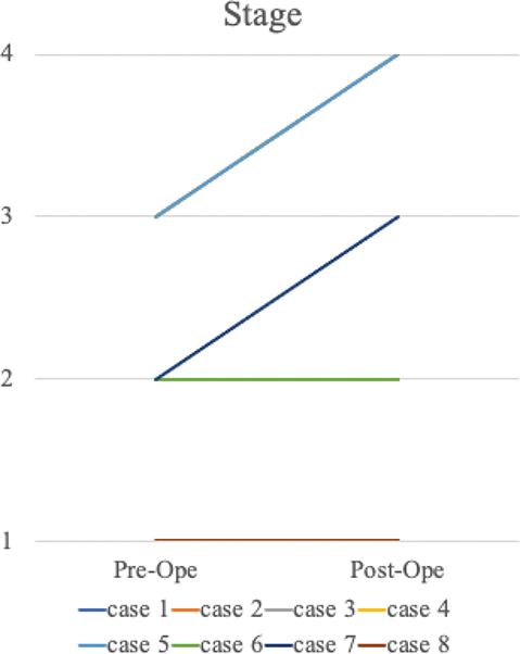
Figure 1. Herbert classification. Pre-Ope = pre-operation, Post-Ope = post-operation.
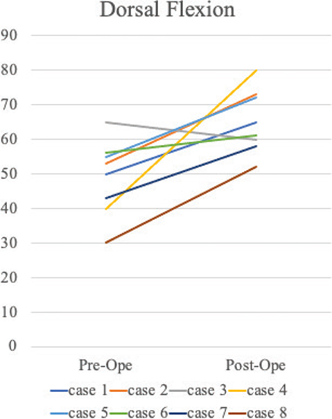
Figure 2. Dorsal flexion (degrees).
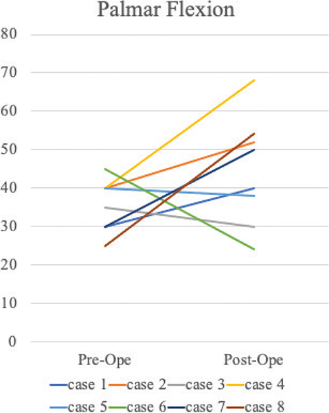
Figure 3. Palmar flexion (degrees).
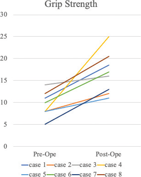
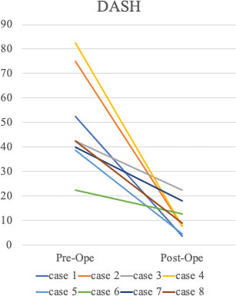
Figure 5. DASH score: disability and symptom. DASH: Disabilities of the Arm, Shoulder, and Hand.
All our patients had contralateral x-rays that could be evaluated. Three of the eight cases exhibited Type 2 navicular bone morphology as described by Morsy et al. [3].
Discussion
Preiser initially reviewed twenty-four cases of scaphoid injury and identified five instances of osteonecrosis without overt fracture, likening them to Kienböck’s and Köhler’s disease. However, a recent reassessment by Kallen et al. concluded that these five cases were secondary to unrecognized fractures rather than examples of idiopathic osteonecrosis [4]. Paradoxically, although Preiser described the condition in the context of scaphoid fracture, ‘Preiser’s disease’ is now commonly used to describe idiopathic necrosis of the entire scaphoid, irrespective of trauma history and excluding cases secondary to scaphoid pseudarthrosis.
Etiologies proposed for Preiser’s disease include trauma, microfractures, systemic disease, smoking, and alcohol use. However, no definitive causative factor has been identified. The prevailing hypothesis implicates a compromised vascular supply to the scaphoid as the underlying pathophysiological mechanism.
Scaphoid vascularity has been well documented. Gelberman et al. demonstrated that the bone receives perfusion from dorsal and palmar vessels [5]. The dorsal scaphoid is supplied primarily by branches of the radial artery, which enter through foramina in the dorsal ridge and nourish 70–80% of the bone. In contrast, palmar branches perfuse only the distal 20–30% of the scaphoid, with minimal intervascular anastomoses. Consequently, disruption of the dorsal supply, especially in the proximal pole, places this region at highest risk for avascular necrosis [6].
Kalainov et al. used MRI to classify Preiser’s disease into two types: Type 1, with necrosis of the entire scaphoid, and Type 2, where necrosis was limited to the proximal pole [7]. A history of wrist trauma was significantly more common in Type 2 cases, and treatment efficacy was better in general in this patient group. They hypothesized that avascular necrosis was more likely to occur due to partial trauma-related hemodynamic changes within or outside the scaphoid bone. Schmitt et al. also evaluated MRI images and reported that the scaphoid bone could be divided into three parts, with only the proximal pole undergoing necrosis in the early stages [8]. These findings, combined with the known vascular architecture, support the theory that interruption of dorsal vascular inflow is central to disease progression.
Buttermann et al. found that, during palmar flexion of the wrist, the extensor carpi radialis brevis tendon exerts compressive force on the site where the dorsal branch of the radial artery enters the scaphoid. They further noted that repetitive manual activity predisposes this region to synovitis, which in turn elevates intracompartmental pressure and consequently impairs arterial perfusion of the scaphoid [9].
Morsy et al. further discussed the relationship between scaphoid morphology and intraosseous vascularity [3]. They classified the scaphoid into two morphotypes, with the slender Type 2 exhibiting reduced vascular networks. Based on these findings, Type 2 navicular bones may be associated with the development of pseudarthrosis and Preiser’s disease. Although Amundsen et al. reported that half of their eight cases could be classified as Type 2, it is unclear whether this condition can lead to Preiser’s disease [10]. Our own cases also demonstrated a mix of morphotypes. All our patients had contralateral x-rays that could be assessed. Previous bilateral measurements for the scaphoids have shown that they are of similar size and shape within the same individual [11]. Therefore, by measuring the scaphoid bone on the contralateral side, we predicted the shape of the scaphoid bone on the affected side before disease onset. As a result, Type 2 morphology was noted in three of eight cases, but no correlation was observed with the stage of Preiser’s disease or postoperative outcomes.
With regard to disease management, conservative treatment has generally yielded poor outcomes. Lenoir et al. reviewed their own cases and found that, even at early stages, the disease progresses in all but the youngest patients [12]. Tomori et al. also followed eight patients treated with conservative therapy, most of whom showed progression of disease stage and lack of symptom improvement [13]. Based on these reports, surgical intervention is recommended even for Stage 1 or 2 Preiser’s disease.
There is no established standard when it comes to surgical treatment, and a variety of techniques and outcomes have been described. These include radial wedge osteotomy for decompression between the radius and scaphoid bones [14], VBG with vascular pattern for revascularization of the scaphoid bone, and salvage procedures such as proximal row carpectomy (PRC) and partial wrist fusion [15].
Tomori et al. reported on Preiser’s disease patients who were treated using wedge osteotomy [14]. Pain was improved in all patients, but more than half of them exhibited progression in disease stage based on plain radiographs, tendon rupture, and arthropathic changes in the distal radial joint. These findings suggest that VBG may be better for the treatment of early Preiser’s disease, such as Stage 1 or 2, where the scaphoid bone has not yet collapsed. Additionally, an osteotomy may improve symptoms but may not prevent the progression of scaphoid bone collapse. As Preiser’s disease progresses, the proximal scaphoid collapses, and the scapholunate (SL) ligament attachment fails, resulting in loss of intermovement with the lunate. Eventually, a dorsal intercalated segmental instability (DISI) deformity occurs, similar to pseudoarthrosis of the scaphoid or injury to the SL ligament. After a prolonged period of time, the carpal alignment is expected to fail, resulting in Scapholunate Advanced Collapse Wrist. In order to avoid this scenario, crushing of the proximal navicular bone must be prevented. In this regard, VBG is considered to be an effective treatment since it can directly revascularize the scaphoid bone and prevent collapse.
Donor sites for VBG have typically included the distal radius. Vascular pedicles that have been described include the 1,2 ICSRA [1], 2,3 ICSRA [16], and pedicled grafts involving the pronator quadratus muscle [17]. We selected the 1,2 ICSRA approach because of its anatomical proximity and favorable surgical exposure following radial styloidectomy.
Kara et al. used the pronator quadratus pedicle in seven Stage 2 patients and observed revascularization on MRI scans after 3 years [17]. Moran et al. reported symptomatic improvement in eight patients using either 1,2 or 2,3 ICSRA VBG and, thus, recommended this approach primarily for Stage 1–2 disease without carpal instability or arthritic changes [18]. In our series, four of five Stage 1–2 cases showed no disease progression, collapse, or the development of DISI deformity.
The use of VBG for Stage 3 disease remains controversial. Moran et al. found no improvement in MRI signal at the proximal pole after VBG in five Stage 3 cases [18]. Amundsen et al. observed that four of seven Stage 3 patients avoided collapse, but two required salvage procedures [10]. They concluded that VBG should be cautiously applied in early Stage 3; however, imaging modalities do not reliably differentiate between early and advanced Stage 3 disease. On the other hand, favorable outcomes have also been reported. Da Gama et al. performed VBG with the 1,2 ICSRA after confirming that the cartilage of the proximal end of the scaphoid bone was intact in Stage 3 Preiser’s disease. Postoperative plain X-ray and MRI images confirmed an improvement in bone necrosis and engraftment of the transplanted bone [19]. In our study, all three Stage 3 patients who underwent VBG showed improvements in DASH scores and Mayo Wrist Scores, but they all had stage progression as well. Even if VBG is performed in Stage 3, the disease may still progress and lead to DISI deformity and carpal instability in the future; thus, it may be more prudent to limit VBG to Stage 2 or apply it cautiously in Stage 3. If VBG is indicated for Stage 3, the patient should be informed preoperatively that the disease may progress and require future surgery. Alternatively, salvage surgery such as wedge osteotomy, proximal carpal row resection, PRC, or partial wrist fusion may be considered to alleviate symptoms [15].
We also noted that certain patients developed reduced palmar flexion following VBG with the 1,2 ICSRA, possibly due to surgical trauma to the dorsal wrist capsule (Table 1: cases 3, 5, and 6). This warrants additional caution when considering VBG in patients with good preoperative wrist mobility. Since it is debatable whether VBG or salvage surgery (e.g. PRC) should be used to treat Stage 3 disease, we believe that clinicians should proceed with caution before performing VBG in Stage 3 patients with no preoperative limitations for wrist range of motion.
There are certain limitations to this study that should be acknowledged. These include the small number of cases, the variable observation period, and the fact that cases classified as Stage 4 prior to surgery were not included. In particular, the variability in the observation period may affect the results of this report, since it is possible that the disease stage may change in the future.
In conclusion, VBG using the 1,2 ICSRA offers a viable treatment option for Stages 1 and 2 Preiser’s disease, effectively preventing proximal pole collapse and preserving carpal stability. Its use in Stage 3 remains controversial and should be reserved for carefully selected patients.
References
[1] Zaidemberg C, Siebert JW, Angrigiani C. A new vascularized bone graft for scaphoid nonunion. J Hand Surg Am. 1991; 16(3): 474–8. https://doi.org/10.1016/0363-5023(91)90017-6
[2] Herbert TJ, Lanzetta M. Idiopathic avascular necrosis of the scaphoid. J Hand Surg Br. 1994; 19(2): 174–182. https://doi.org/10.1016/0266-7681(94)90159-7
[3] Morsy M, Sabbagh MD, van Alphen NA, Laungani AT, Kadar A, Moran SL. The vascular anatomy of the scaphoid: new discoveries using micro-computed tomography imaging. J Hand Surg Am. 2019; 44(11): 928–938. https://doi.org/10.1016/j.jhsa.2019.08.001
[4] Kallen AM, Strackee SD. On the history and definition of Preiser’s disease. J Hand Surg Eur Vol. 2014; 39(7): 770–776. https://doi.org/10.1177/1753193413501099
[5] Gelberman RH, Menon J. The vascularity of the scaphoid bone. J Hand Surg Am. 1980; 5(5): 508–513. https://doi.org/10.1016/S0363-5023(80)80087-6
[6] Bellringer SF, MacLean SBM, Bain GI. Preiser’s disease-current concepts of etiology and management. Hand Clin. 2022; 38(4): 469–77. https://doi.org/10.1016/j.hcl.2022.03.013
[7] Kalainov DM, Cohen MS, Hendrix RW, Sweet S, Culp RW, Osterman AL. Preiser’s disease: identification of two patterns. J Hand Surg Am. 2003; 28(5): 767–78. https://doi.org/10.1016/S0363-5023(03)00260-0
[8] Schmitt R, Frohner S, van Schoonhoven J, Lanz U, Golles A. Idiopathic osteonecrosis of the scaphoid (Preiser’s disease) – MRI gives new insights into etiology and pathology. Eur J Radiol. 2011; 77(2): 228–34. https://doi.org/10.1016/j.ejrad.2010.11.009
[9] Buttermann GR, Putnam MD, Shine JD. Wrist position affects loading of the dorsal scaphoid: possible effect on extrinsic scaphoid blood flow. J Hand Surg Br. 2001; 26(1): 34–40. https://doi.org/10.1054/jhsb.2000.0475
[10] Amundsen A, Oh C, Huang TCT, Cantwell S, Hsu CC, Moran SL. Avascular necrosis of the scaphoid-Preiser disease: outcomes of 39 surgical cases. J Hand Surg Am. 2023; 48(3): 313 e1–e9. https://doi.org/10.1016/j.jhsa.2021.10.023
[11] Heinzelmann AD, Archer G, Bindra RR. Anthropometry of the human scaphoid. J Hand Surg Am. 2007; 32(7): 1005–8. https://doi.org/10.1016/j.jhsa.2007.05.030
[12] Lenoir H, Coulet B, Lazerges C, Mares O, Croutzet P, Chammas M. Idiopathic avascular necrosis of the scaphoid: 10 new cases and a review of the literature. Indications for Preiser’s disease. Orthop Traumatol Surg Res. 2012; 98(4): 390–397. https://doi.org/10.1016/j.otsr.2011.11.013
[13] Tomori Y, Nanno M, Takai S. Clinical outcomes of nonsurgical treatment for Preiser disease. Medicine (Baltimore). 2020; 99(4): e18883. https://doi.org/10.1097/MD.0000000000018883
[14] Tomori Y, Sawaizumi T, Nanno M, Takai S. Closing radial wedge osteotomy for Preiser disease: clinical outcomes. J Hand Surg Am. 2019; 44(10): 896 e1–896 e10. https://doi.org/10.1016/j.jhsa.2018.11.007
[15] Kazemi M, Daliri M, Moradi A. A systematic review on the management of idiopathic avascular necrosis of the scaphoid (Preiser’s disease). Orthop Traumatol Surg Res. 2023; 109(3): 103480. https://doi.org/10.1016/j.otsr.2022.103480
[16] Sheetz KK, Bishop AT, Berger RA. The arterial blood supply of the distal radius and ulna and its potential use in vascularized pedicled bone grafts. J Hand Surg Am. 1995; 20(6): 902–914. https://doi.org/10.1016/S0363-5023(05)80136-4
[17] Kara T, Gunal I. Preiser’s disease treated by pronotor quadratus pedicled bone graft. J Plast Surg Hand Surg. 2014; 48(6): 455–456. https://doi.org/10.3109/2000656X.2014.971803
[18] Moran SL, Cooney WP, Shin AY. The use of vascularized grafts from the distal radius for the treatment of Preiser’s disease. J Hand Surg Am. 2006; 31(5): 705–710. https://doi.org/10.1016/j.jhsa.2006.02.002
[19] da Gama SA, de Rezende MR, Ribak S. Treatment of Preiser disease with vascularized bone graft from the distal radius: a report of two cases. Hand (N Y). 2015; 10(3): 454–60. https://doi.org/10.1007/s11552-014-9733-8