ORIGINAL RESEARCH ARTICLE
Correction of tear trough deformity in young patients without eyebags using orbital fat reposition and release of tear trough ligament
Xianyu Zhoua†, Mengmeng Houb†, Shun Yuc, Jun Yanga, Yucheng Qiua* and Fei Liua*
aDepartment of Plastic & Reconstructive Surgery, The Ninth People’s Hospital Affiliated to Shanghai Jiao Tong University School of Medicine, Shanghai, China; bDepartment of Plastic Surgery, Zhejiang University School of Medicine Second Affiliated Hospital, Hangzhou, China; cDepartment of Burns and Plastic Surgery, Affiliated Hospital of Jiangnan University, Wuxi, China
ABSTRACT
Background: Tear trough deformity (TTD) is a periorbital hollow extending from the medial canthus to the midpupillary line, which is mainly caused by the traction of the tear trough ligament (TTL). This study aimed to present a retroseptal transconjunctival lower-eyelid blepharoplasty with orbital fat reposition and release of TTL for correcting TTD in young patients without eyebags.
Methods: Seventy-nine young patients with TTD without eyebags receiving the described technique were retrospectively reviewed from September 2020 to April 2023. TTD was preoperatively categorized into different Hirmand types. Cosmetic outcomes were evaluated through tear trough rating scale (TTRS), Barton grading system and patient self-assessment.
Results: The average follow-up time was 14.7 months (range, 12–18 months). Average TTRS in Hirmand class I and II were significantly different between pre- and post-operation (p < 0.05), while there was no significant difference in class I (p > 0.05). As to Barton grading system, TTDs in Hirmand class I were all graded as 1 preoperatively and were all shifted to grade 0 postoperatively. In class II, grade 1 (20%), grade 2 (71.4%), and grade 3 (8.6%) TTDs were shifted to grade 0 (85.7%) and grade 1 (14.3%) postoperatively. In class III, grade 2 (54.5%) and grade 3 (45.5%) TTDs were shifted to grade 0 (18.2%), grade 1 (45.5%) and grade 2 (36.3%). The overall patient self-assessed satisfaction was 96.2%.
Conclusions: Retroseptal transconjunctival lower-eyelid blepharoplasty with release of TTL and orbital fat reposition can achieve satisfactory outcomes to correct TTDs without eyebags.
KEYWORDS: Tear trough ligament; tear trough deformity; orbital fat reposition; orbicularis retaining ligament
Citation: Journal of Plastic Surgery and Hand Surgery 2026; 61: 72–77. DOI: https://doi.org/10.2340/jphs.v61.45298.
Copyright: © 2026 The Author(s). Published by MJS Publishing on behalf of Acta Chirurgica Scandinavica. This is an Open Access article distributed under the terms of the Creative Commons Attribution 4.0 International License (http://creativecommons.org/licenses/by/4.0/), allowing third parties to copy and redistribute the material in any medium or format and to remix, transform, and build upon the material, with the condition of proper attribution to the original work.
Received: 23 September 2025; Accepted: 24 November 2025; Published: 21 April 2026
CONTACT: Fei Liu; Yucheng Qiu plasticliu119@163.com; a1rose@sina.com Department of Plastic & Reconstructive Surgery, The Ninth People’s Hospital Affiliated to Shanghai Jiao Tong University School of Medicine, Shanghai, China; Department of Plastic & Reconstructive Surgery, The Ninth People’s Hospital Affiliated with Shanghai Jiao Tong University School of Medicine, Shanghai, China
†These two authors contributed equally to this work and should be listed as the co-first authors.
Competing interests and funding: The authors declare no competing interests.
This study was funded by National Natural Science Foundation of China (NO. 82102354), the Science and Technology Commission of Shanghai Municipality (NO. 22S31904500), and the Specific Research Project for Biomedicine Technology, Agency of Science and Technology, Yunnan Province (NO. 202402AA310072).
Introduction
Lower eyelid rejuvenation surgery is one of the most common cosmetic procedures in plastic surgery nowadays in Eastern Asians. Periorbital aging occurs early in facial aging. The aging of the lower eyelid area is not only manifested as eye bags but also periorbital skin sagging and lower eyelid depression. In 1932, Whitnall first described the tear trough as a fascial gap of the lower eyelid fixed to the bone anchored between the orbicularis oculi and angular head of the quadrates labii superioris muscles [1]. Flowers first introduced the terms tear trough and tear trough deformity (TTD) to describe the oblique depression at the lower eyelid–cheek junction [2]. Anatomically, TTD was defined as a medial depression bounded by the crista lacrimalis anterior and infraorbital rim, while Haddock characterized it as a depression that extended obliquely from the medial canthus to the midpupillary line, in which the orbicularis retaining ligament (ORL) was attached to the orbital rim at the midpupillary line, extending to the lateral canthus, and the deformity caused by ORL was the palpebromalar groove [3–5]. In 2012, Stutman proposed the final definition of palpebromalar groove and TTD [6]. TTD was finally determined as the periorbital hollow extending obliquely from the medial canthus to the midpupillary line, and the depression from the midpupillary line to the outer canthus was defined as the palpebromalar groove. Originating from the maxilla and extending to the inferior orbital rim, the tear trough ligament (TTL) forms a downward traction connected with the skin at the tear trough. At the mid-vertical line of the pupil, TTL extends as the ORL to the outside. Currently, it is believed that the TTL and the ORL are responsible for the deformities of tear trough and palpebromalar groove, respectively [7–11].
Previously, TTD accompanied by lower eyelid fat pads was often corrected by simply removing the bulging lower eyelid fat pads. The aesthetic improvement of TTDs was sometimes not appealing after surgery. TTD without lower eyelid fat pads is often treated with hyaluronic acid (HA) or autologous fat as a dermal filler [12–14]. Codner believes that the volume of lower eyelid orbital fat does not change with age, while the attenuation of the orbital septal structure is the key factor leading to the lower palpebral bulge [15]. The surgical procedure for the preservation and replacement of the lower eyelid fat pads obtains good results in the correction of the eye bags with TTD [16]. Therefore, we believe that TTD without lower eyelid fat pads can also be improved by release of the TTL and orbital fat reposition for the purpose of lower eyelid rejuvenation.
In this study, we retrospectively reviewed patients received surgical correction on TTDs without lower eyelid fat pads using retroseptal transconjunctival lower eyelid blepharoplasty with release of the TTL and orbital fat reposition. Cosmetic outcomes were evaluated based on Tear Trough Rating Scale (TTRS), Barton eyelid and cheek aesthetic grading scale [17], and patient self-assessment. We aimed to reveal the effectiveness of release of the TTL combined with orbital fat reposition on different Hirmand TTDs without lower eyelid fat pads.
Patients and methods
This work was approved by the Medical Ethics Committee of the Ninth People’s Hospital affiliated with Shanghai Jiao Tong University School of Medicine (approval No. SH9H-2020-T341-1) and conducted in accordance with its ethical standards, as well as the Helsinki declaration.
Patients
Outpatients were retrospectively reviewed who consulted about lower eyelid rejuvenation and underwent surgeries from September 2020 to April 2023. Among them, patients who had TTDs but without lower eyelid fat pad bulge (eyebags) and received retroseptal transconjunctival lower eyelid blepharoplasty with TTL release and orbital fat reposition were included. Patients had a history of lower eyelid surgery or obvious eyebags were excluded. Written informed consent was obtained from all patients.
Preoperative evaluations
The front and oblique photographs of the patients were taken with a digital camera. According to Hirmand’s clinical tear trough classifications, patients were divided into three categories (Hirmand I–III) [17]. Patients’ bilateral TTDs were evaluated and scored according to the TTRS created by Sadick considering the four indicators: depth of the trough, hyperpigmentation, prolapse of nasal fat pads/pockets, and lower eyelid skin rhytidosis. A higher score referred to more serious TTDs (Table 2) [4]. According to Barton’s grading scale for aesthetic evaluation of the eyelids and cheeks, lower eyelid regions were preoperatively graded [18]. For those with underlying diseases, especially diabetes and hypertension, the fasting blood glucose should be kept below 10 mmol/L, and the blood pressure should be kept no higher than 140/90 mmHg at least 1 week before surgery. The locations of the tear trough and palpebromalar groove were marked in the seated position. The lower eyelid fat pad volume was determined when the patient gazed upward. The laxity of lower eyelid skin and orbicularis oculi muscle was evaluated in the primary gaze and smiling. Symptoms such as symblepharon separation, ectropion and lower eyelid retraction were examined preoperatively.
Surgical procedure
After patients were placed in the supine position, they were disinfected and draped routinely. The lower lid margin was grasped to expose the fornix conjunctiva with the eye rolling upward. 2 mL of 2% lidocaine solution containing epinephrine (1:100,000) was injected into the conjunctiva and the orbital margin through conjunctiva while the oxybuprocaine hydrochloride eye drops (Benoxil) was applied to the eye for local anesthesia. The surgeon wore an auxiliary light source equipment (Ultra Light Optics, USA). The mucous membrane between the lower edge of the tarsal plate and the conjunctival fornix was cut horizontally about 1.5 cm with an electrocoagulation knife, and the tissue was dissected to the plane of the orbicularis oculi muscle and the anterior orbital space through the conjunctival incision. The central fat pad, the lateral fat pad, and the medial/nasal fat pad were exposed in turn. The capsule was fully loosened to release the orbital septum fat. The pedicle of the lateral fat pad close to the lateral canthus was partly separated so that it could be rotated. The space bed was lifted until the space exposed by the right-angle hook could be illuminated by a head-mounted light when the surgeon was in a sitting position. The shadowless light was also adjusted coordinately. The preorbital space was dissected toward the orbital rim to expose the arcus marginalis and the bony origin of the orbicularis oculi muscle. At the level of the arcus marginalis, the inner eyelid orbicularis oculi muscle was loosened by an electrocoagulation knife to expose TTL, which was then released. Along this plane, the scalpel handle wrapped in a single layer of rough gauze was used to perform blunt separation above the periosteum. The tissue was peeled medially until to the levator labii superioris alaeque nasi to access the anterior maxillary space, and laterally until to the anterior zygomatic space. A proper trim of the orbital fat was made. If the capsule was excessively loosened and the fat pads separated, the capsule could be sewn with a 6–0 absorbable polydioxanone (PDS) suture. Finally, the three masses of fat would be prepared as a rectangular-like pedicled fat flap, and fixed along the horizontal axis at supraperiosteal plane: medial, middle, and lateral parts. These three parts were secured to the medial periosteal point above the levator labii superioris alaeque nasi, the farthest periosteal point and the zygomatic periosteal point in pre-maxillary space, respectively, with a 5–0 absorbable PDS suture. The pedicled fat flap was flattened in the pre-maxillary space, the pre-zygomatic space (to modify the palpebromalar groove), and the preseptal space above the upper plane of the periosteum. The boundary of the pedicled fat flap slightly exceeded to the edge of the tear trough marked preoperatively in a sitting position. During operation, the surgical space was restricted, so the needle was held parallelly rather than at a right angle to facilitate a fine performance. The conjunctival incision left unsutured after surgery. Careful check on whether there was active bleeding was important. The skin surface of surgical area was dressed with sterile gauzes and elastic bandage.
Postoperative care
The dressing was removed about 12 hours after the operation. The cold compression therapy was applied within 72 hours combined with the oral medications of SETUS-M tablets (Melilotus Extract tablets) (three times a day, four tablets/time, 3 days) and dexamethasone tablets (one time a day, 10 mg/time/adult, 3 days), while the patient is placed in low-fowler’s position and the chloramphenicol eye drops are applied to the eye. The patient should avoid prolonged bending, weight-bearing, severe coughing and constipation, and all other factors that may cause much increased abdominal pressure. During the follow-up, the front and 45° oblique profiles were taken with the same digital camera.
Postoperative evaluations
Postoperative complications were recorded, including ecchymosis, swelling, chemosis, hematoma, symblepharon separation, ectropion, dry eyes, epiphora, etc. TTRS and Barton’s aesthetic rating scale were rated to evaluate postoperative outcomes on the eyelids and cheeks. Besides, patients’ self-assessment about their aesthetic perceptions was documented and presented as: satisfied, moderately satisfied, and dissatisfied.
Statistical analysis
SPSS 20.0 software (SPSS Inc., Chicago, IL) was used for TTRS paired-sample t-test statistical analysis. All results were expressed as mean ± standard deviation (Mean ± SD), with p value < 0.05 considered statistically significant.
Results
Seventy-nine patients underwent the described surgery were included. There were 24 men and 55 women, both of whose deformities were bilateral, with an age ranging from 21 to 38 years old (Table 1). Patients had varying degrees of TTDs, some of which were accompanied by palpebromalar groove deformity. Of them, 33 were categorized as Hirmand class I, 35 were Hirmand class II, and 11 were Hirmand class III (Table 1). The average follow-up time was 14.7 months (range, 12–18 months). Barton’s grading scale: 33 cases in Hirmand class I were rated as grade 1 preoperatively, and all were improved to grade 0 after the surgery. In Hirmand class II, cases of grade 1, grade 2, and grade 3 were 7, 25, and 3, respectively, before surgery; and it turned out that there were 30 cases of grade 0 and 5 cases of grade 1 postoperatively. In Hirmand class III, cases of grade 2 were 6, and cases of grade 3 were 5. After operation, there were 2 cases of grade 0, 5 cases of grade 1, and 4 cases of grade 2. As to the TTRS, the scores were 2.82 ± 1.15 in Hirmand class I, 3.83 ± 1.27 in Hirmand class II, and 4.78 ± 1.53 in Hirmand class III before the operation, which decreased to 2.02 ± 1.04, 2.38 ± 1.29 and 2.81 ± 1.48 after surgery, respectively (Table 2). Pre- and post-operative TTRS scores were not statistically significantly different in Hirmand class I (p > 0.05), while it was significantly different in Hirmand class II and III (p < 0.05) (Figure 1). Regarding the self-assessment of patients’ satisfaction, overall, 62 out of 79 were satisfied (78.5%), 14 moderately satisfied (17.7%), and 3 dissatisfied (3.8%). 3 patients who were dissatisfied were from Hirmand class III TTD (Figure 2). Most of the patients with TTDs of various Hirmand classes had early conjunctival edema or hyperemia (≥ 90.9%), and a very small number of patients had one or more of the following complications, such as ecchymosis, hematoma, and malar swelling. No severe complications were reported such as symblepharon separation, ectropion, eyelid retraction, diplopia, strabismus, dry eyes, or epiphora (Table 3). Typical cases were presented in Figures 3–5.
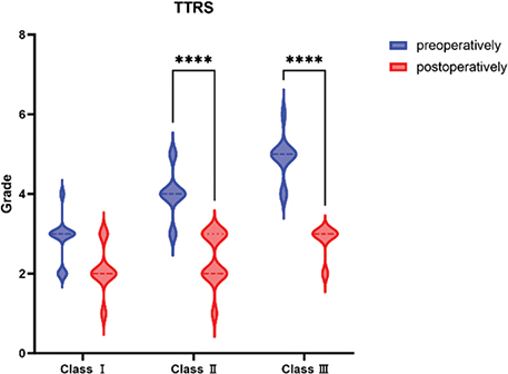
Figure 1. Statistical analysis of TTRS between pre- and post-operation in different Hirmand’s classifications. Pre- and post-operative TTRS scores were significantly different in Hirmand class II and III (p < 0.05). TTRS: Tear trough rating scale.

Figure 2. Self-assessment of patients’ satisfaction.
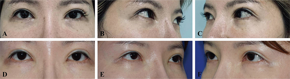
Figure 3. Typical case of a 26-year-old woman. (A–C) The patient was categorized as Hirmand type I, with a concave deformity (depth <1 mm) medially to the tear trough. There was slight palpebromalar pigmentation, but no drooping of the zygomatic fat pads, and no skin loosening in the lower eyelids. The preoperative TTRS score of both sides was 3. (D–F) Twelve months after surgery, the deformity of the bilateral tear trough was significantly improved, and the pigmentation vanished. Tear Trough Rating Scale score of both sides was shifted to 2 with satisfied self-assessment. TTRS: Tear trough rating scale.
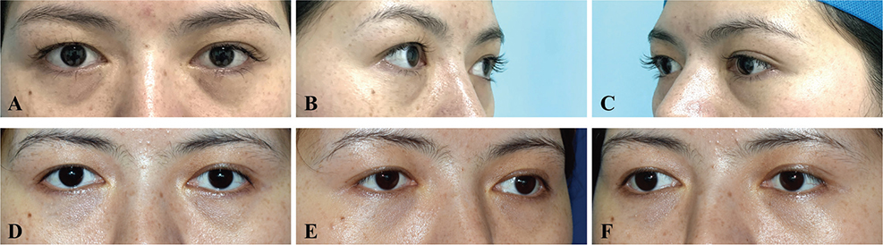
Figure 4. Typical case of a 29-year-old woman. (A–C) The patient was rated as Hirmand class II, with a volume deficiency from the medial orbital area to the middle orbital area. The depth of the tear-trough in the left eye was about 1.5mm, while that was about 1mm in the right eye. For both eyes, there was moderate pigmentation, and slight drooping of zygomatic fat pads without sagging in lower eyelid skin. Preoperative TTRS score was 6 in the left eye while that was 5 in the right one. (D–F) A certain degree of TTD still remained on 12 months after surgery, but the eyelid-cheek junction became smooth. Tear Trough Rating Scale score decreased to 3 on both sides. Self-assessment was recorded satisfied on the right side and moderately satisfied on the left side. TTRS: tear trough rating scale; TTD: tear trough deformity.
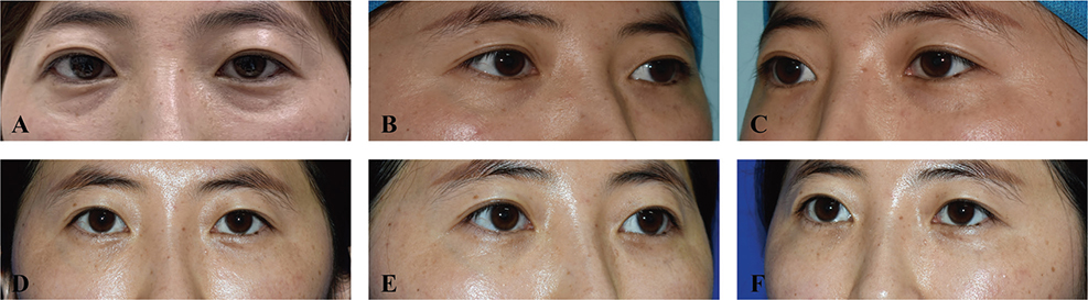
Figure 5. Typical case of a 31-year-old woman. (A–C) The patient was rated as Hirmand class III with obvious depression from the lower eyelid center to the periorbital area on both sides. The depth of the tear trough on both sides was about 2mm with slight pigmentation, no drooping of the zygomatic fat pad, mild folds of the lower eyelid skin, and obvious palpebromalar groove. Preoperative TTRS was scored 5 bilaterally. (D–F) Fourteen months postoperatively, TTD was significantly improved with a smooth and natural eyelid-cheek junction. Tear Trough Rating Scale score was shifted to 2 on both sides after surgery and the patient was very satisfied (self-assessment). TTRS: tear trough rating scale; TTD: tear trough deformity.
Discussion
Rejuvenated lower eyelid is often manifested as the indistinct boundary between eyelid and orbit, smooth transition, and natural extension to the cheek. Frequently, the lower eyelid bag is accompanied by a TTD. The retroseptal transconjunctival approach by Goldberg and the transcutaneous approach by Hamra represented the surgical procedures of preserving lower eyelid fat pads as well as correcting eyebags [19, 20]. Codner believed that the volume of lower eyelid lower eyelid fat pad did not change significantly with aging, however, the degeneration and relaxation of orbital septal tissue was a key factor in bulging of the lower eyelid fat [15]. Nowadays the transconjunctival approach is the first option for young patients with eye bags who do not have significant skin laxity in the lower eyelids. Patients in the present study were 21–38 years old, with varying degrees of TTD while not accompanied by lower eyelid bags. Based on Goldberg, Hamra and Codner’s surgical theories, preservation and reposition of orbital fat could improve eyebags and TTD, we believed that independent TTDs without eyebags could also be corrected using the same strategy. Therefore, in the present study, the retroseptal transconjunctival approach performed by Goldberg was conducted in young patients. Goldberg used a method of arcus marginalis incision and subperiosteal dissection. The pedicled fat flap was then reset under the periosteum for external fixation to the skin. Hidalgo performed the same procedures as that by Goldberg, except that more fat was preserved and the ORL was loosened more laterally [21]. Kawamoto and Wong replanted the fat pad into the preseptal space and pre-maxillary space with supraperiosteal dissection [16, 22]. The most prominent difference of the latter was the full loosening and ORL releasing and TTL. The surgical method used in this study was a combination of the different retroseptal transconjunctival approaches mentioned above. To be specific, with dissection of the pedicled orbital fat flap in upper plane of periosteum and fat repositioning at supraperiosteal plane, the ORL and TTL were fully loosened and released. The upper plane of the periosteum could offer a better fixation for the trimmed orbital fat while maximizing rejuvenation by increasing the volume of the infraorbital soft tissue and the fullness of the anterior zygomatic soft tissue. The vascular density in this plane was higher than that in the subperiosteal plane and could provide a better revascularization for translocated fat.
According to the classifications of tear trough depression proposed by Hirmand in 2010, patients in the present study were classified into type I, II and III [17]. Analysis demonstrated that the retroseptal transconjunctival approach of lower eyelid fat pad reposition and TTL release surgery achieved significant eyelid improvement in different Hirmand classes, especially in class I and II. Although TTRS scores did not change significantly in class I, the Barton aesthetic rating performed the best among these 3 classes. This may be mainly due to the different total periorbital depression in TTD and capacity loss. Hedén compared the effect of lower eyelid fat pad reposition and autologous fat grafting on lower eyelid aesthetics and revealed that insufficient volume of lower eyelid fat pad and restricted pedicle length of fat flap had negative impact on lower eyelid rejuvenation [23]. This was consistent with the conclusion that we had observed, which was that lower eyelid fat pad reposition without eyebags (insufficient orbital fat) was not effective to improve serious TTD in Hirmand class III. The contradiction between the subjective self-assessment results and the objective index analysis in TTD of class I suggested that TTRS was not suitable for evaluation of Hirmand class I, and the three-dimensional digital photographic analysis reported might be an alternative [24].
TTD was a complex phenomenon of lower eyelid aging caused by multiple factors. In addition to the constraint of the tear-trough-ORL system, TTD is also affected by the diversity of the thickness and quality of the lid-orbit, lid–cheek junction, the laxity of the orbital septum, bony atrophy, the skin pigmentation and sagging on the lower eyelid. Therefore, there was no one-size-fits-all surgical method. Hirmand used HA injection to correct different TTDs which achieved good results [17]. HA injection for correcting TTDs has gradually developed into a variety of injection techniques including the Vertical Supraperiosteal Depot Technique. Others have also achieved a good aesthetic appearance by using fat grafting [14]. Elhamaky reported a retroseptal transconjunctival lower blepharoplasty together with TTL and ORL release and resected orbital fat grafting [25]. HA was an absorbable foreign biological material which required repeated injections for the purpose of maintaining a good outcome. As to fat grafting, the biggest problem was unsure and unstable fat absorption. Once too much fat was grafted and the fat survived well, it would be awkward and difficult to deal with the redundant fat. If the fat did not survive well, refilling would be required. Thus, fat grafting was not only time-consuming and laborious but also expensive, preoperative comprehensive consent talk was required.
Conclusion
The retroseptal transconjunctival lower eyelid blepharoplasty with release of TTL and orbital fat reposition is a reliable and safe approach to correct TTDs without eyebags and achieve lower eyelid rejuvenation.
Author contributions
X Zhou performed the surgery, designed the study, analyzed the data, wrote a section of the manuscript and edited the figures. M Hou, S Yu and Y Jun analyzed the data and wrote a section of the manuscript. Y Qiu and F Liu wrote a section of the manuscript and substantively revised the manuscript.
Data availability statement
The datasets used and/or analyzed during the current study are available from the corresponding author on reasonable request.
Informed consent
Written informed consent was obtained from all individual patients for whom identifying information and/or photographs are included in this article. The consent ensures that the patients have reviewed and approved the use of their images for publication purposes. All efforts were made to ensure the privacy and confidentiality of the patients. Consent to publish was obtained from all the individuals participated in the study.
References
[1] Whitnall ES. The Anatomy of the human orbit and accessory organs of vision. 2nd ed. London: Oxford University Press; 1932:119–120
[2] Flowers RS. The tear trough deformity and its correction. Paper presented at: Annual meeting of the California Society of Plastic Surgeons. Monterey, CA, 1970.
[3] Chao Y, Pei-pei Z, Jun-hui L, et al. Anatomic study on the mechanism of tear trough deformity and palpbromalar groove deformity. Chin J Plast Surg. 2010;26:139–142.
[4] Sadick NS, Bosniak SL, Cantisano-Zilkha M, et al. Definition of the tear trough and the tear trough rating scale. J Cosmet Dermatol. 2007;6:218–222. https://doi.org/10.1111/j.1473-2165.2007.00336.x
[5] Haddock NT, Saadeh PB, Boutros S, et al. The tear trough and lid/cheek junction: anatomy and implications for surgical correction. Plast Reconstr Surg. 2009;123:1332–1340. https://doi.org/10.1097/PRS.0b013e31819f2b36
[6] Stutman RL, Codner MA. Tear trough deformity: review of anatomy and treatment options. Aesthet Surg J. 2012;32:426–440. https://doi.org/10.1177/1090820X12442372
[7] Mendelson BC, Muzaffar AR, Adams WP, Jr. Surgical anatomy of the midcheek and malar mounds. Plast Reconstr Surg. 2002;110:885–896; discussion 897–911. https://doi.org/10.1097/00006534-200209010-00026
[8] Wong C-H, Hsieh MKH, Mendelson B. The tear trough ligament: anatomical basis for the tear trough deformity. Plast Reconstr Surg. 2012;129:1392–1402. https://doi.org/10.1097/PRS.0b013e31824ecd77
[9] Muzaffar AR, Mendelson BC, Adams WP. Surgical anatomy of the ligamentous attachments of the lower lid and lateral canthus. Plast Reconstr Surg. 2002;110:873–884; discussion 897–911. https://doi.org/10.1097/00006534-200209010-00025
[10] Hwang K, Nam YS, Kim DJ, et al. Surgical anatomy of retaining ligaments in the periorbital area. J Craniofac Surg. 2008;19:800–804. https://doi.org/10.1097/SCS.0b013e31816b6c5e
[11] Kikkawa DO, Lemke BN, Dortzbach RK. Relations of the superficial musculoaponeurotic system to the orbit and characterization of the orbitomalar ligament. Ophthalmic Plast Reconstr Sur. 1996;12:77–88. https://doi.org/10.1097/00002341-199606000-00001
[12] Kane MAC. Treatment of tear trough deformity and lower lid bowing with injectable hyaluronic acid. Aesthetic Plast Surg. 2005;29:363–367. https://doi.org/10.1007/s00266-005-0071-7
[13] Sharad J. Dermal fillers for the treatment of tear trough deformity: a review of anatomy, treatment techniques, and their outcomes. J Cutan Aesthet Surg. 2012;5:229–238. https://doi.org/10.4103/0974-2077.104910
[14] Miranda SG, Codner MA. Micro free orbital fat grafts to the tear trough deformity during lower blepharoplasty. Plast Reconstr Surg. 2017;139:1335–1343. https://doi.org/10.1097/PRS.0000000000003356
[15] Codner MA. Reduction of lower palpebral bulge by plicating attenuated orbital septa: a technique modification in cosmetic blepharoplasty. Plast Reconstr Surg. 2002;105:2559–2560. https://doi.org/10.1097/00006534-200006000-00041
[16] Wong CH, Mendelson B. Extended transconjunctival lower eyelid blepharoplasty with release of the tear trough ligament and fat redistribution. Plast Reconstr Surg. 2017;140:273–282. https://doi.org/10.1097/PRS.0000000000003561
[17] Hirmand H. Anatomy and nonsurgical correction of the tear trough deformity. Plast Reconstr Surg. 2010;125:699–708. https://doi.org/10.1097/PRS.0b013e3181c82f90
[18] Barton FE, Jr, Ha R, Awada M. Fat extrusion and septal reset in patients with the tear trough triad: a critical appraisal. Plast Reconstr Surg. 2004;113:2115–2121; discussion 2122–2123. https://doi.org/10.1097/01.PRS.0000122409.00716.34
[19] Goldberg RA. Transconjunctival orbital fat repositioning: transposition of orbital fat pedicles into a subperiosteal pocket. Plast Reconstr Surg. 2000;105:743–748; discussion 749–751. https://doi.org/10.1097/00006534-200002000-00044
[20] Hamra ST. Arcus marginalis release and orbital fat preservation in midface rejuvenation. Plast Reconstr Surg. 1995;96:354–362. https://doi.org/10.1097/00006534-199508000-00014
[21] Hidalgo DA. An integrated approach to lower blepharoplasty. Plast Reconstr Surg. 2011;127:386–395. https://doi.org/10.1097/PRS.0b013e3181f95c66
[22] Kawamoto HK, Bradley JP. The tear ‘TROUF’ procedure: transconjunctival repositioning of orbital unipedicled fat. Plast Reconstr Surg. 2003;112:1903–1907; discussion 1908–1909. https://doi.org/10.1097/01.PRS.0000091238.72843.7E
[23] Hedén P, Fischer S. Comparison of fat repositioning versus onlay segmental fat grafting in lower blepharoplasty. Aesthet Surg J. 2021;41:NP717–NP727. https://doi.org/10.1093/asj/sjab070
[24] Donath AS, Glasgold RA, Meier J, et al. Quantitative evaluation of volume augmentation in the tear trough with a hyaluronic acid-based filler: a three-dimensional analysis. Plast Reconstr Surg. 2010;125:1515–1522. https://doi.org/10.1097/PRS.0b013e3181d70317
[25] Elhamaky TR. Orbital fat graft retroseptal transconjunctival blepharoplasty for treatment of groove in the infraorbital region. Int Ophthalmol. 2024;44(1):217. https://doi.org/10.1007/s10792-024-03148-z