ORIGINAL RESEARCH ARTICLE
Endoscopic cubital tunnel release: a modified surgery and clinical application
Yinglu Zhaoa,b,c,d, Jing Xua,b,c,d, Jie Laoa,b,c,d and Jianyun Yanga,b,c,d 
aDepartment of Hand Surgery, Huashan Hospital, Fudan University, Shanghai, China; bNHC Key Laboratory of Hand Reconstruction, Shanghai, China; cShanghai Key Laboratory of Peripheral Nerve and Microsurgery, Shanghai, China; dInstitute of Hand Surgery, Shanghai, China
ABSTRACT
Background: Varied surgical alternatives for treating cubital tunnel syndromes have been used. A trend of endoscopic ulnar nerve release is emerging. The purpose of this study is to introduce a modified technique of endoscopic ulnar nerve decompression in association with anterior transposition of the ulnar nerve and to assess the feasibility and efficacy of the surgery.
Materials and Methods: We introduced a modified method of endoscopic release and subfascial anterior transposition of the ulnar nerve. The technique was applied to six patients who presented signs, symptoms, and abnormal neurophysiological studies of cubital tunnel syndrome. The patients were classified according to the Dellon classification preoperatively. The Bishop rating system was used to evaluate the postoperative outcomes. Trial registration number for the study is MR-31-25-090620.
Results: Preoperatively, all six patients were classified as severe according to the Dellon classification. The endoscopic cubital tunnel release and subfascial anterior ulnar nerve transposition surgeries were performed with no difficulty in all patients. All the patients had improvement in symptoms and scored excellent or good according to the modified Bishop Rating System postoperatively.
Conclusions: The modified endoscopic cubital tunnel release and subfascial anterior ulnar nerve transposition technique is technically feasible with satisfactory outcomes in six patients in this study.
KEYWORDS: Cubital tunnel syndrome; endoscopy; transposition; ulnar nerve
Citation: Journal of Plastic Surgery and Hand Surgery 2026; 61: 115–120. DOI: https://doi.org/10.2340/jphs.v61.45986.
Copyright: © 2026 The Author(s). Published by MJS Publishing on behalf of Acta Chirurgica Scandinavica. This is an Open Access article distributed under the terms of the Creative Commons Attribution 4.0 International License (http://creativecommons.org/licenses/by/4.0/), allowing third parties to copy and redistribute the material in any medium or format and to remix, transform, and build upon the material, with the condition of proper attribution to the original work.
Received: 19 February 2026; Accepted: 12 April 2026; Published: 12 May 2026.
CONTACT: Jianyun Yang bellystraw@163.com Department of Hand Surgery, Huashan Hospital, 12 Middle Wulumuqi Road, Shanghai, 200040, China
Competing interests and funding: The authors report there are no competing interests to declare.
The authors received no financial support for the research, authorship, and/or publication of this article.
Introduction
Cubital tunnel syndrome (CuTS) is the second most frequent entrapment neuropathy of the upper extremity. The pathophysiology of CuTS is believed to be a combination of compression and traction, both of which can be aggravated by elbow flexion. Potential anatomical sites of entrapment include the arcade of Struthers, Osborne’s ligament, and the Flexor Carpi Ulnaris (FCU) muscle fascia [1]. Surgical treatment alternatives for treating CuTSs vary from simple decompression, medial epicondylectomy [2] to forms of complete neurolysis with anterior transposition of the ulnar nerve in subcutaneous [3], submuscular [4], or intramuscular [5] position. Each procedure has its advantages in certain patients, whereas all approaches involve extensive soft-tissue dissection and may result in substantial postoperative scarring [6]. Recent advancements have introduced minimally invasive and endoscopic techniques, enabling surgeons to achieve adequate visualization and intervention through smaller incisions compared to traditional techniques.
The essential goal of surgery for CuTS is to correct the etiological factors contributing to nerve entrapment on the ulnar nerve at the cubital tunnel. This involves removing compressive pathology, correcting subluxation, and reducing strain on the ulnar nerve. Several studies have reported on endoscopic-assisted cubital tunnel release (Hoffmann and Siemionow [7], Tsai [8], Mirza [9], and Cobb [10]), focusing on decompressing the ulnar nerve at the entrapment sites. These techniques alone may be insufficient for patients with severe elbow osteoarthritis characterized by restricted range of motion and radiographic evidence of medial osteophyte formation. Simple in-situ decompression or endoscopic release is also contraindicated in cases of cubitus valgus or clinically symptomatic ulnar nerve subluxation. In such circumstances, anterior transposition of the ulnar nerve is necessary to achieve adequate symptomatic relief while maintaining a low recurrence rate.
There are few reports of endoscopic release in association with anterior transposition of the ulnar nerve, especially in treating CuTS patients with elbow osteoarthritis and ulnar nerve subluxation. Therefore, we introduce a modified technique of endoscopic cubital tunnel release in association with subfascial anterior transposition of the ulnar nerve. The purpose of this paper is to present our surgical technique and share our experience in six patients with this technique.
Materials and methods
Our technique was applied to a consecutive series of six patients with primary CuTS, including four males and two females, between May 2024 and August 2024, involving the right side in five patients and the left in one patient. All patients involved gave their informed consent. All work was conducted with the formal approval of Institutional Review Board of Huashan Hospital, Fudan University. Trial registration number for the study is MR-31-25-090620. Diagnosis was based on history and clinical examination (sensory loss, positive Tinel sign, weakness and atrophy of the muscles innervated by the ulnar nerve, and positive elbow flexion test). It was furthermore confirmed by abnormal neurophysiological studies. Exclusion criteria include recurrent CuTS and severe cervical spine or shoulder diseases. All surgery was standardized and performed by the senior author to minimize performance bias. Data were collected including age, preoperative Dellon scale of severity of symptoms [11] (Table 1), and motor nerve conduction velocity (MNCV) across the elbow.
Based on preoperative Dellon classification, all six patients were classified as severe. All patients manifested as numbness and partial sensory loss on the little finger and ulnar side of the ring finger. These symptoms had persisted for an average of 21 months (6 to 48 months). All patients had positive elbow flexion test and Tinel sign along the course of ulnar nerve at the elbow. The preoperative electrophysiological studies were abnormal in all cases. All patients presented with atrophy of the muscles innervated by the ulnar nerve. Five patients presented with marked medial osteophyte on preoperative X-ray, and one patient had subluxation of the ulnar nerve.
The mean follow-up time was 12.33 (12 to 14) months, and the outcome was evaluated according to the Bishop Rating System [12], which includes subjective information (severity of the residual deficits, postoperative subjective improvement, and postoperative work status) and objective parameters (pinch strength and sensory two-point discrimination). The Bishop score is defined as poor, 0 to 2; fair, 3 to 4; good, 5 to 7; and excellent, 8 to 9 (Table 2). Pinch strength was measured with a Pinch Gauge (North Coast). Static two-point discrimination testing was carried out preoperatively and postoperatively. Postoperative electrophysiological studies were conducted additionally.
Instruments
Surgery was performed using instruments shown in Figure 1.

Figure 1. Surgical instruments used in this study: (A) L-shaped retractor, (B) speculum, (C) plastic plate, (D) dissection scissors, (E) tunneling forceps, and (F) 4.0-mm 30-degree endoscope.
Surgical technique
The operation is carried out under regional block anesthesia. A tourniquet is applied as high as possible on the upper arm. The arm is positioned in 90-degree shoulder abduction on a hand table, the elbow joint flexed and supinated.
An incision measuring 25 mm in length is made along the retrocondylar groove (Figure 2). Atypical musculature (anconeus epitrochlearis muscle), though rare, can hinder dissection and needs to be resected. The cubital tunnel roof is transected, and the ulnar nerve is identified.

Figure 2. An incision is made along the retrocondylar groove.
The space required for endoscopic intervention is then created. Tunneling forceps are inserted underneath the deep fascia of the forearm. A space sufficiently large to accommodate the instruments is created by gently spreading the tunneling forceps distally. A speculum (blade length approximately 10 cm) retractor is introduced into the space. At this time, the medial antebrachial cutaneous nerve (MACN) is lifted up above the speculum. Endoscope is placed in, and Osborne’s ligament is divided. Both heads of the FCU and its covering fascia are then endoscopically visible. A plastic plate (length 10 cm, width 10 mm) is placed above the ulnar nerve. The FCU muscle and its covering fascia are divided using an ultrasonic scalpel (Figure 3) to a point approximately 10 cm from the midpoint of the retrocondylar groove. Once the FCU muscle is divided, the speculum is reinserted in between the two heads of FCU muscle. Spreading open the speculum, the ulnar nerve and the covering submuscular membrane are revealed. The submuscular membrane should be thoroughly divided using scissors (Figure 4). Any muscle branches that come off from the ulnar nerve must be protected. The nerve is then dissected from its surroundings in a 360-degree manner for subsequent anterior transposition.

Figure 3. The FCU muscle and its covering fascia are divided: (A) Speculum, (B) plastic plate, (C) the FCU muscle and its covering fascia, and (D) ultrasonic scalpel.

Figure 4. The submuscular membrane is divided using scissors: (A) Speculum, (B) the ulnar nerve. (C) submuscular membrane, and (D) scissors.
The surgical procedure is carried out similarly in a proximal direction. A space is created above the layer of the intermuscular septum. After placing the speculum retractor, the intermuscular septum is divided (Figure 5), for up to a length of 10 cm proximal to the midpoint of the retrocondylar groove. If the triceps has an aponeurotic edge, it is divided as well. Care is taken to protect the MACN. The speculum is then removed, and a L-shaped retractor is inserted. The medial intermuscular septum and the Struther’s arcade are resected (Figure 6) to avoid consequent compression after anterior transposition of the ulnar nerve. The ulnar nerve is freed from its attachments in a 360-degree manner.
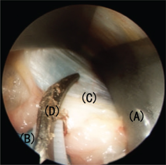
Figure 5. The intermuscular septum is divided. (A) Speculum, (B) plastic plate, (C) intermuscular septum, and (D) ultrasonic scalpel.

Figure 6. The medial intermuscular septum and the Struther’s arcade are resected. (A) L-shaped retractor, (B) plastic plate, (C) intermuscular septum, and (D) scissors.
Following these steps, subfascial anterior transposition of the ulnar nerve is to be performed. The nerve is carefully lifted from its bed. Atraumatic dissection of the motor branches from the ulnar nerve needs to be performed if necessary, allowing adequate anterior nerve transposition without tension. A flap of flexor pronator fascia based on the medial epicondyle is created (Figure 7). The ulnar nerve is moved anteriorly and examined to ensure that there is no iatrogenic compression and kinking of the ulnar nerve. The flap is then sutured to the overlying subcutaneous tissue to create a barrier that keeps the ulnar nerve in its transposed position. Passive mobilization of the elbow is made to confirm that there is no subluxation of the ulnar nerve after anterior transposition.

Figure 7. A flap of flexor pronator fascia based on the medial epicondyle is carved.
Vessels at potential bleeding sites are cauterized under the endoscopic vision. The tourniquet is released. The wound is closed, and a compression dressing is applied. A long-arm splint is recommended for 3 weeks of immobilization.
Ethics
All patients involved in this study gave their informed consent. Ethical approval for this study was obtained from Institutional Review Board of Huashan Hospital, Fudan University (2025-HSYY-1288).
Results
All patients reported improvement of symptoms after surgery (Table 3). The clinical and electrophysiological follow-up lasted an average of 12.33 (12 to 14) months. At the latest follow-up, excellent result (8 or 9 points on the Bishop scale) was observed in 1 patient (Patient No. 6), who had nerve subluxation preoperatively. The other five patients, who presented with medial osteophyte on preoperative X-ray, also showed good results (5 to 7 points on the Bishop scale). On average, these patients reached 6.00 points on the Bishop scale. An objective improvement in the static two-point discrimination, pinch strength and MNCV across the elbow occurred in all the patients at their latest follow-up.
| Patient number | Age (years) | Gender | Dellon classification | Duration of symptoms (month) | Pre-operative data | Follow-up (month) | Post-operative data | ||||||
| MNCV (m/s) | Pinch strength (%) * | 2-PD (mm) | Atrophy | MNCV (m/s) | Pinch strength (%) * | 2-PD (mm) | Bishop score | ||||||
| 1 | 56 | M | Severe | 12 | 12.2 | 45 | 12 | +++ | 14 | 31.3 | 60 | 8 | 6 |
| 2 | 65 | M | Severe | 48 | 17.2 | 30 | 15 | +++ | 12 | 34.6 | 40 | 10 | 5 |
| 3 | 59 | M | Severe | 24 | 24 | 60 | 10 | ++ | 12 | 32 | 65 | 8 | 6 |
| 4 | 74 | F | Severe | 24 | 23.3 | 50 | 10 | +++ | 12 | 35.3 | 60 | 6 | 6 |
| 5 | 77 | F | Severe | 6 | 13.6 | 60 | 8 | ++ | 12 | 30.2 | 65 | 6 | 5 |
| 6 | 34 | M | Severe | 12 | 37.9 | 81 | 8 | + | 12 | 45.9 | 90 | 4 | 8 |
| Mean | 60.83 | 21.00 | 54.33 | 10.50 | 12.33 | 63.33 | 7.00 | 6.00 | |||||
| MNCV: motor nerve conduction velocity *Compared with the contralateral side. |
|||||||||||||
The mean operative time was 80 min. The mean tourniquet time was 50 min. The endoscopic cubital tunnel release was not necessary to convert to open surgery in our cases. There were no injuries to nerves or vessels as a result of employing this technique. No complications of infection, painful scarring, hypodermic pneumatosis, superficial hematoma, or sensory loss caused by MACN injury occurred in our cases. There has been no recurrence during the follow-up period. All the patients were satisfied with the results. They all returned to their previous jobs or daily activities.
Discussion
There is a trend to minimize the incision for cubital tunnel surgery to avoid extensive tissue dissection, allowing for quicker recovery and a less troublesome scar [13]. Advances in technology and improved understanding of anatomy have led to the development of endoscopic ulnar nerve release. The concept of endoscopic ulnar nerve release was pioneered by Tsai in 1992, using a custom-made glass tube as means of guidance. He later reported on a series of 112 patients in 1999 [8] with his endoscopic approach. However, the glass tube was too bulky and was not easy to be placed into the tight cubital tunnel. It was also not readily available [14]. Another endoscopic technique was reported by Nakao in 2001 [6], involving three separate 5-mm incisions to enable a fine tape to be inserted subcutaneously and ‘lifted’ up. This produced a space above the ulnar nerve for dissection with a retrograde knife. However, this technique has the potential risk to injure cutaneous nerves. As the retrograde knife is outside the cubital retinaculum and the deep edge of the knife can’t be seen, the ulnar nerve and its motor branches could be injured as well [14]. In 2006, tunneling forceps and illuminated specula were used by Hoffmann and Siemionow to release the ulnar nerve for up to 17 cm in length through a 28 mm longitudinal skin incision [7]. Krishnan later described his technique of building a pocket in the subcutaneous tissue with the use of a large retractor. The scope he used was a combination of the optical system and long retractor, which was believed to be not cost-effective [15]. Flores reduced the costs by applying a neuro-endoscopic technique originally designed for ventricular endoscopy, in which the scope and the retractor were used separately [16]. Merolla reported an endoscopic technique with arthroscopic shaver and blunt retractor using elbow arthroscopic portals [17]. All techniques above focus on decompression of the ulnar nerve by dividing the structures at entrapment sites, with the ulnar nerve remained in situ.
There have been several studies comparing the results of techniques whether to transpose the ulnar nerve anteriorly or not for CuTS, but the ideal solution has not been universally acknowledged yet. Macadam reviewed ten studies including 449 simple decompressions, 342 subcutaneous transpositions, and 115 submuscular transpositions [18]. The studies did not show any statistically significant differences between the groups. However, a trend could be seen favoring transposition of the nerve [18]. Mackinnon reviewed the trend for treating CuTSs in the US and found that most surgeons used more than one operative procedure in their treatment of patients with CuTS [19]. The selection of the surgical procedure was influenced by evidence of muscle atrophy, abnormal nerve conduction studies, and failed nonoperative treatment [20]. Correction of the etiological factors and reducing strain on the nerve are the key points to obtain satisfactory results. In our center, anterior transposition of the ulnar nerve is a standard procedure when dealing with clinically severe CuTS. It is also performed for CuTS patients with severe osteoarthritis, cubitus valgus, and clinically symptomatic ulnar nerve subluxation.
Very few literatures have addressed endoscopic cubital tunnel release in association with anterior transposition of the ulnar nerve. Konishiike used the tube of the ‘USE system’ to guide the endoscope [21]. After transposing the ulnar nerves anteriorly, the cubital tunnel was closed by suturing the divided Osborne’s ligament to the medial epicondyle. According to their study, excellent outcomes were obtained in nine patients and good results in eight patients, while three patients had fair results. However, the operation space inside a tube of USE system could be narrow, causing difficulties in observing and dissecting. Meanwhile, insertion of the USE tube into the cubital tunnel could compress the nerve directly and cause iatrogenic nerve palsy. Vessels and MACN outside the cubital tunnel are difficult to observe and prone to be injured. Jiang used CO2 insufflation through an endoscopic approach to produce a subcutaneous pocket for fascia release for a distance of 20 cm around the medial epicondyle [22]. At the follow-up of 15 months in average, excellent results were observed in ten out of 12 patients (83.3%), with the other two patients showing good results (16.7%). However, insufflation of CO2 is comparatively not cost-effective and requires additional equipments. Management of suitable pressure for gas insufflation can be challenging, for higher pressure would cause complications such as hypodermic pneumatosis and lower pressure would difficult adequate visualization to perform endoscopic neurolysis. Wong designed a zigzag incision and optic speculum instrument sets to create a space for endoscopic neurolysis and further subfascial nerve transposition [23]. Improvements of symptoms were seen in 19 of 21 patients, and no complications were noted. However, their design of a zigzag incision seems redundant and could be replaced by a simple longitudinal incision. Additionally, patients in their study were specifically included only if subluxation of the nerve occurred after the initial decompression of the ulnar nerve.
In our study, we introduce the speculum forceps to create an operation cavity approximately 10 cm in length and 3 cm in width. The cavity is stable and spacious, accommodating the surgical instruments and exposing all entrapment structures from outside the cubital tunnel under endoscopic view. Once the speculum forceps are inserted, the MACN is lifted up and hence protected. We did not injure any MACN in our case series. Hematoma is the main complication seen at an increased rate after endoscopic cubital tunnel releases. For this reason, we use an ultrasonic scalpel for dividing the FCU muscle and the fibrous fascia overlying the ulnar nerve. The ultrasonic scalpel has a strong hemostatic ability, as evidenced by the absence of bleeding upon releasing the tourniquet during the final stage of the surgical procedure. Furthermore, the ultrasonic scalpel is capable of achieving an adequate length and effectively cauterizing blood vessels at sites of latent hemorrhage. To insulate the heat generated by the ultrasonic scalpel, a plastic plate is placed above the ulnar nerve. The plastic plate serves to isolate the nerve, providing operational space and safeguarding the nerve against physical injuries. Using the ultrasonic scalpel and the plastic plate also facilitates the endoscopic resection of the fibrous band of the intermuscular septum and the Struther’s arcade, which prevents consequent compression after anterior transposition of the ulnar nerve. We create a flap of flexor pronator fascia, utilizing the medial epicondyle as its base and subsequently suture it to the overlying subcutaneous tissue to secure the anterior transposition of the ulnar nerve. The flap features a strong and relatively broad base with a width of approximately 3 cm, which effectively prevents iatrogenic compression of the ulnar nerve and ensures the stability of the flap. Instruments in our technique are easy to acquire.
In this study, we showed that our modified endoscopic release and subfascial anterior transposition of the ulnar nerve are technically feasible. The ulnar nerve can be sufficiently released, safely transposed, and securely maintained in its transposed position by a subfascial flap. The procedure has a relatively short learning curve. Satisfactory relief of symptoms in our CuTS patients with nerve instability and osteoarthritis is achieved. No complications or recurrence occurred during the follow-up period. The limitation of our study is that more clinical cases are needed. Results after long-term follow-up are required. Future studies are planned to address these issues.
Conclusions
The modified technique of endoscopic cubital tunnel release and subfascial anterior ulnar nerve transposition is technically feasible with satisfactory outcomes in six patients in this study.
Acknowledgments
None.
References
[1] Gabel GT, Amadio PC. Reoperation for failed decompression of the ulnar nerve in the region of the elbow. J Bone Joint Surg Am. 1990;72(2):213–219. https://doi.org/10.2106/00004623-199072020-00008
[2] Kuschner SH. Cubital tunnel syndrome. Treatment by medial epicondylectomy. Hand Clin. 1996;12(2):411–419. https://doi.org/10.1016/S0749-0712(21)00325-5
[3] Eaton RG, Crowe JF, Parkes JC, 3rd. Anterior transposition of the ulnar nerve using a non-compressing fasciodermal sling. J Bone Joint Surg Am. 1980;62(5):820–825. https://doi.org/10.2106/00004623-198062050-00019
[4] Rayan GM. Proximal ulnar nerve compression. Cubital tunnel syndrome. Hand Clin. 1992;8(2):325–336. https://doi.org/10.1016/S0749-0712(21)00718-6
[5] Kleinman WB, Bishop AT. Anterior intramuscular transposition of the ulnar nerve. J Hand Surg Am. 1989;14(6):972–979. https://doi.org/10.1016/S0363-5023(89)80046-2
[6] Nakao Y, Takayama S, Toyama Y. Cubital tunnel release with lift-type endoscopic surgery. Hand Surg. 2001;6(2):199–203. https://doi.org/10.1142/S0218810401000679
[7] Hoffmann R, Siemionow M. The endoscopic management of cubital tunnel syndrome. J Hand Surg Br. 2006;31(1):23–29. https://doi.org/10.1016/J.JHSB.2005.08.008
[8] Tsai TM, Chen IC, Majd ME, et al. Cubital tunnel release with endoscopic assistance: results of a new technique. J Hand Surg Am. 1999;24(1):21–29. https://doi.org/10.1053/jhsu.1999.jhsu25a0021
[9] Mirza A, Reinhart MK, Bove J, et al. Scope-assisted release of the cubital tunnel. J Hand Surg Am. 2011;36(1):147–151. https://doi.org/10.1016/j.jhsa.2010.10.016
[10] Cobb TK. Endoscopic cubital tunnel release. J Hand Surg Am. 2010;35(10):1690–1697. https://doi.org/10.1016/j.jhsa.2010.07.030
[11] Dellon AL. Techniques for successful management of ulnar nerve entrapment at the elbow. Neurosurg Clin N Am. 1991;2(1): 57–73. https://doi.org/10.1016/S1042-3680(18)30757-5
[12] Nouhan R, Kleinert JM. Ulnar nerve decompression by transposing the nerve and Z-lengthening the flexor-pronator mass: clinical outcome. J Hand Surg Am. 1997;22(1):127–131. https://doi.org/10.1016/S0363-5023(05)80192-3
[13] Watts AC, Bain GI. Patient-rated outcome of ulnar nerve decompression: a comparison of endoscopic and open in situ decompression. J Hand Surg Am. 2009;34(8):1492–1498. https://doi.org/10.1016/j.jhsa.2009.05.014
[14] Bain GI, Bajhau A. Endoscopic release of the ulnar nerve at the elbow using the Agee device: a cadaveric study. Arthroscopy. 2005;21(6):691–695. https://doi.org/10.1016/j.arthro.2005.02.018
[15] Krishnan KG, Pinzer T, Schackert G. A novel endoscopic technique in treating single nerve entrapment syndromes with special attention to ulnar nerve transposition and tarsal tunnel release: clinical application. Neurosurgery. 2006;59(1 Suppl 1):ONS89–ONS100. https://doi.org/10.1227/01.NEU.0000219979.23067.5C
[16] Flores LP. Endoscopically assisted release of the ulnar nerve for cubital tunnel syndrome. Acta Neurochir (Wien). 2010;152(4): 619–625. https://doi.org/10.1007/s00701-009-0578-9
[17] Merolla G, Staffa G, Paladini P, et al. Endoscopic approach to cubital tunnel syndrome. J Neurosurg Sci. 2008;52(3):93–98.
[18] Macadam SA, Gandhi R, Bezuhly M, et al. Simple decompression versus anterior subcutaneous and submuscular transposition of the ulnar nerve for cubital tunnel syndrome: a meta-analysis. J Hand Surg Am. 2008;33(8):1314.e1–e12. https://doi.org/10.1016/j.jhsa.2008.03.006
[19] Lowe JB, 3rd, Maggi SP, Mackinnon SE. The position of crossing branches of the medial antebrachial cutaneous nerve during cubital tunnel surgery in humans. Plast Reconstr Surg. 2004;114(3):692–696. https://doi.org/10.1097/01.PRS.0000130966.16460.3C
[20] Novak CB, Mackinnon SE. Selection of operative procedures for cubital tunnel syndrome. Hand (N Y). 2009;4(1):50–54. https://doi.org/10.1007/s11552-008-9133-z
[21] Konishiike T, Nishida K, Ozawa M, et al. Anterior transposition of the ulnar nerve with endoscopic assistance. J Hand Surg Eur Vol. 2011;36(2):126–129. https://doi.org/10.1177/1753193410381675
[22] Jiang S, Xu W, Shen Y, et al. Endoscopy-assisted cubital tunnel release under carbon dioxide insufflation and anterior transposition. Ann Plast Surg. 2012;68(1):62–66. https://doi.org/10.1097/SAP.0b013e318211913c
[23] Wong JK, Hsu CC, Lin CH, et al. Endoscopy-assisted subfascial anterior transposition of the ulnar nerve for the treatment of cubital tunnel syndrome. J Plast Reconstr Aesthet Surg. 2016;69(12): 1704–1710. https://doi.org/10.1016/j.bjps.2016.09.004