ORIGINAL RESEARCH ARTICLE
The influence of patient and surgical factors on prognosis in repeated flexor tenorrhaphy
Jong Ju Park  , Si Young Roh , Sung Hoon Koh , Dong Chul Lee , Jin Soo Kim and Kyung Jin Lee
Department of Plastic and Reconstructive Surgery, Gwangmyeong Sungae General Hospital, Gwangmyeong, Republic of Korea
ABSTRACT
Purpose: This study aimed to investigate factors influencing outcome in patients who experienced re-rupture after flexor tenorrhaphy.
Background: Flexor tendon re-rupture following tenorrhaphy remains a significant complication that can adversely affect functional outcomes and require repeated surgical intervention. This study aimed to investigate factors influencing prognosis in patients who experienced re-rupture after flexor tenorrhaphy.
Materials and Methods: We retrospectively analyzed patients who underwent flexor tendon tenorrhaphy on little fingers between January 2011 and September 2024 and subsequently experienced re-rupture followed by a primary repair. We analyzed factors such as re-rupture timing, rupture to surgery interval, patient demographics, medical history, and participation in rehabilitation therapy using a simple linear regression model. Patients with underlying diseases, concurrent fractures, thumb or extensor tendon ruptures, or follow-up periods less than 6 months were excluded. The Adjusted Strickland and Whitaker methods were used for evaluation.
Results: A total of 17 patients out of 313 who underwent flexor tendon repair experienced re-rupture and met the inclusion criteria. Age and re-rupture period significantly affected prognosis. Older patients showed poorer final range of motion outcomes. In contrast, the initiation date, frequency, and duration of rehabilitation therapy did not demonstrate statistically significant correlations with prognosis.
Conclusions: Older age and delayed re-rupture were associated with poorer postoperative finger range of motion following repeated flexor tenorrhaphy. Therefore, patients should be advised to avoid excessive hand use until adequate tendon healing is achieved, and thorough postoperative education may be particularly important in elderly patients.
KEYWORDS: Finger; tendon injuries; rupture; reinjuries; reoperation; prognosis
Citation: Journal of Plastic Surgery and Hand Surgery 2026; 61: 237–242. DOI: https://doi.org/10.2340/jphs.v61.46464.
Copyright: © 2026 The Author(s). Published by MJS Publishing on behalf of Acta Chirurgica Scandinavica. This is an Open Access article distributed under the terms of the Creative Commons Attribution 4.0 International License (http://creativecommons.org/licenses/by/4.0/), allowing third parties to copy and redistribute the material in any medium or format and to remix, transform, and build upon the material, with the condition of proper attribution to the original work.
Received: 9 March 2026; Accepted: 24 June 2026; Published: 10 September 2026.
CONTACT: Si Young Roh psczero@gmail.com Department of Plastic and Reconstructive Surgery, Gwangmyeong Sungae General Hospital, 36 Digital-ro, Gwangmyeong 14241, Korea
Competing interests and funding: The authors declare no conflicts of interest.
The authors have nothing to disclose.
None.
Introduction
Flexor tendon injuries are a complex area of hand surgery that require precise repair techniques and postoperative care to achieve optimal functional recovery. Despite advances in surgical methods and rehabilitation protocols, re-rupture remains a significant complication that undermines patient outcomes and necessitates further intervention [1]. This prolongs the recovery process and imposes additional physical and emotional burdens on patients. Previous studies have also reported rare mechanisms of delayed or spontaneous flexor tendon rupture, highlighting the complexity of tendon healing and structural vulnerability after injury repair [2]. Recent cadaveric studies have demonstrated anatomical variability within the flexor tendon system, highlighting the importance of detailed anatomical understanding during flexor tendon surgery [3].
The reported re-rupture rate following flexor tendon repair ranges from 3% to 9% in the literature, with contributing factors including overly aggressive therapy [4, 5]. Previous studies have demonstrated that secondary tendon rupture may result from multiple mechanisms, including postoperative mechanical irritation and structural compromise of the repaired tendon [6]. In cases of tendon rupture, immediate exploration and repair within 48 h is recommended [7], with 50% of patients achieving ‘Good’ or ‘Excellent’ based on the adjusted Strickland criteria [8, 9]. However, there is still no consensus on the necessity of immediate repair for re-ruptured flexor post-tenorrhaphy [10]. Therefore, we investigated the relationship between the time interval from re-rupture to surgery, the extent of participation in physical therapy, and the final prognosis.
In addition, there has been a lack of research investigating the relationship between the timing of tendon rupture and the final prognosis. Tendon healing progresses through three phases: an inflammatory phase (week 1), a proliferative phase (weeks 1–3), and a remodeling phase (weeks 3–8) [4, 11]. We considered the timing of re-rupture to be associated with the stage of tendon healing and sought to evaluate the impact of re-rupture timing on the ultimate clinical outcome.
Materials and methods
Patient selection
This retrospective study included 324 patients who underwent flexor tendon repair on little fingers at our facility between January 2011 and September 2024. Sixteen patients experienced re-rupture following flexor tenorrhaphy in zone 1 (from the fingertip to the proximal interphalangeal (PIP) joint) [12]. The study included individuals who underwent repeated tendon repair, not tendon graft nor tendon transfer, for re-rupture [13]. Patients with concomitant fractures or extensor ruptures were excluded owing to differences in post-surgical rehabilitation methods and splinting. Thumb injuries were excluded because their prognostic evaluation methods differed from those of the other fingers. Patients with severe systemic diseases other than diabetes mellitus and smoking history that could significantly interfere with tendon healing were excluded from the study. Assuming that tendon recovery takes approximately 2 months, only patients who were available for a follow-up period of ≥ 3 months were included in this study [14].
Data selection
We investigated the patients’ demographics, date of initiation, frequency and duration of physical therapy. The interval from the initial operation to re-rupture and the interval from re-rupture to exploratory surgery were evaluated. Outcomes were assessed using the Strickland evaluation method, which calculates the ratio of the sum of active flexion at the proximal interphalangeal (PIP) and distal interphalangeal (DIP) joints minus extension loss of the digit, divided by 175° [9].
Operation method
Patients suspected to have tendon re-rupture after flexor tenorrhaphy underwent immediate exploratory surgery and repair. However, some patients visited the outpatient clinic late or refused immediate surgery and opted for a delayed surgery. The core suture methods employed were the Modified Kessler suture method with 4-0 polydioxanone sutures (PDS) [15, 16]. Peritendinous suture was performed using continuous-running methods [17].
Rehabilitation method
A dorsal splint was applied with the wrist positioned at approximately 30°, the metacarpophalangeal (MP) joint at 70° of flexion, and the interphalangeal (IP) joint angled to prevent extension [8]. The patients commenced self-directed exercises by passively flexing the IP joints within 3–4 days to reduce adhesion risks [18]. Approximately 1–2 weeks postoperatively, the patients commenced structured physical therapy under the supervision of the physical therapist. The splint was removed between the 3rd and 4th weeks to prevent tendon adhesions [19].
Statistical analysis
To verify the characteristics of the subjects according to Strickland Score 2, the frequency and percentage of categorical variables were presented, and a chi-square test was conducted. A linear regression analysis determined the statistical association between independent variables and Strickland scores, with statistical significance set at p < 0.05. All statistical analyses were conducted using the International Business Machine Statistical package for the social sciences (IBM SPSS version 22.0; IBM Corp., Armonk, NY, USA).
This study adhered to the principles of the Declaration of Helsinki. Written informed consent was obtained from all participants. This study design was approved by the Institutional Review Board of Gwangmyeong Sungae General Hospital (No. KIRB-2024-N-002).
Results
Of the 313 patients who underwent flexor tenorrhaphy, 18 (5.75%) met the inclusion criteria for this study. Following the Strickland criteria, outcomes were classified as follows: excellent (22.2%), good (27.8%), fair (38.9%), and poor (11.1%) (Table 1). The average age of patients was 43.94 years, with older patients generally showing poorer outcomes. The poor and excellent outcome groups had average ages of 59 and 38 years, respectively. Males comprised 72.2% of the cohort and had better outcomes than females. Smoking showed no consistent impact on outcomes, as smokers and non-smokers were distributed across the Strickland classifications. Diabetes mellitus was a significant factor, with all diabetic patients in the poor or fair outcome groups. The most injured finger was the little finger (61.1%). Surgical interventions, such as arteriorrhaphy and neurorrhaphy, were performed in 50% and 72.2% of patients, respectively. However, these did not show a significant impact on outcomes. The average operation period was 13.33 days, with longer periods seen in the poor group and shorter periods in the excellent group. The re-rupture period averaged 43.56 days but did not significantly affect outcomes. The average number of core strands was 4.00. Physical therapy began for an average of 6.83 days, lasting 2.90 days per week for an average of 6.17 weeks. Splints were removed after an average of 22.56 days. Figure 1 shows a case of reoperation in patients with re-rupture.
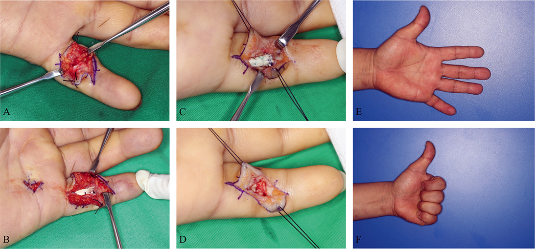
Figure 1. (A) A 42-year-old male presented with a flexor tendon rupture. (B) The tendon was initially repaired using the Modified Kessler method. (C) Re-rupture occurred 36 days later during physical therapy, necessitating exploratory surgery. (D) Tendon repair was performed again using the same technique. (E, F) Follow-up photographs taken 2 years postoperatively.
In the single-variate linear regression analysis, age and diabetes mellitus significantly influenced prognosis (Table 2). Age negatively correlated with outcomes (B = –0.69, p = 0.041), with older age predicting poorer results. Diabetes mellitus had a similarly negative impact (B = –32.45, p = 0.011), strongly correlating with worse outcomes. Other factors, including sex, smoking, Body mass index (BMI), injured finger, surgical technique (arteriorrhaphy and neurorrhaphy), operation period, re-rupture period, and the number of strands, were not statistically significant. In multivariate linear regression, no statistically significant differences were observed in all variables.
Discussion
Various studies have explored the mechanisms underlying tendon recovery and rupture [20]. Strickland et al. describe tendon repair as a process that unfolds in three distinct phases [14]. During the initial inflammatory phase for a week, fibroblasts and macrophages migrate to the injury site, removing necrotic tissue and clots. The proliferation phase (approximately 2 weeks) follows, marked by increased fibroblast activity, accumulation of immature collagen, and commencement of neovascularization. The final remodeling phase, lasting up to 8 weeks post-injury, involves collage fiber realignment to restore the tendon’s original structure [21, 22]. reported that the tendon integrity is notably compromised between 6 and 18 days post-repair, with the highest rupture risk during this period, although ruptures can occur up to 6–7 weeks post-repair.
Various studies have investigated factors affecting tendon suture prognosis. Svingen [23] suggested that age is a factor that influences re-rupture because aging delays the tendon healing process. Supporting this, Ackerman [24] observed in an animal study that with increasing age, fewer bridging tendon collagen fibers were present at the repair site, leading to more frequent ruptures. Our univariate analysis identified a negative correlation between age and patient prognosis. Given that age affects not only the likelihood of re-rupture but also the functional outcome of the finger, it is essential to consider age when developing a treatment plan and discussing the surgical prognosis with the patient. Older patients should be monitored for long-term functional outcomes, and rehabilitation strategies should accommodate age-related declines in tissue elasticity and muscle strength, ensuring that functional recovery is optimized over the long term. Moreover, providing clear information about the potential challenges and expected outcomes based on age can help to manage patient expectations and improve satisfaction with the treatment process.
Svingen [23] claimed that sex and re-rupture rates are statistically correlated, with males being at higher risk than women, owing to non-adherence to treatment guidelines. Sandford [25] suggested that a higher frequency of splint removal in males leads to an increased risk of rupture. However, our study did not show a statistically significant correlation between sex and prognosis of re-rupture. In other words, the assertion that men remove the splint more quickly and do not adhere to the treatment guidelines compared to women, leading to poor prognosis, was not supported by the data from our institution. An independent sample t-test (p = 0.482) showed no significant differences in splint removal or physical therapy adherence between genders. These variables did not significantly affect prognosis, contradicting earlier studies that linked non-adherence in males to poor outcomes. Moreover, these variables themselves did not demonstrate a direct statistical causality with the actual prognosis.
Smoking is known to negatively affect the musculoskeletal system, contributing to higher incidences of tendon rupture, soft tissue infections, wound healing complications, and adverse clinical outcomes [24, 26]. Despite these associations, smoking did not significantly affect the finger range of motion following tendon repair [27, 28]. One potential explanation is that while smoking impairs overall healing, it may not directly impact the flexibility or functional recovery of tendons once they have healed.
Recent studies highlight a significant correlation between diabetes and poor tenorrhaphy outcomes owing to the adverse effects of diabetes on tendon structure and healing [29]. Diabetes induces several pathological changes in tendons, including collagen disorganization, tendon thickening, and the accumulation of advanced glycation end-products, which reduce tendon elasticity and mechanical strength. Our univariate analysis confirmed a statistically significant correlation between diabetes and worse post-tenorrhaphy outcomes, consistent with previous research.
Immediate primary repair is recommended for re-ruptures with scar tissue <1 cm [7]. It has been suggested that repairs within a week post-rupture do not negatively influence prognosis [8]. However, these studies did not provide specific details on the permissible delay in suturing that would not detrimentally affect clinical outcomes. Gorriz [10], Kasashima [30], and Tottenham [31] all argued that the timing of surgery is a crucial prognostic factor. We found that the relationship between delayed repair and prognosis is not statistically significant. As we did not observe a statistically significant correlation between the timing of surgery and prognosis, this may indicate some flexibility for clinicians in scheduling without adversely impacting outcomes. This could potentially reduce the pressure to perform immediate repairs, allowing for better preparation and patient stabilization.
Moreover, we hypothesized that earlier re-ruptures might result in better outcomes because of less scar tissue formation. Some studies suggest that early remodeling processes, such as those modulated by matrix metalloproteinase-9 (MMP9), may influence tissue healing and scar tissue formation [11, 32]. However, our analysis did not reveal a significant relationship between the stage of tendon recovery at which a re-rupture occurs and the patient’s prognosis, suggesting that the timing of re-rupture may not be as critical as previously thought, allowing for greater flexibility in planning rehabilitation and repair without compromising recovery outcomes.
Lin [33] asserted that re-ruptures typically occur at a previous knot and are positively correlated with the number of strands and repair strength. Similarly, Hardwicke [34] reported fewer ruptures in patients with more strands. Therefore, a greater number of strands is anticipated to result in increased tendon strength and a reduction in rupture incidence, thereby significantly impacting prognosis. However, no significant statistical relationship was found between the number of core strands and prognosis.
Adequate mechanical stimulation is a well-known critical prognostic factor [30, 35]. Protocols incorporating early motion, as opposed to strict immobilization, improve tensile strength and tenocyte proliferation, augmenting collagen synthesis [36, 37]. However, our study showed no statistically significant differences in the final range of motion related to the timing, frequency, or duration of rehabilitation therapy. Our findings contrast with expectations that early and frequent physical therapy would improve outcomes. This discrepancy may be attributed to several factors. Firstly, we did not account for patients who did not fully engage in physical therapy from the outset due to an early achievement of adequate finger range of motion. In other words, the reasons patients discontinued physical therapy were not considered. Secondly, the amount of self-directed exercise performed by patients, in addition to hospital-based rehabilitation, is an important factor that should have been considered; however, this aspect was not sufficiently explored in our study. Thirdly, the timing of physical therapy initiation may be inherently linked to poorer prognosis. For instance, in patients with significant inflammation, physical therapy is often delayed until the inflammation subsides, resulting in a later start. In these cases, the underlying reason for the delayed initiation of physical therapy may be inherently related to the prognosis.
Our analysis did not reveal a statistically significant impact of the timing of re-rupture or the interval between re-rupture and surgery on patient prognosis. While these factors may seem crucial, they may not be as important as previously thought. Clinicians can focus on optimizing the patient’s condition before surgery rather than rushing into immediate repair, potentially improving overall outcomes. Our data identified age and diabetes as the only statistically significant predictors of prognosis, underscoring the need for careful management of these factors in patients undergoing tendon repair. These findings align with existing literature emphasizing the importance of patient-specific factors such as age and underlying comorbidities in predicting surgical outcomes. The primary limitation of this study was the small sample size of patients who experienced re-ruptures after flexor tenorrhaphy, which prevented the development of a multivariate linear regression model. In addition, we could not present a clear analysis of the difference in prognosis between patients with single or multiple rupture cases. As a result, we could not determine if prognostic factors differ between these groups. Further research with a larger cohort and a more detailed examination of these variables is needed to confirm these findings and refine prognostic models. Moreover, since age-related finger stiffness was not compared to normal people, this factor could not be considered [38].
Conclusion
This study identifies age and diabetes as significant predictors of prognosis in patients undergoing repeated flexor tenorrhaphy. These findings highlight the need for careful management of these factors to improve surgical outcomes. Notably, the timing of re-rupture and the interval between re-rupture and surgery were not found to significantly affect prognosis, suggesting greater flexibility in surgical timing. Given the limitations of our study, including a small sample size and the absence of comparisons between single and multiple rupture cases, further research is needed to validate these results and enhance prognostic models. Future studies should also consider age-related changes in finger stiffness to provide a more comprehensive understanding of recovery factors.
Acknowledgements
None.
References
[1] Taras JS, Gray RM, Culp RW. Complications of flexor tendon injuries. Hand Clin. 1994;10:93–109. https://doi.org/10.1016/S0749-0712(21)01037-4
[2] Stenekes MW, Rüttermann M, Werker PMN. Rare causes of closed rupture of the flexor tendon. J Plast Surg Hand Surg. 2014;48(3): 219–221. https://doi.org/10.3109/2000656X.2013.777226
[3] Faccio D, Montanari S, Maschio N, et al. The origins of the camper’s chiasm asymmetry: a cadaveric study and clinical implications for flexor tendons surgery. J Plast Surg Hand Surg. 2023;57(1–6):471–475. https://doi.org/10.1080/2000656X.2022.2158844
[4] Lilly SI, Messer TM. Complications after treatment of flexor tendon injuries. J Am Acad Orthop Surg. 2006;14:387–396. https://doi.org/10.5435/00124635-200607000-00001
[5] Netscher DT, Badal JJ. Closed flexor tendon ruptures. J Hand Surg. 2014;39:2315–2323. https://doi.org/10.1016/j.jhsa.2014.04.005
[6] Rubensson CC, Ydreborg K, Boren L, et al. Flexor tendon repair after rupture caused by volar plate fixation of the distal radius. J Plast Surg Hand Surg. 2015;49(2):112–115. https://doi.org/10.3109/2000656X.2014.951050
[7] Leddy JP. Operative hand surgery , 4th edition. In: Green DP, editor. New York, Edinburgh, London, and Melbourne: Churchill Livingstone; 1982. p. 1360–1363.
[8] Allen BN, Frykman GK, Unsell RS, et al. Ruptured flexor tendon tenorrhaphies in zone II: repair and rehabilitation. J Hand Surg. 1987;12:18–21. https://doi.org/10.1016/S0363-5023(87)80153-3
[9] LaSalle WB, Strickland JW. An evaluation of the two-stage flexor tendon reconstruction technique. J Hand Surg. 1983;8:263–267. https://doi.org/10.1016/S0363-5023(83)80155-5
[10] Gorriz GJA, Cooke J. Assessment of the influence of the timing of repair on flexor tendon injuries in chickens. Br J Plast Surg. 1976;29:82–84. https://doi.org/10.1016/0007-1226(76)90099-0
[11] Galatz LM, Gerstenfeld L, Heber-Katz E, et al. Tendon regeneration and scar formation: the concept of scarless healing. J Orthop Res. 2015;33:823–831. https://doi.org/10.1002/jor.22853
[12] Kleinert HE, Verdan C. Report of the committee on tendon injuries. J Hand Surg. 1983;8:794–798. https://doi.org/10.1016/S0363-5023(83)80275-5
[13] Paneva-Holevich E. Two-stage reconstruction of the flexor tendons. Int Orthop. 1982;6:133–138. https://doi.org/10.1007/BF00268657
[14] Strickland JW. Delayed treatment of flexor tendon injuries including grafting. Hand Clin. 2005;21:219–243. https://doi.org/10.1016/j.hcl.2004.12.003
[15] Lee KC, Lee DC, Kim JS, et al. Loop suture technique for flexor digitorum profundus tendon repair in the insertion site. J Korean Soc Plast Reconstr Surg. 2010:650–658.
[16] Uslu M, Isik C, Ozsahin M, et al. Flexor tendons repair: effect of core sutures caliber with increased number of suture strands and peripheral sutures. A sheep model. Orthop Traumatol Surg Res. 2014;100:611–616. https://doi.org/10.1016/j.otsr.2014.05.009
[17] Elliot D, Giesen T. Primary flexor tendon surgery. Hand Clin. 2013;29:191–206. https://doi.org/10.1016/j.hcl.2013.03.001
[18] Rrecaj S, Martinaj M, Murtezani A, et al. Physical therapy and splinting after flexor tendon repair in zone II. Med Arch. 2014;68: 128–131. https://doi.org/10.5455/medarh.2014.68.128-131
[19] Tang JB. Recent evolutions in flexor tendon repairs and rehabilitation. J Hand Surg Eur Vol. 2018;43:469–473. https://doi.org/10.1177/1753193418773008
[20] Nichols AEC, Best KT, Loiselle AE. The cellular basis of fibrotic tendon healing: challenges and opportunities. Transl Res. 2019;209:156–168. https://doi.org/10.1016/j.trsl.2019.02.002
[21] Voleti PB, Buckley MR, Soslowsky LJ. Tendon healing: repair and regeneration. Ann Rev Biomed Eng. 2012;14:47–71. https://doi.org/10.1146/annurev-bioeng-071811-150122
[22] Zhao C, Amadio PC, Momose T, et al. Effect of synergistic wrist motion on adhesion formation after repair of partial flexor digitorum profundus tendon lacerations in a canine model in vivo. J Bone Joint Surg Am. 2002;84:78–84. https://doi.org/10.2106/00004623-200201000-00012
[23] Svingen J, Wiig M, Turesson C, et al. Risk factors for reoperation after flexor tendon repair: a registry study. J Hand Surg Eur Vol. 2022;47: 1071–1076. https://doi.org/10.1177/17531934221101563
[24] Ackerman JE, Bah I, Jonason JH, et al. Aging does not alter tendon mechanical properties during homeostasis, but does impair flexor tendon healing. J Orthop Res. 2017;35:2716–2724. https://doi.org/10.1002/jor.23580
[25] Karousou E, Ronga M, Vigetti D, et al. Collagens, proteoglycans, MMP-2, MMP-9 and TIMPs in human Achilles tendon rupture. Clin Orthop Relat Res. 2008;466:1577–1582. https://doi.org/10.1007/s11999-008-0255-y
[26] Kasashima T, Kato H, Minami A. Factors influencing prognosis after direct repair of the flexor pollicis longus tendon: multivariate regression model analysis. Hand Surg. 2002;07:171–176. https://doi.org/10.1142/S0218810402001126
[27] Tottenham VM, Wilton-Bennett K, Jeffrey J. Effects of delayed therapeutic intervention following zone II flexor tendon repair. J Hand Ther. 1995;8:23–26. https://doi.org/10.1016/S0894-1130(12)80152-9
[28] Gelberman RH, Woo SL, Lothringer K, et al. Effects of early intermittent passive mobilization on healing canine flexor tendons. J Hand Surg Am. 1982;7:170–175. https://doi.org/10.1016/S0363-5023(82)80083-X
[29] Bishop AT, Cooney WPI, Wood MB. Treatment of partial flexor tendon lacerations: the effect of tenorrhaphy and early protected mobilization. J Trauma Acute Care Surg. 1986;26:301. https://doi.org/10.1097/00005373-198604000-00001
[30] Boyer MI, Strickland JW, Engles D, et al. Flexor tendon repair and rehabilitation: state of the art in 2002. Instr Course Lect. 2003;52:137–161.
[31] Hurley CM, Reilly F, Callaghan S, et al. Negative predictors of outcomes of flexor tendon repairs. Cureus. 2019;11:e4303. https://doi.org/10.7759/cureus.4303
[32] Santiago-Torres J, Flanigan DC, Butler RB, et al. The effect of smoking on rotator cuff and glenoid labrum surgery: a systematic review. Am J Sports Med. 2015;43:745–751. https://doi.org/10.1177/0363546514533776
[33] Lee JJ, Patel R, Biermann JS, et al. The musculoskeletal effects of cigarette smoking. J Bone Joint Surg. 2013;95:850–859. https://doi.org/10.2106/JBJS.L.00375
[34] Sandford F, Barlow N, Lewis J. A study to examine patient adherence to wearing 24-hour forearm thermoplastic splints after tendon repairs. J Hand Ther. 2008;21:44–53. https://doi.org/10.1197/j.jht.2007.07.004
[35] Vaidya R, Lake SP, Zellers JA. Effect of diabetes on tendon structure and function: not limited to collagen crosslinking. J Diabetes Sci Technol. 2023;17:89–98. https://doi.org/10.1177/19322968221100842
[36] Lin TW, Cardenas L, Soslowsky LJ. Biomechanics of tendon injury and repair. J Biomech. 2004;37:865–877. https://doi.org/10.1016/j.jbiomech.2003.11.005
[37] Hardwicke JT, Tan JJ, Foster MA, et al. A systematic review of 2-strand versus multistrand core suture techniques and functional outcome after digital flexor tendon repair. J Hand Surg. 2014;39:686.e2–695.e2. https://doi.org/10.1016/j.jhsa.2013.12.037
[38] Chung M-J, Wang M-JJ. The effect of age and gender on joint range of motion of worker population in Taiwan. Int J Ind Ergon. 2009;39:596–600. https://doi.org/10.1016/j.ergon.2007.11.004