ORIGINAL RESEARCH ARTICLE
Venous flaps to repair the soft tissue defects of the hand
Ali Murat Başaka  , Çağla Çiçekb , Arzu Türkseven Topaçoğluc , Hande Akdenizd and Derya Özçelike
aDepartment of Orthopaedics and Traumatology, University of Health Sciences, Gülhane Research and Education Hospital, Ankara, Turkiye; bDepartment of Plastic Reconstructive and Aesthetic Surgery, Kartal Dr. Lütfi Kirdar Şehir Hastanesi Plastik Rekonstrüktif ve Estetik Cerrahi Kliniği, Kartal, İstanbul, Turkiye; cPlastic Reconstructive and Aesthetic Surgeon, Private Practice, Kadiköy, İstanbul, Turkiye; dDepartment of Plastic Reconstructive and Aesthetic Surgery, Selçuk Üniversitesi Tip Fakültesi, Selçuklu, Konya, Turkiye; ePlastic Reconstructive and Aesthetic Surgeon, Private Practice, Nişantaşi Şişli, İstanbul, Turkiye
ABSTRACT
Aim: The venous flaps provide a versatile, easy flap choice to repair the dorsal skin defects of the fingers and web spaces. Its thin and pliable nature provides natural coverage for the important delicate structures of the hand such as bone, tendon, joint or neurovascular bundle.
Material and methods: From 2010 to 2025, we used the venous flaps to cover exposed bone, tendon and joint located on the distal phalanx, proximal phalanx, web space and dorsum of the hand in 10 patients. Flaps were unipedicled distally based in nine cases. It was a free venous flap in one case. During the flap elevation, the pedicle of the flap included one superficial vein of the hand and its perivenous areolar tissue.
Results: The size of the flaps ranged from 4 cm2 to 9 cm2. The mean length of the vascular pedicles was 3.1 cm (min: 2 cm, max: 10 cm). In all cases, the flaps survived completely. The donor sites were on the dorsal surface of the hand in nine cases, the volar surface of the wrist in one case. The donor sites were closed primarily in eight cases; skin graft was used in two cases.
Conclusion: The aim of this study was to share our experience regarding the venous flap usage in the hand. Another aim is to remind us of this useful flap, which was not popularized enough despite its versatility and being described 40 years ago.
KEYWORDS: Venous flap; hand; skin defect
Citation: Journal of Plastic Surgery and Hand Surgery 2026; 61: 189–196. DOI: https://doi.org/10.2340/jphs.v61.46466.
Copyright: © 2026 The Author(s). Published by MJS Publishing on behalf of Acta Chirurgica Scandinavica. This is an Open Access article distributed under the terms of the Creative Commons Attribution 4.0 International License (http://creativecommons.org/licenses/by/4.0/), allowing third parties to copy and redistribute the material in any medium or format and to remix, transform, and build upon the material, with the condition of proper attribution to the original work.
Received: 14 May 2026; Accepted: 24 June 2026; Published: 3 August 2026.
CONTACT: Derya Özçelik deryaozcelik34@gmail.com Plastic Reconstructive and Aesthetic Surgeon, Private Practice, Poyracik Sokak 47/5 Nişantaşi Şişli, İstanbul, Turkiye
Supplemental data for this article can be accessed online at https://doi.org/10.2340/jphs.v61.46466
Competing interests and funding: None.
This research did not receive any specific grant from funding agencies in the public, commercial, or not-for-profit sectors.
Introduction
The use of the venous flap to cover the soft tissue defects has been documented in the literature for over 40 years. First described in 1981, Nakayama et al. [1] demonstrated that a vein and overlying skin, a venous flap, may be transposed from one area of the body to another with complete survival of the flap. By using the abdominal skin flap of the rats, they showed that the arterial inflow through the venous system nourished the distal part of the flap [1]. Venous flap was prepared both as a pedicled island flap and a free flap [1]. In 1985, Baek et al. [2] studied the vascular morphology and blood gas changes of venous flaps in a dog model. They observed that venous flaps without arterial inflow survived with normal hair growth and wound healing [2]. Therefore, they suggested that capillary diffusion can occur without the continuous flow of blood through a capillary [2]. In 1987, Tsai et. al. used venous flap as a free flap to cover dorsal skin defect of fingers that underwent replantation/revascularization in 15 patients, with a survival rate of 100% [3].
In the 1990s, experimental studies [4–8] have shown that a rich perivenous areolar tissue into the pedicle and the presence of a vascular recipient bed played important roles for the viability of the venous flaps. Then, clinical use of the venous flaps for the coverage of traumatic finger skin defects has been reported [9–12].
The aim of this study is to share our experience regarding the venous flap usage to cover dorsal skin defects in the hand and remind us of venous flaps as a versatile alternative.
Material and methods
From 2010 to 2025, we used the venous flaps on the hand to repair the dorsal skin defects in 10 patients. The indication for the surgery was the presence of the dorsal skin defect exposing bone, tendon, or joint in the hand. The location of the defects was the hand dorsum in four patients, the web space in three patients, the proximal phalanx in one patient, and the distal phalanx in two patients. Eight patients (80%) were male, and two patients (20%) were female. The mean age was 45 (min: 16, max: 87). The etiologic factors of the defects included acute trauma injury, skin contracture caused by injury and burn, benign lesions such as hyperkaratosis, keratoacanthoma and hemangioma; and pre-malign and malign lesions such as Bowen’s disease and squamous cell carcinoma (Table 1).
Before surgery, patient consents in written form were provided. Data gathered from the patients adhere to local/national regulations. The principles outlined in the Declaration of Helsinki have also been followed.
Pedicled venous flaps can be prepared both as proximally and distally based flaps. In our series, the venous flap was prepared as a distally based unipedicled flap in nine patients and as a free-flap in one patient. Venous flap was not planned in the patients who had injury/scar at the potential donor area, which would interfere with the flap preparation.
Surgery was performed under axillary block anesthesia in nine patients and under general anesthesia by using a laryngeal mask airway in one patient.
Operative technique
In order to select an appropriate vein, a tourniquet is applied to the upper arm and inflated without draining the venous blood flow. The pedicled venous flap is planned on the metacarpal or carpal area of the hand dorsum by tracing a significant superficial vein. A dorsal vein trace and its pivot point (rotational arc) are marked on the dorsum of the hand (Figure 1) (Supplemental Video). While choosing the correct vein, the distance between the defect and pivot point should match with the pedicle length of the flap. The skin flap is centered on the selected dorsal vein and sized a little bit larger than the size of the defect. After vein selection and flap positioning, tourniquet is deflated by draining the venous blood flow and re-inflated. Following the skin incision, the selected dorsal vein is divided proximally and ligated with 3/0 silk suture. The skin flap is elevated by preserving the vein and its perivenous areolar tissue and dissected distally until the pivot point. Care is taken to include as much perivenous areolar tissue in the pedicle as possible. Mostly a subcutaneous tunnel (Figures 2–4) is created to be able to transfer the island flap into the recipient area. After tourniquet release, viability of the elevated flap is checked by observation. Then, the flap is adapted into the defect with 5/0 polypropylene suture under no tension. The donor site can be closed primarily or with a skin graft depending on the defect size. Postoperatively, the light wound dressing is put on the flap, and a volar static splint is placed to keep the wrist at rest and fingers at the extension position for 10 days.
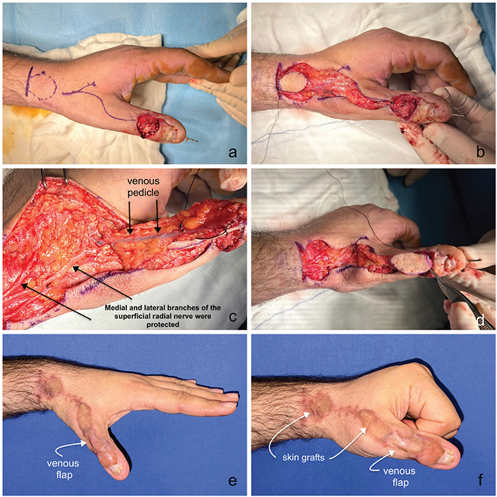
Figure 1. A 38-year-old man presented with a 1.5 × 2 cm skin defect located at the dorsal surface of the interphalangeal joint (IPJ) of his thumb of the left hand. The etiology of the injury was a crush trauma. The defect was reconstructed with a 2.5 × 2 cm distally based pedicled venous flap. (a) The defect exposed an intraarticular fracture of the distal phalanx, which was stabilized using Kirschner wire and a repaired extansor pollicis longus (EPL) tendon. In planning of the flap, the vein was marked first, then a venous flap island was marked accordingly. (b) A unipedicled distally based island venous flap, 3 × 2 cm in size, was planned on the carpal area by tracing a significant superficial vein. (c) During flap elevation, medial and lateral branches of the superficial radial nerve (arrows at the bottom) were protected. Continuation of the superficial vein (arrows on top) is easily seen inside the venous pedicle of the flap, which is surrounded by a large amount of periareolar tissue. (d) Transfer and inset of the venous flap into the defect. The pedicle length is 10 cm. The pivot point of the pedicle is located at the level of the first metacarpophalangeal joint. (e, f) Two months after the surgery, the flap and the thumb healed well. From an aesthetic point of view, the venous flap adapted to the area very well. Because of the intra-articular fracture of the distal phalanx and the crush injury, the range of motion of the IPJ of the thumb was approximately 200.

Figure 2. A 63-year-old man presented with a 2 × 1 cm hyperkeratotic lesion located at the head part of the second metacarpal area of his left hand. After excision of the lesion, the defect was reconstructed with a 3 × 2 cm venous flap. (a) A 2 × 1 cm hyperkeratotic ulcerated lesion located at the head part of the second metacarpal area. (b) A unipedicled venous flap, 2.5 × 2 cm in size, was planned on the third metacarpal area by tracing a significant superficial vein. The vein was marked (arrow) first, then the venous flap was marked accordingly. (c) After excision of the lesion, extensor tendons were exposed. (d) The flap was elevated based on its vein, including a large amount of periareolar tissue (arrow), and transferred through the subcutaneous tunnel. (e) The flap was inset to the defect. The proximal end of the superficial vein used as a pedicle is clearly seen here (arrows). (f) The flap healed well functionally and cosmetically. The donor area was closed primarily (arrows).

Figure 3. A 41-year-old man presented with a 2 × 1 cm hemangiomatous lesion located in the fourth web space of his right hand. After excision of the lesion, the defect was reconstructed with a 2 × 2 cm distally based pedicled venous flap. (a, b) Perioperative appearance: The flap was harvested from the fourth intermetacarpal area, transferred through the subcutaneous tunnel and inset into the defect. (c, d) Postoperative 7th day appearance: The flap survived completely. The donor area was closed primarily.

Figure 4. A 16-year-old man presented with a scar contracture at the third web space in his left hand. After contracture release, the defect was reconstructed with a 3 × 2 cm distally based pedicled venous flap. (a) Scar contracture at the third web space. Markings of the distally based venous flap. (b) Elevation of the venous flap from the third intermetacarpal area and contracture release. (c) Transfer of the venous flap through the subcutaneous tunnel. (d) Inset of the venous flap into the defect. No venous congestion detected. (e) Complete flap survival was achieved. The hand function of the patient improved. (f) The skin flap shows perfect color and texture match with its new place since it is obtained from the area having the same characteristics.
When we prepare the venous flap as a free flap, we need to choose the superficial veins, which have similar diameter with that of the recipient vessels (Figure 5).

Figure 5. A 20-year-old man presented with a 1.5 × 1 cm skin defect located at the dorsal surface of the distal interphalangeal joint of his 5th finger of the right hand. (a) The skin defect exposing the intraarticular fracture of the distal phalanx and the separated extensor tendon from its insertion on the distal phalanx (mallet finger deformity) is seen. (b) A free venous flap, 2 × 2 cm in size, was planned on the volar surface of the wrist by tracing a superficial vein. (c) Harvestment of the free venous flap. (d) After fracture stabilization and tendon repair using the palmaris longus tendon graft, the free venous flap was inset to the defect, vein anastomosis was achieved, and the skin defect was reconstructed. Since it is a free venous flap, the color of the flap intraoperatively and in the early postoperative period was a little bit bluish. It healed with a superficial marginal tissue loss (5%). In late follow-up, the flap and the finger healed well functionally and cosmetically.
Case presentations
Case 1
A 38-year-old man presented with a 1.5 × 2 cm skin defect (Figure 1a, b) located at the dorsal surface of the interphalangeal joint (IPJ) of his thumb of the left hand. Etiology was crush injury. The defect was exposing an intraarticular fracture of the distal phalanx and a crushed extensor pollicis longus (EPL) tendon. After debridement, the fractured distal phalanx was fixed using 1.0 mm percutaneous intramedullary Kirschner wire (K-wire) following open reduction. The EPL tendon was repaired. A unipedicled distally based island venous flap, 2.5 × 2 cm in size, was planned on the carpal area by tracing a significant superficial vein (Figure 1a, b). The flap was harvested based on its vein, including a large amount of periareolar tissue, transferred, and inset to the defect (Figure 1c, d). The pedicle length was 10 cm. The donor area was repaired with a skin graft. In the postoperative period, neither significant edema nor venous congestion appeared, only superficial epidermolysis was observed. In the follow-up, the flap and the thumb healed well. Aesthetically, the venous flap adapted to the area very well. The range of motion of IPJ was approximately 20° (Figure 1e, f) (Video 1).
Case 2
A 63-year-old man presented with a 2 × 1 cm hyperkeratotic lesion (Figure 2a, b) located at the head part of the second metacarpal area of his left hand. After excision of the lesion, extensor tendons were exposed (Figure 2c, d). A unipedicled distally based venous flap, 2.5 × 2 cm in size, was planned on the third metacarpal area by tracing a significant superficial vein (Figure 2b–d). The flap was harvested based on its vein including a large amount of periareolar tissue (Figure 2d). Then, it was transferred through a subcutaneous tunnel and inset to the defect (Figure 2c–e). The pedicle length was 2.5 cm. Neither significant edema nor venous congestion appeared in the postoperative period. The flap healed well functionally and cosmetically (Figure 2f). Postoperative pathological examination confirmed hyperkeratosis.
Case 3
A 41-year-old man presented with a 2 × 1 cm hemangiomatous lesion located in the fourth web space of his right hand. After excision of the lesion, the defect was reconstructed with a 2 × 2 cm distally based pedicled venous flap. The flap was harvested from the fourth intermetacarpal area, transferred through a subcutaneous tunnel, and inset into the defect. Pedicle length was 2 cm. Flap survived completely (Figure 3a–d). Postoperative pathological examination confirmed hemangioma.
Case 4
A 16-year-old man presented with a third web space skin contracture in his left hand caused by a trauma (Figure 4a). After contracture release (Figur 4b), the defect was reconstructed with a 3 × 2 cm venous flap that was harvested from the third intermetacarpal area, transferred through the subcutaneous tunnel, and inset into the defect (Figure 4b–d). Pedicle length was 3 cm. No venous congestion occurred. Complete flap survival with appealing cosmetic result and functional improvement was achieved (Figure 4e, f).
Case 5
A 20-year-old man presented with a 1.5 × 1 cm skin defect (Figure 5a, b) located at the dorsal surface of the distal IPJ of his 5th finger of the right hand. Etiology was crush injury. The defect was exposing intraarticular fracture of the distal phalanx and the separated extensor digitorum communis (EDC) tendon from its insertion. As a result, the patient had a mallet finger deformity (Figure 5a, b). After debridement, the fractured and displaced distal phalanx was fixed using K-wire following open reduction. Then, a free venous flap, 2 × 2 cm in size, was planned on the volar surface of the wrist by tracing a superficial vein (Figure 5b, c). The small diameter of the veins located on the volar surface of the wrist was found suitable to be able to make an anastomosis to a digital vein. The flap was prepared based on its vein (Figure 5c, d). At the same time, the palmaris longus tendon graft obtained from the same area was used to adapt the ruptured EDC tendon to the distal phalanx. Then, the flap was transferred and inset to the defect. Vein-to-vein, end-to-end anastomosis was achieved by using 11/0 polypropylene suture. The donor area was closed primarily. Venous congestion was observed in the early postoperative period (Figure 5d). It healed with a superficial marginal tissue loss (5%). A splint was used for 6 weeks. The K-wire was removed at 6th week and motion started. In late follow-up, the flap and the finger healed well functionally and cosmetically.
Results
Venous flaps were transferred to the dorsal skin defects located on the hand, web space, and proximal and distal phalanx of the fingers in 10 patients. In nine patients, the pedicled venous flap, based on a significant superficial vein, was raised from the carpal or metacarpal areas of the hand dorsum. In one patient, a free venous flap prepared from the volar surface of the wrist was transferred to the skin defect located on the dorsal surface of the distal phalanx of the 5th finger.
The size of the defects ranged from 1.5 cm2 to 9 cm2 (mean: 3.9 cm2). The size of the venous flaps (skin paddles) ranged from 4 cm2 to 9 cm2 (mean: 5.7 cm2). The mean length of the vascular pedicles was 3.1 cm (min: 2 cm, max: 10 cm). The skin paddle was island-type in nine patients and rotational-type in one patient. Flap sizes, flap types, and pedicle lengths are shown in Table 1.
In the early postoperative period, venous congestion was observed only in the free venous flap. Neither venous congestion nor significant edema occurred in none of the distally based pedicled flaps. Among the distally based pedicled flaps, only superficial epidermolysis was observed in the flap having a pedicle length of 10 cm. All flaps survived completely. The donor site was closed primarily in eight cases, skin graft was used in two cases. Arterial inflow was not used in any of the flaps.
The mean follow-up period of the flaps was 6 months. In follow-ups, all flaps healed well functionally and aesthetically. Aesthetic results were evaluated by patients and found good or excellent. Sensory recovery was observed in five of 10 flaps during the follow-up.
Discussion
Small skin defects located on the phalanges, web spaces of the hand, and hand dorsum can cause exposure of important structures such as bones, tendons, joints, and neurovascular bundles. In some cases, these important structures may also be damaged and repaired. In such cases, skin graft applications are often not possible. Local skin flaps, such as advancement, rotation, or transposition flaps, can be a choice for small defects. However, they have limited capacity to be able to provide a tension-free coverage of the defect in that specific area and often require multiple incisions. In addition, local flaps cannot be designed when the surrounding tissue damaged because of the injury. Repair with a cross-finger flap [13] or thenar flap may not always be possible, especially when the finger has a bone fixation or a tendon repair. Furthermore, the cross-finger and distant flaps (abdominal and groin flaps) are the flaps dependent on the recipient blood-supply, contrary to the venous flaps which bring blood supply to the damaged area. Metacarpal arterial flaps [14] may be an option in these cases. However, the decision to prepare a flap based on an important blood supply of the hand, such as metacarpal artery, should be evaluated for each patient carefully. Furthermore, the pedicles of the third or fourth metacarpal arterial flaps consist of small arterial branches [15]. Contrary to the metacarpal arterial flaps, venous flaps create almost no morbidity regarding the blood supply of the hand. The venous flap uses skin and superficial veins for the inflow and outflow of its blood supply. Because of the laxity of the dorsal skin, the donor area is usually closed primarily. The chief advantages offered by the venous flaps are single-stage reconstruction, minimal donor site morbidity, thin pliable skin replacement thus eliminating the need for debulking procedures, early mobilization hence decreased risk of stiffness, and cost saving by avoiding 2nd or more surgical procedures [16]. The distant and heterodigital flaps require a second surgical procedure to divide and inset the flap, longer immobilization of the hand for about 2–3 weeks, and hence more hospitalization and delay in recovery of the hand functions (about 5–6 weeks). The distant flaps sometimes require more than two stages to adjust the flaps and to make the flaps thinner. In our cases, the patients who had no bone fixation or tenorrhaphy routinely mobilized their hands at a week, and gained full range of motion within 10 days and returned to work within 2–3 weeks.
The reverse dorsal hand flaps described by Quaba and Davison are based on a direct cutaneous branch of the dorsal metacarpal artery [17]. It is described as an axial pattern metacarpal artery island flap for web spaces as well as dorsal metacarpal and phalangeal skin defects [17]. Lai-jin and Xu [18] suggested that the dissection of the pedicle should be done cautiously to keep sufficient surrounding adipose tissue when the flap is based on the fourth dorsal metacarpal artery. During the flap preparation, however, involvement of the digital or metacarpal arteries may impair the vascular supply to the related digits. In comparison with the third or fourth dorsal metacarpal artery flaps, dissection of the dorsal venous flaps in the hand is easier since interosseous muscle fascia is not pierced with the veins. Shi et al. performed a meta-analysis to compare the effectiveness and safety of the first dorsal metacarpal artery flaps (FDMAF) and reverse homodigital dorsal flaps (RHDF) for thumb reconstruction [19]. The two surgical procedures were found both safe and reliable since no significant differences were detected in flap necrosis between the two groups [19]. In our opinion, venous flaps prevent sacrification of these important arterial sources and these important flaps.
Suh et al. [20] performed the unipedicled venous island flap procedure on the hand in 16 cases of 14 patients. Han et al. [21] treated 31 skin defects on the hand of 29 patients with unipedicled venous flaps. Both Suh et al. [20] and Han et al. [21] demonstrate that proximally pedicled venous flaps of the hand can survive regardless of pedicle length. Distally pedicled venous flaps can also survive completely when pedicle length is <5 cm. Therefore, distally pedicled venous flaps with pedicle lengths ≥5 cm should be used with caution [20, 21].
Cil Y et al. used a distally based venous flap to correct syndactyly in nine patients to provide skin coverage without a skin graft. The sizes of the flaps ranged from 10 cm2 (5 × 2 cm) to 16 cm2 (8 × 2 cm). All flaps survived successfully [22].
Davami et al. [23] used a reverse distally based venous flap on 18 patients to repair dorsal skin defects of the proximal and the middle phalanges. A total of 17 cases healed successfully; one case experienced superficial necrosis at the margin of the flap that was treated conservatively [23].
In our series, the pedicled venous flaps completely survived with no circulation problem, including the distally based flap having a pedicle length of 10 cm. In the free venous flap, venous congestion developed in the early stage (Figure 5) disappeared completely without any intervention. The reason of the high survival rate is the preparation of the pedicle with a bulky periareolar tissue surrounding the vein (Figures 1c, d, 2c, and 4b, c). Various studies confirmed that small arterial channels surrounding the vein were to be in the perivenous areolar tissue [5–7, 24]. According to Chang [25], adding this perivenous tissue into the pedicle developed a flap to become a random pattern flap, which includes a significant vein. Furthermore, if another large superficial vein is left in continuity with the distally based pedicle, it can cause congestion and jeopardize the flap viability [26]. It should be carefully ligated at 1–2 cm distal to the pedicle [26]. The other study showed that the venous flap perfusion occurs along the axis of the subcutaneous vein passing through the flap [27]. Murata et al. [11] found that two conditions should be satisfied for venous flap survival. Firstly, the pedicle should include a vein having perivenous areolar tissue, and secondly, the recipient bed should have at least moderate vascularization [11]. Tang et al. [28] demonstrated that the venous flap was supplied by its axial vein during the first 3 days until neovascularization. In case of venous insufficiency development, medicinal leeches, hyperbaric oxygen therapy, thrombolytic agents and subcutaneous low molecular weight heparin (LMWH) administration can be used to diminish congestion by the time of neovascularization (usually between the fifth and seventh postoperative day) [29]. Bayir et al. showed that LMWH therapy alone was not adequate to save venous congested flaps. They detected that combining this method with anticoagulant dressing is critical in maintaining venous perfusion and rescuing the congested flap [29].
Since the perfusion area of the venous flaps is less than that of traditional arterial flaps, their use is limited in small-to-moderate defects [11]. To overcome this disadvantage, Ueda et al. [30] investigated the possibility of increasing the survival area of a venous island flap by a delay procedure in an experimental study. In a proximally based venous island flap, the survival area increased when a delay procedure was used, especially with ligation of the distal vein. In a distally based venous island flap, the survival area increased when a delay procedure was used without ligation of the proximal vein. They thought that if a stable and reliable venous island flap can be elevated, it may enable us to make various flaps everywhere on the body surface by using subcutaneous veins [30].
Conclusion
A well-planned distally based venous flap or a free venous flap, both are useful options to repair the complicated dorsal skin defects in the phalanx, web space and dorsum of the hand, since flap options are limited in these places. They create almost an identical appearance with a minimal donor area morbidity.
Authors’ contribution
Dr Başak gathered the data of the patients and wrote the article and operated the patients.
Dr Çiçek operated the patients.
Dr Türkseven operated the patients.
Dr Akdeniz operated the patients.
Dr Ozcelik operated the patients.
References
[1] Nakayama Y, Soeda S, Kasai Y. Flaps nourished by arterial inflow through the venous system: an experimental investigation. Plast Reconstr Surg. 1981;67:328–334. https://doi.org/10.1097/00006534-198103000-00009
[2] Baek SM, Weinberg H, Song Y, et al. Experimental studies in the survival of venous island flaps without arterial inflow. Plast Reconstr Surg. 1985;75:88–95. https://doi.org/10.1097/00006534-198501000-00020
[3] Tsai TM, Matiko JD, Breidenbach W, et al. Venous flaps in digital revascularization and replantation. J Reconstr Microsurg. 1987;3:113–119. https://doi.org/10.1055/s-2007-1006973
[4] Fukui A, Tamai S, Maeda M, et al. The pedicled venous flap. An experimental study. Br J Plast Surg. 1993;46:116–121. https://doi.org/10.1016/0007-1226(93)90141-w
[5] Noreldin AA, Fukuta K, Jackson IT. Role of perivenous areolar tissue in the viability of venous flaps: an experimental study on the inferior epigastric venous flap of the rat. Br J Plast Surg. 1992;45:18–22. https://doi.org/10.1016/0007-1226(92)90108-a
[6] Smith RJ, Fukuta K, Wheatley M, et al. Role of perivenous areolar tissue and recipient bed in the viability of venous flaps in the rabbit ear model. Br J Plast Surg. 1994;47:10–14. https://doi.org/10.1016/0007-1226(94)90110-4
[7] Adamo C, Rubino C. Venous flaps and perivenous areolar tissue: an experimental study in rats. J Reconstr Microsurg. 1996;12:179–181. https://doi.org/10.1055/s-2007-1006473
[8] Murata K, Tamai S, Inada Y, et al. Transfer of a pedicled venous flap containing perivenous areolar tissue and nerve: an experimental study. Br J Plast Surg. 1999;52:223–229. https://doi.org/10.1054/bjps.1997.3039
[9] Chavoin JP, Rouge D, Vachaud M, et al. Island flaps with an exclusively venous pedicle. A report of eleven cases and a preliminary haemodynamic study. Br J Plast Surg. 1987;40:149–154. https://doi.org/10.1016/0007-1226(87)90187-1
[10] Foucher G, Norris RW. The venous dorsal digital island flap or the ‘neutral’ flap. Br J Plast Surg. 1988;41:337–343. https://doi.org/10.1016/0007-1226(88)90069-0
[11] Murata K, Inada Y, Fukui A, et al. Clinical application of the reversed pedicled venous flap con- taining perivenous areolar tissue and/or nerve in the hand. Br J Plast Surg. 2001;54: 615–620. https://doi.org/10.1054/bjps.2001.3673
[12] Roberts JM, Carr LW, Haley CT, et al. Venous flaps for revascularization and soft-tissue coverage in traumatic hand injuries: a systematic review of the literature. J Reconstr Microsurg. 2020;36:104–109. https://doi.org/10.1055/s-0039-1695053
[13] Gurdin M, Pangman WJ. The repair of surface defects of fingers by trans-digital flaps. Plast Reconstr Surg (1946). 1950;5: 368–371. https://doi.org/10.1097/00006534-195004000-00011
[14] Earley MJ, Milner RH. Dorsal metacarpal flaps. Br J Plast Surg. 1987;40:333–341. https://doi.org/10.1016/0007-1226(87)90035-x
[15] Rezende MR, Mattar R Jr, Cho AB, et al. Anatomic study of the dorsal arterial system of the hand. Rev Hosp Clin Fac Med Sao Paulo. 2004;59:71–76. https://doi.org/10.1590/s0041-87812004000200005
[16] Cil Y, Yapici AK, Kocman AE, et al. Distally based venous flap for proximal phalangeal soft tissue burn defect and web space burn contracture. Br J Plast Surg. 2009;30:643–647. https://doi.org/10.1097/BCR.0b013e3181abffe7
[17] Quaba AA, Davison PM. The distally-based dorsal hand flap. Br J Plast Surg. 1990;43:28–39. https://doi.org/10.1016/0007-1226(90)90042-x
[18] Lai-jin L, Xu G. The reverse dorsal metacarpal flap: experience with 153 cases. Ann Plast Surg. 2006;56:614–617. https://doi.org/10.1097/01.sap.0000205775.41405.fa
[19] Shi H, Huang Y, Shen Y, et al. The first dorsal metacarpal artery flaps versus reverse homodigital dorsal flaps for thumb reconstruction: a systematic review and meta-analysis. J Plast Surg Hand Surg. 2024;26:24–31. https://doi.org/10.2340/jphs.v59.12435
[20] Suh IS, Han SK, Lee BI, et al. Venous Island flap for reconstruction in the hand. J Korean Soc Plast Reconstr Surg. 2003;30:293–299.
[21] Han S-K, Kim S-Y, Gu JH, et al. Influence of the pedicle orientation and length on viability of unipedicled venous island flaps. Microsurgery. 2014;34:197–202. https://doi.org/10.1002/micr.22161
[22] Cil Y, Kocman AE, Yapici AK. Distally based venous flap: a new technique for the correction of syndactyly without skin graft in adult patients. Chir Organi Mov. 2009;93:123–129. https://doi.org/10.1007/s12306-009-0044-0
[23] Davami B, Arasteh E, Pourkhamene G. Versatility of venous flap for coverage of proximal and middle phalanges of fingers. Tech Hand Up Extrem Surg. 2012;16:23–26. https://doi.org/10.1097/BTH.0b013e31822d5e1c
[24] Shalaby HA, Saad MA. The venous island flap: is it purely venous? Br J Plast Surg. 1993;46:285–287. https://doi.org/10.1016/0007-1226(93)90003-t
[25] Chang SM. The pedicle of neurocutaneous island flaps. Plast Reconstr Surg. 1996;98:374–376. https://doi.org/10.1097/00006534-199608000-00036
[26] Chang SM, Gu YD, Li JF. The role of the large superficial vein in survival of proximally based versus distally based sural veno-neuro-fasciocutaneous flaps in a rabbit model. Plast Reconstr Surg. 2005;115:213–218. https://doi.org/10.1097/01.PRS.0000138255.89687.F4
[27] Krishnan KG. The venous flaps: an experimental study of the microvascular architecture, the area of perfusion and their correlation. Br J Plast Surg. 2002;55:340–350. https://doi.org/10.1054/bjps.2002.3842
[28] Tang YB, Simchon S, Chen HC. Microcirculation of a venous flap: an experimental study with microspheres in rabbits. Scand J Plast Reconstr Surg Hand Surg. 2000;34:207–212. https://doi.org/10.1080/02844310050159774
[29] Bayir İ, Kemaloğlu CA, Deniz K. Management of venous flap insufficiency: the role of low molecular weight heparin in salvage. J Plast Surg Hand Surg. 2022;56:285–290. https://doi.org/10.1080/2000656X.2021.1968417
[30] Ueda K, Tajima S, Tanaka Y, et al. Experimental study of delay of venous island flaps. Ann Plast Surg. 1994;33:519–523; discussion 523–524. https://doi.org/10.1097/00000637-199411000-00009