ORIGINAL RESEARCH ARTICLE
Ultrasound improves preoperative Wassel classification of thumb polydactyly in children
aDepartment of Orthopaedics, National Clinical Research Center for Children and Adolescents’ Health and Diseases, Ministry of Education Key Laboratory of Child Development and Disorders, Children’s Hospital of Chongqing Medical University, Chongqing, China; bChongqing Key Laboratory of Structural Birth Defect and Reconstruction, Chongqing, China
ABSTRACT
Background: The Wassel classification for thumb polydactyly relies on radiographs; however, invisible unossified cartilage in young children can lead to misclassification and suboptimal surgical planning. This study evaluated whether ultrasound improves the accuracy of polydactyly classification compared with radiographs.
Materials and methods: This prospective study included 31 cases of radial polydactyly initially classified as Wassel type IV or type II based on radiographs. All patients underwent preoperative ultrasound to identify potential cartilaginous connections between the proximal and distal phalanges. The ultrasound findings were compared against intraoperative exploration, which served as the gold standard for final classification.
Results: Ultrasound correctly identified cartilaginous connections in the majority of cases, achieving an overall diagnostic accuracy of 93.5%. Thirty eight per cent of radiologic type IV cases were reclassified as type III, and 10% of radiologic type II cases were reclassified as type I after ultrasound detection of hidden connections. The accuracy was 100% for types IV-C and II, whereas type IV-D presented a diagnostic challenge with 75% accuracy. Overall diagnostic accuracy of ultrasound for polydactyly classification was high, with only a small number of misclassifications.
Conclusions: Radiograph-based Wassel classification may misclassify thumb polydactyly in children due to unossified cartilage. Ultrasound allows direct visualization of cartilaginous connections and improves preoperative classification accuracy. Combined use of ultrasound and radiographs provides a more reliable anatomical assessment and may help optimize surgical planning and outcomes.
KEYWORDS: Ultrasound; Wassel classification; thumb polydactyly
Citation: Journal of Plastic Surgery and Hand Surgery 2026; 61: 203–206. DOI: https://doi.org/10.2340/jphs.v61.46504.
Copyright: © 2026 The Author(s). Published by MJS Publishing on behalf of Acta Chirurgica Scandinavica. This is an Open Access article distributed under the terms of the Creative Commons Attribution 4.0 International License (http://creativecommons.org/licenses/by/4.0/), allowing third parties to copy and redistribute the material in any medium or format and to remix, transform, and build upon the material, with the condition of proper attribution to the original work.
Received: 28 May 2026; Accepted: 28 June 2026; Published: 4 August 2026
CONTACT: Bo He, PhD hebov7@126.com Children’s Hospital of Chongqing Medical University, Yuzhong District Zhongshan 2 road 136#, Chongqing 400014, China
Competing interests and funding: All named authors hereby declare that they have no conflicts of interest to disclose.
This work was supported by the [Chongqing medical scientific research project (Joint project of Chongqing Health Commission and Science and Technology Bureau)] under Grant [number 2025MSXM144].
Introduction
Polydactyly is the most common congenital deformity of the hand in children, with an estimated incidence of approximately 1% [1]. Thumb polydactyly represents the predominant subtype, accounting for nearly 90% of all cases [2]. Surgical correction remains the only effective treatment, with the primary goals of restoring function and improving appearance. However, the wide spectrum of morphological variations, the existence of multiple classification systems, and the complexity of underlying anatomical abnormalities make surgical management technically challenging, and postoperative outcomes are not always satisfactory. Given the high clinical prevalence of this condition, there is a clear need to improve diagnostic accuracy and treatment planning in order to achieve optimal functional and esthetic results. Clinical classification plays a central role in guiding surgical decision-making. A precise preoperative understanding of the osseous and tendinous development of thumb polydactyly, together with accurate classification, is therefore essential for ensuring a favourable surgical outcome.
Because of the wide morphological variability of polydactyly, multiple classification systems are used in clinical practice. At present, the most widely accepted system is the Wassel classification [3–4], which is based on radiographic findings and categorizes thumb polydactyly into seven types according to the level and pattern of phalangeal duplication. However, previous studies have shown that only approximately 60–80% of polydactyly cases can be reliably classified using the Wassel system [5–8]. In our clinical experience, some cases classified according to radiographs alone were inaccurate (Figure 1). This limitation arises primarily because most affected children are young, and incomplete ossification renders cartilaginous structures invisible on radiographs, leading to misclassification. In such cases, the phalanges are not truly separate but remain connected by cartilage at their base, which gradually ossifies with age (Figure 2). This represents a fundamental limitation of the Wassel classification when applied to paediatric patients. Inaccurate classification may result in inappropriate preoperative surgical planning, adversely affecting surgical outcomes and potentially leading to severe postoperative complications, residual deformities, or irreversible damage (Figure 3). Therefore, accurate preoperative classification and a clear understanding of the true developmental anatomy of polydactyly are essential for achieving satisfactory surgical results. Musculoskeletal ultrasound, which is well suited for the evaluation of ligaments, muscles, bone surfaces, and cartilage, offers several advantages, including high resolution, ease of use, avoidance of sedation, and absence of ionising radiation. Ultrasound has previously been reported in the prenatal diagnosis of polydactyly [9–11]. The present study describes our experience using ultrasound to improve the classification of polydactyly.
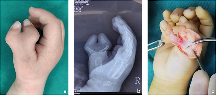
Figure 1. Inaccuracy of Wassel classification based solely on radiographs. (a, b) Clinical appearance and radiograph suggestive of Wassel type IV-D. (c) Intraoperative exploration confirming Wassel type III anatomy with cartilaginous connections between the phalanges.

Figure 2. Demonstration of cartilage ossification over time. (a, b) Initial appearance suggestive of Wassel type IV-D. (c, d) Subsequent ossification reveals the true anatomy corresponds to Wassel type III.
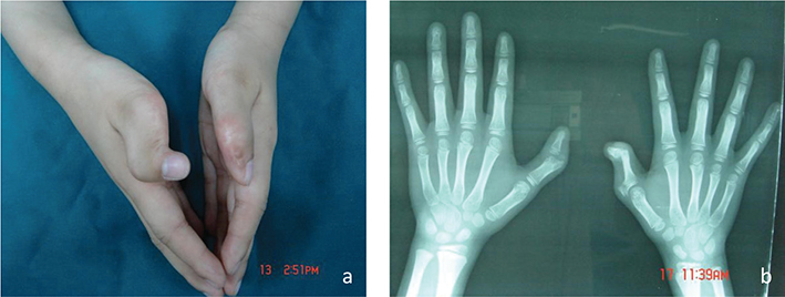
Figure 3. Secondary deformity resulting from incorrect preoperative classification. (a) Clinical appearance of severe postoperative deformity. (b) Intraoperative view showing incomplete cartilage resection caused by the initial misclassification.
Materials and methods
This prospective descriptive study was conducted in the pediatric orthopedic department of our hospital between May 2023 and May 2025. Preoperative ultrasound examinations were performed in patients initially classified as Wassel type IV or Wassel type II polydactyly to assess the presence of cartilaginous connections between the proximal phalanges in Wassel type IV and between the distal phalanges in Wassel type II, thereby allowing more precise classification. All ultrasound findings were subsequently verified intraoperatively to confirm their accuracy. According to the Wassel classification, the study cohort comprised 21 cases of Wassel type IV polydactyly, including subtypes IV-C and IV-D, and 10 cases of Wassel type II polydactyly.
Ethics
The study protocol was approved by the ethics committee of our hospital, and written informed consent was obtained from the parents or legal guardians of all participants.
Results
The baseline patient characteristics are summarised in Table 1. Based on preoperative radiographic findings and the Wassel classification system, ultrasound identified cartilaginous connections between the proximal phalanges in 10 of the 21 cases initially classified as Wassel type IV polydactyly. Intraoperative exploration confirmed the absence of cartilaginous connections in 2 of these 10 cases, whereas the remaining 8 cases were accurately reclassified as Wassel type III, representing 38.1% of the total cohort. Of these 8 cases, 3 were preoperatively classified as Wassel type IV-C and 5 as Wassel type IV-D. Among the 10 cases initially classified as Wassel type II polydactyly, ultrasound detected a cartilaginous connection between the distal phalanges in 1 case. This finding was confirmed intraoperatively, and the case was appropriately reclassified as Wassel type I (Figure 4), accounting for 10% of the Wassel type II group (Table 2). Overall, ultrasound misclassified only 2 cases, resulting in a diagnostic accuracy rate of 93.5% (Table 3).
| Preoperative classification (Wassel) | Number of patients (n) | Mean age (months) | Sex | |
| M (n) | F (n) | |||
| Wassel IV | ||||
| IV-C | 13 | 14 ± 3 | 6 | 7 |
| IV-D | 8 | 17 ± 4 | 5 | 3 |
| Wassel II | 10 | 13 ± 3 | 7 | 3 |

Figure 4. Reclassification of a case initially diagnosed as Wassel type II. (a, b) Clinical appearance and radiographs are consistent with Wassel type II thumb polydactyly. (c) Intraoperative findings confirmed Wassel type I, revealing a cartilaginous connection between the duplicated distal phalanges.
Discussion
The primary purpose of clinical disease classification is to guide treatment by informing preoperative assessment, surgical planning, intraoperative decision-making and expectations regarding postoperative outcomes. Ultimately, accurate classification aims to improve the effectiveness of surgical intervention. The Wassel classification, which categorises thumb polydactyly into seven distinct types, has long served as a foundational framework for surgical decision-making and has contributed substantially to improvements in the treatment of polydactyly [3].
The Wassel classification is primarily based on radiographic assessment. However, because cartilaginous structures are not fully ossified in childhood, radiographs often fail to reveal cartilaginous fusion between phalanges. In our clinical experience, some cases classified as Wassel type IV exhibit cartilaginous connections between the proximal phalanges, while some cases classified as Wassel type II show cartilaginous connections between the distal phalanges. These findings highlight inherent limitations of the Wassel classification when applied to pediatric patients. Misclassifying Wassel type III as type IV, or Wassel type I as type II, can lead to inappropriate surgical strategies, particularly with respect to osteotomy level selection, collateral ligament reconstruction, and joint capsule repair. Such errors may compromise postoperative joint stability, alter the force transmission axis of the thumb, and ultimately impair thumb function, resulting in suboptimal surgical outcomes in children. Accurate preoperative classification and a precise understanding of the true developmental anatomy of polydactyly are therefore fundamental to achieving satisfactory surgical results. Magnetic resonance imaging can clearly delineate cartilage and soft-tissue structures, and previous studies have demonstrated its value in assessing shared joints and tendon anatomy in duplicated thumbs, thereby aiding classification [12, 13]. However, magnetic resonance imaging is associated with high cost and prolonged examination time, and limited patient cooperation often necessitates sedation in younger children, increasing both the clinical burden and procedural risk. Consequently, there is an urgent need for a low-cost, convenient imaging modality capable of reliably visualising cartilaginous and tendinous structures.
This study introduces an ultrasound-based approach to improve the accuracy of polydactyly classification. Using ultrasound, a substantial proportion of cases initially classified as Wassel type IV-C and IV-D were reclassified as Wassel type III, accounting for 23 and 62.5% of cases, respectively, while 10% of cases initially classified as Wassel type II were reclassified as Wassel type I. Ultrasound demonstrated complete diagnostic accuracy for Wassel type IV-C and Wassel type II, whereas accuracy for Wassel type IV-D was lower, at 75%, resulting in an overall diagnostic accuracy of 93.5%. The reduced accuracy observed in Wassel type IV-D cases may be attributable to two factors. First, diagnostic performance may depend on the operator’s experience and proficiency in paediatric musculoskeletal ultrasound. Second, the close apposition of the proximal ends of the proximal phalanges in Wassel type IV-D can obscure cartilaginous connections, increasing the likelihood of misclassification.
Conclusions
Accurate preoperative classification is essential for preventing secondary deformities following surgery for polydactyly. In paediatric patients, incomplete cartilage ossification limits the reliability of radiograph-based Wassel classification and may lead to misclassification that adversely affects surgical outcomes. Ultrasound, when used in combination with radiographs, provides a valuable adjunct by improving the accuracy of Wassel classification and supporting more informed surgical planning.
Acknowledgements
Not applicable.
Ethical approval declaration
This study was approved by the Chongqing Medical University Institutional Review Board for all data collection.
References
[1] Cheng JC, Chan KM, Ma GF, et al. Polydactyly of the thumb: a surgical plan based on ninety-five cases. J Hand Surg Am. 1984;9(2): 155–164. https://doi.org/10.1016/S0363-5023(84)80133-1
[2] Townsend DJ, Lipp EB, Jr, Chun K, et al. Thumb duplication, 66 years’ experience – a review of surgical complications. J Hand Surg Am. 1994;19(6):973–976. https://doi.org/10.1016/0363-5023(94)90099-X
[3] Wassel HD. The results of surgery for polydactyly of the thumb. A review. Clin Orthop Relat Res. 1969;64:175–193.
[4] Hung L, Cheng JC, Bundoc R, et al. Thumb duplication at the metacarpophalangeal joint. Management and a new classification. Clin Orthop Relat Res. 1996;Feb;(323):31–41. https://doi.org/10.1097/00003086-199602000-00005
[5] Yao Y, Zhou H, Li L, et al. Epidemiological statistics of congenital thumb duplication in the Chinese population. J Orthop Surg Res. 2021;16(1):481. https://doi.org/10.1186/s13018-021-02567-3
[6] Islam S, Oka I, Fujita S. Anatomical study of preaxial polydactyly in 158 hands. Tohoku J Exp Med. 1992;168(3):459–466. https://doi.org/10.1620/tjem.168.459
[7] Al-Qattan MM. The distribution of the types of thumb polydactyly in a Middle Eastern population: a study of 228 hands. J Hand Surg Eur Vol. 2010;35(3):182–187. https://doi.org/10.1177/1753193409352417
[8] Dijkman RR, van Nieuwenhoven CA, Selles RW, et al. A multicenter comparative study of two classification systems for radial polydactyly. Plast Reconstr Surg. 2014;134:991–1001. https://doi.org/10.1097/PRS.0000000000000590
[9] Zhang SJ, Lin HB, Jiang QX, et al. Prenatal diagnosis of triphalangeal thumb-polysyndactyly syndrome by ultrasonography combined with genetic testing: a case report. World J Clin Cases. 2021;9(23):6832–6838. https://doi.org/10.12998/wjcc.v9.i23.6832
[10] Zhu Y, Fu Y, Lu K, et al. Use of skeletal and soft tissue imaging to guide the surgical repair of misclassified Wassel type VI thumb duplication with convergent deformity. J Plast Reconstr Aesthet Surg. 2025;103:354–362. https://doi.org/10.1016/j.bjps.2025.02.006
[11] Zhou L, Lv M, Zeng M, et al. Metacarpal bone plane examination by ultrasonography for the diagnosis of fetal forearm and hand deformity. Sci Rep. 2017;7:42161. https://doi.org/10.1038/srep42161
[12] Wang A, Ding J, Wang L, et al. MRI analysis and surgical treatment of Wassel Type IV duplicated thumbs. J Hand Surg Eur Vol. 2021;46(4):360–366. https://doi.org/10.1177/1753193420983213
[13] Du M, Jiang L, Wang A, et al. MR imaging provides practical information for preoperative assessment of radial polydactyly with bifurcation at the metacarpophalangeal joint level. Int J Gen Med. 2022;15:2163–2169. https://doi.org/10.2147/IJGM.S352501