ORIGINAL RESEARCH ARTICLE
Clinical outcomes of a defect-adapted strategy for free fibular great toe flaps in finger reconstruction: a retrospective series of 35 cases
Yangyang Liua, Huajian Zhoua, Haoran Jianga, Jun Zhua, Renjie Lia and Mingyu Jiaa
aDepartment of Hand and Foot Microsurgery, The First Affiliated Hospital of Bengbu Medical University, Bengbu, China
ABSTRACT
Background: To evaluate the clinical outcomes of a personalized surgical strategy for small-to-medium finger defects, utilizing either free dorsal toe or plantar proper digital artery flaps.
Methods: We retrospectively analyzed 35 patients with finger defects (March 2021–January 2025). Patients were assigned by defect characteristics to Group A (pulp defects, n = 21; plantar proper digital artery flap) or Group B (dorsal/lateral defects, n = 14; modified dorsal toe artery flap preserving the plantar proper neurovascular bundle). We evaluated operative time, flap survival, Disabilities of the Arm, Shoulder and Hand (DASH), Michigan Hand Outcomes Questionnaire (MHQ), static two-point discrimination (s2PD), and American Orthopaedic Foot & Ankle Society (AOFAS) scores.
Results: All 35 flaps survived. Group B had a significantly shorter mean operative time than Group A (124.00 ± 6.42 vs. 146.57 ± 9.61 minutes, p < 0.001). At final follow-up (6–13 months), Group B exhibited slightly better hand function (DASH: 5.89 ± 0.52 vs. 7.17 ± 1.00, p < 0.001; MHQ: 86.89 ± 2.82 vs. 83.61 ± 4.39, p = 0.018). Conversely, Group A achieved superior sensory recovery (s2PD: 7.48 ± 0.93 vs. 8.71 ± 0.83 mm, p < 0.001). Group B showed superior donor-site preservation without painful calluses (AOFAS: 96.08 ± 1.44 vs. 84.31 ± 2.19, p < 0.001).
Conclusions: A tailored approach to donor vessel selection reliably guides personalized finger reconstruction. The plantar proper system optimizes pulp reconstruction for superior sensory recovery, whereas the modified dorsal system suits dorsal/lateral defects by minimizing operative time and maximizing donor-site protection.
KEYWORDS: Great toe fibular flap; plantar proper artery; dorsal toe artery; dual-system; soft tissue defect; microsurgery
Citation: Journal of Plastic Surgery and Hand Surgery 2026; 61: 224–228. DOI: https://doi.org/10.2340/jphs.v61.46668.
Copyright: © 2026 The Author(s). Published by MJS Publishing on behalf of Acta Chirurgica Scandinavica. This is an Open Access article distributed under the terms of the Creative Commons Attribution 4.0 International License (http://creativecommons.org/licenses/by/4.0/), allowing third parties to copy and redistribute the material in any medium or format and to remix, transform, and build upon the material, with the condition of proper attribution to the original work.
Received: 10 July 2026; Accepted: 30 July 2026; Published: 27 August 2026.
CONTACT: Yangyang Liu 18375539028@163.com Department of Hand and Foot Surgery, The First Affiliated Hospital of Bengbu Medical University No. 287 Changhuai Road, Bengbu 233004, Anhui, China
Supplemental data for this article can be accessed online at https://doi.org/10.2340/jphs.v61.46668
Competing interests and funding: The authors declare that they have no relevant financial or non-financial interests to disclose.
This work was supported by the Natural Science Key Project of Bengbu Medical University (Grant No. 2023byzd069).
Introduction
The reconstruction of small-to-medium-sized digital defects remains a significant challenge, requiring both functional restoration and minimal donor-site compromise [1–3]. Free toe-based flaps, particularly the great toe fibular flap, are established ‘gold standards’ due to their anatomical consistency [4, 5]. However, conventional harvest techniques often sacrifice the proper plantar neurovascular bundle, leading to long-term donor-site morbidity such as painful calluses and altered gait [6–8]. Based on the microanatomical findings of Gu et al. and Strauch [9, 10], we evaluate a specific tailored pathway that differentiates between the plantar proper and dorsal systems. Rather than introducing a novel surgical entity, this retrospective study aims to report the clinical outcomes and reliability of this targeted, personalized decision-making process in maximizing functional recovery and minimizing donor-site morbidity.
Materials and methods
General information
This study was approved by the Institutional Ethics Committee, and informed consent was obtained from all patients. We retrospectively analyzed 35 patients who underwent free fibular great toe flap reconstruction between March 2021 and January 2025. Patients were divided into two groups based on defect characteristics:
Group A (n = 21)
Finger pulp defects requiring high-precision pinch sensation. Reconstructed using the free plantar proper digital artery flap.
Group B (n = 14)
Dorsal or lateral defects with lower demands for two-point discrimination. Reconstructed using the modified free dorsal toe artery flap.
Baseline characteristics, including age, sex, injured side, affected finger, injury mechanism, and defect area, showed no statistically significant differences between the two groups in Table 1 (p > 0.05).
Preoperative preparation and recipient site
Preoperatively, an 8 MHz handheld Doppler was used to map recipient and donor vessels. Under brachial plexus block, thorough debridement was performed under a microscope (10–15x magnification). The recipient digital artery, palmar proper digital nerve, and dorsal superficial veins were dissected. A sterile template of the defect was created.
Flap harvest technique
Group A (plantar proper system)
The flap was designed on the plantar-fibular aspect. Following dorsal incision to identify veins, deep dissection isolated the proper plantar digital artery and main nerve trunk, preserving deep fat. The neurovascular bundle was dissected proximally. Once perfusion was confirmed, the pedicle was divided distal to the transverse metatarsal artery.
Group B (modified dorsal system)
The flap was designed on the dorsal-fibular aspect. During deep dissection, the main plantar proper neurovascular bundle was deliberately avoided. Only the dorsal toe artery and terminal branches of the deep peroneal nerve were harvested. The paratenon of the extensor hallucis longus was meticulously preserved to ensure a viable bed for subsequent skin grafting.
Microsurgical anastomosis and closure
Under the microscope, flap arteries and veins were anastomosed to recipient vessels using 10-0 or 11-0 nylon sutures (artery to vein ratio of 1:1 or 1:2). For Group A, the plantar proper nerve was coapted to the digital nerve. For Group B, harvested cutaneous nerves were coapted appropriately. For the donor site, Group A wounds healed by secondary intention with petroleum gauze dressings. Group B wounds received a full-thickness skin graft from the ipsilateral calf.
Postoperative management and evaluation
Postoperative protocols included bed rest, warmth, anticoagulation, and antispasmodics. Follow-up ranged from 6 to 13 months. To ensure objective evaluation, upper extremity function was assessed using the Disabilities of the Arm, Shoulder and Hand (DASH) questionnaire [11] and the Michigan Hand Outcomes Questionnaire (MHQ) [12]. For donor-site evaluation, the American Orthopaedic Foot & Ankle Society (AOFAS) score was utilized [13]. Static two-point discrimination (s2PD) was measured as the primary indicator of sensory recovery, a metric previously validated for its correlation with patient-reported outcomes [14]. Continuous variables were analyzed using the independent samples t-test, and categorical variables via chi-square test (SPSS 26.0). P < 0.05 was considered significant.
Results
The surgical outcomes and postoperative functional follow-up results are summarized in Table 2.
Flap survival and operative time
All 35 flaps survived completely. Group A experienced two cases of vascular crisis (salvaged post-exploration) and two cases of partial marginal necrosis (healed via dressing changes). Group B had 100% smooth survival. Operative time in Group B was significantly shorter than in Group A (124.00 ± 6.42 minutes vs. 146.57 ± 9.61 minutes, p < 0.001).
Functional and sensory recovery
At final follow-up, Group B exhibited slightly better overall hand function than Group A (DASH: 5.89 ± 0.52 vs. 7.17 ± 1.00, p < 0.001; MHQ: 86.89 ± 2.82 vs. 83.61 ± 4.39, p = 0.018). Conversely, Group A achieved significantly better sensory recovery (s2PD: 7.48 ± 0.93 mm vs. 8.71 ± 0.83 mm, p < 0.001).
Donor site morbidity
Group B donor sites demonstrated near-normal function with a mean AOFAS score of 96.08 ± 1.44, significantly higher than Group A’s 84.31 ± 2.19 (p < 0.001). No patients in Group B developed painful calluses.
One representative case of a digital pulp defect repaired with a plantar proper digital artery flap (Group A) is illustrated in Figure 1 (see also Supplemental Video 1).
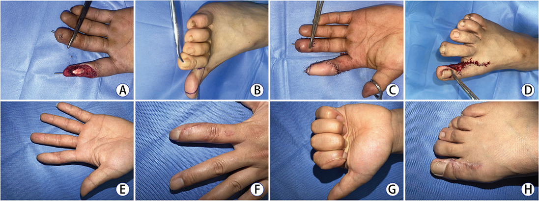
Figure 1. Representative case of Group A: Reconstruction of a high-sensitivity finger pulp defect. (A) Preoperative view of a 28-year-old male with a crush injury to the left index finger, resulting in a 2.5 × 4.0 cm pulp defect with tendon exposure. (B) Flap design on the fibular-plantar aspect of the great toe, based on the plantar proper digital artery system. (C) Immediate postoperative view showing a well-perfused flap after anastomosis of the digital artery and nerve. (D) Immediate postoperative view of the donor site, managed with secondary intention healing (wet dressing). (E, F) Volar and dorsal views of the index finger 8 months postoperatively, demonstrating excellent flap contour and tissue match. (G) Functional recovery of the finger at the 8-month follow-up; the static two-point discrimination (s2PD) reached 7 mm. (H) Appearance of the donor site at 8 months, showing an intact web space with no reported gait impairment or pain.
A typical case from Group B involving a dorsal defect is shown in Figure 2 (see also Supplemental Video 2).
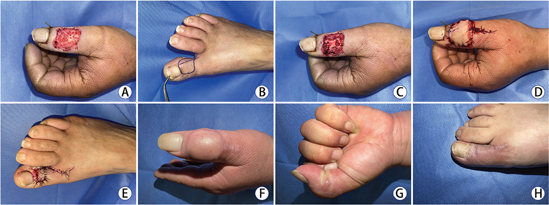
Figure 2. Representative case of Group B: Minimal-invasive reconstruction of a dorsal finger defect. (A) Preoperative view of a 56-year-old male with a saw injury to the dorsal aspect of the left thumb, involving a 2.0 × 2.5 cm soft tissue defect and partial extensor tendon loss. (B) Flap design on the dorsal-fibular aspect of the great toe, targeting the modified dorsal toe artery system. (C) Intraoperative view of the composite flap harvest, including a segment of the extensor hallucis longus tendon for extensor apparatus reconstruction. The plantar proper neurovascular bundle and paratenon were strictly preserved. (D) Immediate postoperative appearance of the thumb with a well-vascularized flap. (E) Immediate postoperative view of the donor site after full-thickness skin grafting. (F) Dorsal view of the thumb at the 6-month follow-up, showing an aesthetically pleasing outcome. (G) Functional recovery at 6 months, demonstrating normal range of motion in extension and flexion. (H) Appearance of the donor site at 6 months; the skin graft survived completely with preserved donor-toe sensation and no signs of tendon adhesion.
Discussion
The goal of hand reconstruction is to optimize recipient function while minimizing donor-site morbidity. The selection of reconstructive options relies on tailored decision-making processes that hand surgeons utilize to address specific clinical problems. The objective of our study is to report the empirical clinical outcomes of a personalized subset of this reconstructive strategy. By systematically differentiating between the plantar proper and modified dorsal systems, our data demonstrate that this established clinical pathway yields highly reproducible outcomes when meticulously matched to specific defect types.
Maximizing donor site function
Our findings demonstrate that the modified dorsal system (Group B) yields near-perfect donor-site preservation (AOFAS 96.08). This is crucial as donor-site morbidity remains a primary concern in hallux-based transfers [15]. By preserving the plantar proper bundle, we achieved results superior to conventional harvests reported in recent systematic reviews. This minimal-invasive strategy aligns with the tailored approach suggested by Ekin et al., favoring dorsal systems for non-pulp reconstructions. Additionally, this shallower dissection significantly reduced operative time (p < 0.001).
Balancing tissue match and sensation
Despite the donor-site advantages of the dorsal system, the plantar proper system (Group A) remains vital for finger pulp defects [16]. The pulp requires exquisite tactile gnosis. Group A achieved superior s2PD (7.48 ± 0.93 mm), justifying the sacrifice of the plantar nerve for high-quality sensory recovery [17]. As recent literature has shown [18], the inclusion of subcutaneous adipose tissue is vital for late-phase sensory remodeling, a technique we strictly followed in Group A harvest. This sensory advantage directly contributes to lower DASH scores, reinforcing the link between tactile gnosis and overall hand utility as evidenced in recent comparative studies [19]. Group B’s better overall hand function scores likely reflect the less severe nature of dorsal/lateral defects, which leave the native pulp intact.
Surgical nuances for donor protection and recipient optimization
Beyond the macro-anatomical selection of the vascular system, several meticulous surgical techniques are imperative to ensure flap survival and minimize donor-site morbidity:
Meticulous hemostasis
Complete hemostasis is the ‘lifeline’ for preventing hematoma and subsequent vascular crisis. We strongly advocate deflating the pneumatic tourniquet prior to pedicle division. This allows for the exact identification and management of bleeding points at the flap margins and recipient bed under normal physiological perfusion. Forceful closure with a risk of hematoma inevitably leads to pedicle compression and irreversible ischemia.
Triangular flap decompression
Designing a triangular skin flap at the pedicle inset effectively prevents circumferential constriction of the transposed vascular pedicle. This ‘geometric decompression’ breaks the linear scar trajectory, avoiding long-term cicatricial band contracture and yielding a more natural contour [20]. As demonstrated in Figure 1B, the geometric decompression provided by the triangular flap prevented pedicle constriction.
Web space integrity
During flap harvest, the structural integrity of the first web space must be strictly preserved [21]. Inappropriate incision design can lead to web space contracture (iatrogenic syndactyly), severely compromising foot esthetics, shock absorption, and gait biomechanics.
Donor hallux remodeling
In Group A, deliberately preserving the deep fat pad beneath the neurovascular bundle facilitates flap thinning while maintaining donor toe volume. By managing the donor site with secondary intention healing and initiating weight-bearing ambulation at 2 weeks postoperatively, repeated mechanical pressure promotes pulp remodeling and skin crease formation, effectively preventing severe scar deformities or toe atrophy. Furthermore, in Group B, meticulous preservation of the extensor hallucis longus paratenon remains an absolute prerequisite for successful skin graft take [22].
Indications boundary and salvage strategies
While the great toe fibular flap excels in small-to-medium defects, its indications must not be recklessly expanded. For extensive or multi-digit palmar defects, the medial plantar flap is preferred due to its larger surface area and concealed nonweight-bearing donor site [23]. Conversely, if a larger dorsal flap with a longer pedicle is required to achieve tension-free anastomosis, proximal dissection to include the main trunk of the first dorsal metatarsal artery (FDMA) is often necessary [24].
Surgeons must also be prepared for the notorious anatomical variability of the FDMA [25]. If an absent or hypoplastic dorsal toe artery is encountered intraoperatively during a planned Group B harvest, the flap design must be shifted distally to rely on cutaneous perforators arising from the plantar proper digital artery. In such salvage scenarios, the primary surgical plan must be converted to harvest the main trunk of the plantar proper artery (Group A approach) to guarantee flap viability [26].
Limitations
This study is a single-center retrospective review with a relatively small sample size (n = 35). Long-term follow-up is required to fully assess gait dynamics and definitive sensory remodeling.
Level of evidence
Therapeutic, Level III.
Ethical approval
This retrospective study was approved by the Institutional Review Board (IRB) and Ethics Committee of the First Affiliated Hospital of Bengbu Medical University (Approval Number: 2020-154). All procedures performed in studies involving human participants were in accordance with the ethical standards of the institutional and/or national research committee and with the 1964 Helsinki Declaration and its later amendments or comparable ethical standards.
Informed consent
Written informed consent was obtained from all individual participants included in the study. Furthermore, explicit written informed consent was obtained from the patients (specifically those depicted in Figures 1 and 2, as well as the accompanying Supplemental Digital Content videos) for the publication of any potentially identifiable clinical photographs, dynamic functional assessment recordings, and medical data included in this article.
Author contributions
Yangyang Liu contributed to the study conception and design. Surgical procedures were performed by Yangyang Liu. Material preparation, data collection, and analysis were performed by Jun Zhu, Renjie Li, Mingyu Jia, Huajian Zhou, and Haoran Jiang. The first draft of the manuscript was written by Yangyang Liu, and all authors commented on previous versions of the manuscript. All authors read and approved the final manuscript.
Data availability
The datasets generated during and/or analyzed during the current study are available from the corresponding author on reasonable request.
References
[1] Lin CH, Lin YT, Sassu P,et al. Functional assessment of the reconstructed fingertips after free toe pulp transfer. Plast Reconstr Surg. 2007 Oct;120(5):1315-1321. https://doi.org/10.1097/01.prs.0000279476.29055.5f
[2] Evin N, Evin SG, Guneren E. Composite classification and algorithmic reconstruction of fingertip defects with free lateral great toe flaps. J Plast Reconstr Aesthet Surg. 2023 May;80:133-144. Epub 2023 Feb 11. https://doi.org/10.1016/j.bjps.2023.02.010
[3] Wang J, Xue M, Lu H ,et al. Functional and aesthetic recovery of the second toe defect using a wrap-around pedicled flap from the great toe. J Hand Surg Eur Vol. 2023 Oct;48(9):926-929. Epub 2023 Jun 19. https://doi.org/10.1177/17531934231181329
[4] Agnihotri I, Nayak BB, Lakhotia P,et al. Comparative Study of Degree of Great Toe Movement after Complete and Partial Flexor Hallucis Longus Harvest in Free Fibula Flap. Indian J Plast Surg. 2024 May 30;57(3):192-200. https://doi.org/10.1055/s-0044-1786989
[5] Zavala A, Ore JF, Broggi A,et al. Pediatric Mandibular Reconstruction Using the Vascularized Fibula Free Flap: Functional Outcomes in 34 Consecutive Patients. Ann Plast Surg. 2021 Dec 1;87(6):662-668. https://doi.org/10.1097/SAP.0000000000002963
[6] Sosin M, Lin CH, Steinberg J,et al. Functional Donor Site Morbidity After Vascularized Toe Transfer Procedures: A Review of the Literature and Biomechanical Consideration for Surgical Site Selection. Ann Plast Surg. 2016 Jun;76(6):735-42. https://doi.org/10.1097/SAP.0000000000000591
[7] Tang SFT, Tang ACW, Chen CK,et al. Foot Plantar Pressure Profile Alteration after Microsurgical Great Toe-to-thumb Transfer. Plast Reconstr Surg Glob Open. 2023 Sep 1;11(9):e5228. https://doi.org/10.1097/GOX.0000000000005228
[8] Lin PY, Sebastin SJ, Ono S,et al. A systematic review of outcomes of toe-to-thumb transfers for isolated traumatic thumb amputation. Hand (N Y). 2011 Sep;6(3):235-43. Epub 2011 May 26. https://doi.org/10.1007/s11552-011-9340-x
[9] Gu YD, Wu MM, Zheng YL,et al. Vascular variations and their treatment in toe transplantation. J Reconstr Microsurg. 1985 Jan;1(3):227-32. https://doi.org/10.1055/s-2007-1007079
[10] Strauch B, Yu HL. Atlas of Microvascular Surgery: Anatomy and Operative Techniques. 2nd ed. New York, NY: Thieme Medical Publishers; 2006.
[11] Hudak PL, Amadio PC, Bombardier C. Development of an upper extremity outcome measure: the DASH (disabilities of the arm, shoulder and hand) [corrected]. The Upper Extremity Collaborative Group (UECG). Am J Ind Med. 1996 Jun;29(6):602-8. https://doi.org/10.1002/(SICI)1097-0274(199606)29:6%3C602::AID-AJIM4%3E3.0.CO;2-L Erratum in: Am J Ind Med 1996 Sep;30(3):372.
[12] Chung KC, Pillsbury MS, Walters MR,et al. Reliability and validity testing of the Michigan Hand Outcomes Questionnaire. J Hand Surg Am. 1998 Jul;23(4):575-87. https://doi.org/10.1016/S0363-5023(98)80042-7
[13] Kitaoka HB, Alexander IJ, Adelaar RS,et al. Clinical rating systems for the ankle-hindfoot, midfoot, hallux, and lesser toes. Foot Ankle Int. 1994 Jul;15(7):349-53. https://doi.org/10.1177/107110079401500701
[14] Rosén B, Lundborg G. A model instrument for the documentation of outcome after nerve repair. J Hand Surg Am. 2000 May;25(3):535-43. https://doi.org/10.1053/jhsu.2000.6458
[15] Sosin M, Lin CH, Steinberg J,et al. Functional Donor Site Morbidity After Vascularized Toe Transfer Procedures: A Review of the Literature and Biomechanical Consideration for Surgical Site Selection. Ann Plast Surg. 2016 Jun;76(6):735-42. https://doi.org/10.1097/SAP.0000000000000591
[16] Kwon HJ, Yoon S, Han HH,et al. Partial second-toe pulp free flap for fingertip reconstruction: Experience and surgical tips to minimize complications. Microsurgery. 2021 Oct;41(7):629-636. Epub 2021 Sep 4. https://doi.org/10.1002/micr.30804
[17] Lin CH, Lin YT, Sassu P,et al. Functional assessment of the reconstructed fingertips after free toe pulp transfer. Plast Reconstr Surg. 2007 Oct;120(5):1315-1321. https://doi.org/10.1097/01.prs.0000279476.29055.5f
[18] Unglaub F, Langer MF, Unglaub JM,et al. Defektdeckung an den Fingern und am Daumen : Indikation und Therapie [Defect coverage of fingers and thumb : Indications and treatment]. Unfallchirurg. 2018 Apr;121(4):321-334. German. https://doi.org/10.1007/s00113-018-0469-8
[19] Bott SM, Rachunek K, Medved F,et al. Functional outcome after digit replantation versus amputation. J Orthop Traumatol. 2022 Jul 27;23(1):35. https://doi.org/10.1186/s10195-022-00654-7
[20] Mahmoud M, Mohsen I. Revision Surgery for Scar Contracture and Web Creep after Syndactyly Release. J Hand Surg Asian Pac Vol. 2024 Dec;29(6):594-600. Epub 2024 Nov 22. https://doi.org/10.1142/S2424835524970026
[21] Sosin M, Lin CH, Steinberg J,et al. Functional Donor Site Morbidity After Vascularized Toe Transfer Procedures: A Review of the Literature and Biomechanical Consideration for Surgical Site Selection. Ann Plast Surg. 2016 Jun;76(6):735-42. https://doi.org/10.1097/SAP.0000000000000591
[22] do Amaral SA, de Carvalho BLF, Andrade AC,et al. DORSALIS PEDIS NEUROVASCULAR FLAP, OUR EXPERIENCE. Acta Ortop Bras. 2023 Sep 8;31(spe3):e267572. https://doi.org/10.1590/1413-785220233103e267572
[23] Li J, Luo X, Zou Y. Reconstruction of the Palmar Defect of the Hand with a Sensory Medial Plantar Artery Flap. Front Surg. 2022 May 6;9:874629. https://doi.org/10.3389/fsurg.2022.874629
[24] Hallock GG. The First Dorsal Metatarsal Artery Perforator Propeller Flap. Ann Plast Surg. 2016 Jun;76(6):684-7. https://doi.org/10.1097/SAP.0000000000000264
[25] Saboye M, Majchrzak A, d’Andréa G,et al. The First Dorsal Metatarsal Artery Perforator Flap: A Description and Anatomical Study. J Clin Med. 2025 Jun 11;14(12):4136. https://doi.org/10.3390/jcm14124136
[26] Wu Y, Chen B, Dong Z,et al. Repair of distal phalanx finger wound with modified great toe fibular flap with distal artery pedicle as reflux vein. Zhongguo Xiu Fu Chong Jian Wai Ke Za Zhi. 2021 Feb 15;35(2):206-209. Chinese. https://doi.org/10.7507/1002-1892.202009022