ORIGINAL RESEARCH ARTICLE
Suturing and knot-tying techniques in microsurgery: anastomosis time, learning curve and scanning electron microscopic assessment
Çağhan Benli, MD  and Mustafa Akyürek, MD
Department of Plastic, Reconstructive, and Aesthetic Surgery, Faculty of Medicine, Çanakkale Onsekiz Mart University, Çanakkale, Turkey
ABSTRACT
Background: Various suturing and knotting techniques exist to enhance microsurgical efficiency, yet a comprehensive comparison is lacking. This study evaluates two suturing (interrupted vs. open-loop) and two knot-tying (conventional vs. airborne) techniques to determine how anastomosis time is apportioned between them.
Methods: Seventy-two femoral artery anastomoses were performed on 36 female Wistar-albino rats by nine microsurgeons, stratified into three groups (<1, 1–4, >4 years experience). The four combinations were interrupted or open-loop suture with conventional or airborne knot (IS-CK, IS-AK, OLS-CK, OLS-AK). Each surgeon performed all four before and after a standardised practice session, making technique and practice within-subject factors and experience a between-subject factor, analysed by split-plot repeated-measures ANOVA. Twelve anastomoses from the most experienced group were examined by scanning electron microscopy (SEM), blind to technique and operator.
Results: The knot-tying method, rather than the suturing technique, was the principal factor associated with anastomosis time. Conventional knotting was approximately 11 min faster than airborne knotting (666 s; p < 0.001), whereas the two suturing techniques differed by 18.6 s within conventional knotting (p = 0.46) and 82.3 s within airborne knotting (p = 0.005) – approximately 1% and 4% of total time. Experience dominated (51.2 vs. 13.3 min; 3.85-fold; p < 0.001). All techniques improved after practice, and the improvement depended on the technique (F(3,18) = 8.30, p = 0.001): approximately 15% for the airborne-knot techniques vs. 5–10% for the conventional-knot techniques. SEM showed a dissociation: suture spacing was determined by the suturing technique (open-loop superior, p = 0.001) and knot quality by the knot-tying technique (airborne superior, p < 0.001); neither affected suture–tissue interaction (p = 0.77).
Conclusion: In elective, uncomplicated anastomoses the knot-tying technique, rather than the suturing technique, was most strongly associated with anastomosis time, while surgical experience outweighed both. The airborne knot costs approximately 11 min but yields better knot quality and improves most with practice; the open-loop suture gives more regular suture spacing at negligible time cost. Technique selection is therefore a trade-off between ischaemia time and technical refinement, informed primarily by the operator’s experience.
Grant sponsor: Çanakkale Onsekiz Mart University Scientific Research Projects Coordination Unit.
Grant number: TTU-2024-4668.
KEYWORDS: Microsurgery; suture techniques; anastomosis; surgical speed; free flap; replantation
Citation: Journal of Plastic Surgery and Hand Surgery 2026; 61: 229–236. DOI: https://doi.org/10.2340/jphs.v61.46794.
Copyright: © 2026 The Author(s). Published by MJS Publishing on behalf of Acta Chirurgica Scandinavica. This is an Open Access article distributed under the terms of the Creative Commons Attribution 4.0 International License (http://creativecommons.org/licenses/by/4.0/), allowing third parties to copy and redistribute the material in any medium or format and to remix, transform, and build upon the material, with the condition of proper attribution to the original work.
Received: 20 December 2025; Accepted: 19 August 2026; Published: 10 September 2026.
CONTACT: Çağhan Benli caghanbenli@gmail.com Department of Plastic, Reconstructive and Aesthetic Surgery, Faculty of Medicine, Çanakkale Onsekiz Mart University, Barbaros Mah. Prof. Dr. Sevim Buluc Sk. 17100 Merkez/Çanakkale, Turkey
Supplemental data for this article can be accessed online at https://doi.org/10.2340/jphs.v61.46794
Competing interests and funding: All named authors at this moment declare that they have no conflicts of interest to disclose.
This work was supported by the Scientific Research Projects Coordination Unit of Çanakkale Onsekiz Mart University under Grant TTU-2024-4668. The funder had no role in the design of the study, the collection or analysis of data, or the preparation of the manuscript.
Introduction
Advances in instrumentation, microscopes and suture materials have enabled microsurgeons to perform patent anastomoses on vessels less than 1 mm in diameter, raising the success rate of free tissue transfer above 99% [1]. Where ischaemia time is limiting – finger replantation, intestinal flaps, reconstructions involving multiple flaps or vein grafts – anastomosis time becomes a determinant of success in its own right, so the ideal technique should minimise operative time as well as ensuring long-term patency [2, 3].
The simple interrupted suture remains the gold standard for many surgeons [4]. However, its time-consuming nature has prompted the development of alternative methods. Techniques such as the continuous open-loop suture were introduced to reduce the number of knots, though they carry risks of luminal stenosis and a purse-string effect [5, 6]. The ‘airborne’ knot-tying technique was developed to expedite knot tying by keeping the suture end suspended, avoiding entanglement with surrounding tissues [7].
Comprehensive comparisons of these techniques across surgeon experience levels are lacking, particularly with regard to learning curves and the quality of the resulting anastomosis [4]. We therefore evaluated two suturing techniques (interrupted and open-loop) combined with two knotting methods (conventional and airborne), assessing the influence of experience and of practice on speed, and the technical quality of the anastomoses by scanning electron microscopy (SEM).
Materials and methods
All animal procedures and care complied with the institution’s established guidelines for the care and use of laboratory animals.
Experimental design and subjects
Thirty-six healthy adult female Wistar albino rats weighing 250–300 g were obtained from the Experimental Research Application and Research Centre of Çanakkale Onsekiz Mart University. Animals were housed at 21 ± 2°C under a 12-h light/dark cycle with ad libitum access to standard chow and water. Anaesthesia and analgesia were provided by intramuscular ketamine 50 mg/kg with xylazine 15 mg/kg, with supplementary doses as required to maintain a surgical plane, as the two anastomoses performed in each animal could together exceed 2 h in the least experienced group. Animals were euthanised by anaesthetic overdose without recovery from anaesthesia; humane endpoints were therefore not applicable. No animals were lost and no anastomoses were excluded from the analysis. A total of 72 femoral artery anastomoses were performed, using both femoral arteries of each rat. The mean femoral artery diameter was 1.6 mm, comparable to that of a human digital artery.
Surgeon and technique groups
Nine microsurgeons who routinely perform microsurgery participated in the study. They were stratified into three equal groups based on their years of active clinical microsurgery experience. Two of the three surgeons in Group C had completed a microsurgery fellowship, and none of those in Groups A or B had:
- Group A: < 1 year (n = 3)
- Group B: 1–4 years (n = 3)
- Group C: > 4 years (n = 3)
Surgeons in the least experienced group (Group A) were required to have completed the basic microsurgery course of the Reconstructive Microsurgery Society, to have served at least 6 months on plastic surgery emergency rotations, and to have performed at least three successful independent microsurgical anastomoses in clinical practice. Group A therefore represents surgeons early in independent practice rather than complete beginners. No previously validated stratification was available for this purpose, and the three strata were defined pragmatically from the distribution of experience among the participants.
Four different technique combinations were established for evaluation:
1. IS-CK: Interrupted Suture with Conventional Knot
Stay sutures were placed at 0° and 180° to align the vessel edges. Simple interrupted sutures were then applied and secured with conventional knots; after three further stitches completed the anterior wall, the clamp was rotated 180° and the posterior wall sutured in the same manner, giving eight sutures in total (Figure 1).
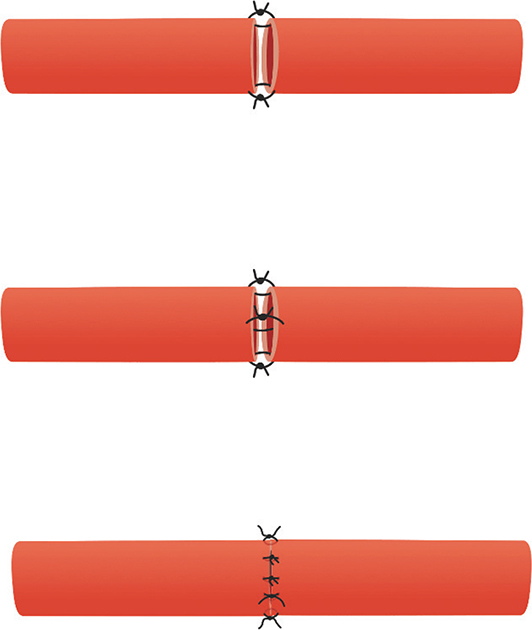
Figure 1. Illustration of Interrupted Suture with Conventional Knot.
2. IS-AK: Interrupted Suture with Airborne Knot
This technique differed from IS-CK only in the knot-tying method, which used the airborne technique previously described [7]. The suture is drawn so that the leading strand remains approximately three times longer than the free end, and the knot is then formed and tightened without the free end contacting surrounding tissue (Figure 2).
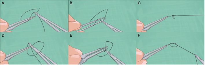
Figure 2. Step-by-step airborne knotting. (A) The short suture end is grasped with the right forceps, tip pointing up, and the long end with the left. (B) The long end is circled around the right forceps. (C) The short end is regrasped through the loop and pulled through. (D) The short end is regrasped with the forceps tip pointing down. (E) A loop of the long end is pushed under the right forceps. (F) The short end is regrasped from above and pulled through to complete the knot.
3. OLS-CK: Open-Loop Suture with Conventional Knot
Stay sutures were placed and tied at 0° and 180°. Open-loop suturing was performed between them, with three passes along the anterior wall; each loop was then cut and tied individually with a conventional knot. The clamp was rotated 180° and the posterior wall sutured in the same fashion (Figure 3).
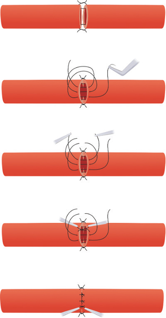
Figure 3. Illustration of Open-Loop Suture with Conventional Knot.
4. OLS-AK: Open-Loop Suture with Airborne Knot
This technique was identical to OLS-CK except that the loops were progressively cut and all eight knots secured with the airborne technique.
Surgical procedure and data collection
Each surgeon performed every technique combination twice, before and after a standardised training session. This session served to quantify the learning curve for the less familiar alternative techniques and to allow adaptation to the study microscope, whose focus and zoom are manual rather than foot-pedal operated. It comprised eight repetitions of each technique on an in vitro model (plastic tubes of similar calibre) and three in vivo on rat carotid arteries or femoral veins. Anastomoses were performed on the femoral artery with 10/0 monofilament nylon (ETHILON®, 3/8 circle, 3.8 mm, Ethicon, Somerville, NJ) under a surgical microscope (Leica M651, Leica Microsystems, Germany). Total anastomosis time was recorded from the first needle pass through the arterial wall to completion of the eighth and final suture, from video recordings, by a single assessor who performed every measurement and was not blinded to technique. The four combinations were performed in the same fixed, non-randomised order (IS-CK, IS-AK, OLS-CK, OLS-AK) in both sessions to standardise the protocol. Vessel allocation was randomised: for each animal a computer-generated draw determined which femoral artery was operated on first. No a priori power calculation was performed. The number of animals was instead set at the smallest number judged likely to demonstrate the anticipated difference, in accordance with the 3Rs principle of reduction. No interim analysis or sample-size re-estimation was carried out; all analyses were performed once, on the complete dataset. Patency was confirmed immediately and 1 h after the procedure by the milking test. Where leakage was seen on completion, haemostasis was obtained by 1 min of compression; no anastomosis required revision, additional sutures or a repeat attempt, and none failed.
Scanning electron microscopy evaluation
Every post-practice anastomosis performed by Group C – 12 in total – was harvested, fixed, dehydrated and gold-coated for SEM. Evaluation was restricted to Group C in order to characterise the quality attainable by each technique when executed proficiently rather than by an average operator. The 12 specimens comprised four anastomoses – one per technique – from each of the three Group C surgeons. All nine participating surgeons assessed all 12 specimens and awarded a single score to each technique on each criterion, having viewed the three specimens of that technique together. Images (60×, 250× and 575×) were randomised and coded, and assessors were blind to technique and operator. Assessors were not calibrated on the scale before scoring. Three criteria (Table 1) were scored on a 5-point Likert scale (1 = poor, 5 = excellent):
- Suture Spacing: Uniformity of distance between sutures.
- Knot Quality: Proper seating and absence of crossing.
- Suture–Tissue Interaction: Proper eversion and absence of backwalling.
Statistical analysis
Data were analysed using SPSS version 27.0 (IBM, Armonk, NY, USA). Because each surgeon performed all four technique combinations both before and after practice, technique and practice were within-subject factors and surgeon experience a between-subject factor; anastomosis time was therefore analysed by split-plot repeated-measures analysis of variance using the appropriate error term for each effect. The experimental unit was the surgeon, the anastomosis the unit of observation. Standard errors of group means derive from the pooled error term and, the design being balanced, are identical across techniques. Homogeneity of the between-subject error was assessed by Levene’s test and sphericity by Mauchly’s test, with Greenhouse–Geisser correction where violated; no correction applies to effects with a single within-subject degree of freedom. Effect sizes are reported as partial η². Pairwise comparisons used Tukey’s Honestly Significant Difference test with the within-subject error term, and specific technique pairs additionally by paired t-tests, which use the error variance specific to that contrast. The effect of practice within each technique was tested by paired t-tests with Bonferroni correction for four comparisons and Cohen’s dz. As residuals departed from normality (Shapiro–Wilk p = 0.002), all analyses were repeated on log-transformed times as a sensitivity check. The untransformed scale was retained as primary because the clinical question concerns absolute ischaemia time; log-scale results are given wherever the two scales differ. SEM scores, being ordinal and obtained from raters who assessed all four techniques, were compared by the Friedman test with Conover post hoc comparisons and Bonferroni correction; interobserver agreement was quantified by intraclass correlation coefficients (two-way random effects, absolute agreement) for a single assessor and for the mean of the nine, with 95% confidence intervals, and concordance by Kendall’s coefficient of concordance. Statistical significance was set at p < 0.05.
Ethics
Ethical approval for this study was obtained from Çanakkale Onsekiz Mart University Animal Experiments Ethics Committee (07/07/2023, No: 2023/06-02). This study was not preregistered.
Results
Anastomosis time
All 72 anastomoses were patent and were included. Anastomosis time differed markedly between techniques (F(3,18) = 45.48, p < 0.001, Greenhouse–Geisser ε = 0.344; partial η² = 0.883), but almost entirely because of the knot-tying method: an orthogonal contrast comparing conventional with airborne knotting accounted for 99.2% of the between-technique sum of squares (F(1,18) = 135.35, p < 0.001), the residual variation being non-significant (F(2,18) = 0.54, p = 0.59; Supplementary Table S3). Mean times were 1487.0 s (24.8 min) for IS-CK, 1468.4 s (24.5 min) for OLS-CK, 2184.6 s (36.4 min) for IS-AK and 2102.3 s (35.0 min) for OLS-AK (Table 2), a knot-related difference of 666 s (11.1 min). Within conventional knotting the two suturing techniques differed by 18.6 s (95% CI −210.1 to 247.3; p = 0.46) and within airborne knotting by 82.3 s (p = 0.005), approximately 1% and 4% of total anastomosis time (Table 3). The open-loop suture therefore confers a small but consistent advantage that is an order of magnitude smaller than that of the knot-tying method.
Thrombosis rates
All 72 anastomoses were patent at 1 min and 1 h after completion, in every technique group. As the outcome showed no variance, no comparative test was applicable.
Impact of surgeon experience
Surgeon experience was the most influential determinant of anastomosis time (F(2,6) = 120.85, p < 0.001; partial η² = 0.976). Group C completed anastomoses in approximately one quarter of the time required by Group A: 798 s (13.3 min) versus 3,072 s (51.2 min), a 3.85-fold difference of approximately 38 min (Supplementary Table S1). All three strata differed from one another (A vs. B 1,510 s, p = 0.001; B vs. C 763 s, p = 0.005; A vs. C 2,273 s, p = 0.001), each more experienced stratum completing anastomoses in approximately half the time of the next. The penalty associated with airborne knotting varied across strata in absolute terms, from approximately 19 additional minutes in Group A to 3.5 min in Group C (technique × experience interaction F(6,18) = 7.61, p < 0.001; Greenhouse–Geisser p = 0.021); in relative terms it was more nearly comparable (+46%, +51% and +30%) and did not persist after log transformation (p = 0.33), indicating a largely multiplicative effect.
Effect of practice on learning curve
Practice reduced anastomosis time for all four techniques, and the size of the reduction depended on the technique (technique × practice interaction F(3,18) = 8.30, p = 0.001; Greenhouse–Geisser p = 0.012; partial η² = 0.580). Reductions were 162.2 s (2.7 min; 10.3%) for IS-CK, 350.0 s (5.8 min; 14.8%) for IS-AK, 77.4 s (1.3 min; 5.1%) for OLS-CK and 330.1 s (5.5 min; 14.6%) for OLS-AK (Supplementary Table S2). After Bonferroni correction only the airborne-knot improvements remained individually significant (IS-AK p = 0.031, dz = 1.18; OLS-AK p = 0.014, dz = 1.36). The improvement was therefore approximately 1.5- to 3-fold larger for the airborne-knot techniques, and the significant interaction confirms that this difference is itself reliable rather than an artefact of comparing significant with non-significant simple effects.
Scanning electron microscopy findings
Blinded evaluation revealed a dissociation between the two components of technique (Table 4, Figure 4). Suture spacing was determined by the suturing technique, both open-loop techniques scoring higher than both interrupted techniques (Friedman χ²(3) = 15.93, p = 0.001; all four cross-comparisons p ≤ 0.017), and knot quality by the knot-tying technique, both airborne techniques scoring higher than both conventional ones (χ²(3) = 24.68, p < 0.001; all four p < 0.001); neither pair differed internally. Suture–tissue interaction did not differ between techniques (χ²(3) = 1.13, p = 0.77). Interobserver agreement across the nine assessors was moderate for suture spacing (ICC(2,1) = 0.52, 95% CI 0.15 to 0.94) and good to excellent for knot quality (0.81, 0.47 to 0.98), but absent for suture–tissue interaction (0.00, −0.12 to 0.16); for the mean of the nine assessors the corresponding values were 0.91, 0.97 and 0.00. The confidence intervals are wide because only four techniques were compared. These coefficients describe agreement on the relative standing of the four techniques, not the reliability of scoring an individual anastomosis.
| Criterion | Statistic | IS-CK | IS-AK | OLS-CK | OLS-AK |
| Suture spacing | Mean (SD) | 3.22 (0.44) | 3.11 (0.33) | 4.00 (0.71) | 4.22 (0.44) |
| Median [IQR] | 3 [3–3] | 3 [3–3] | 4 [4–4] | 4 [4–4] | |
| Knot quality | Mean (SD) | 2.56 (0.53) | 4.56 (0.53) | 2.67 (0.50) | 4.22 (0.44) |
| Median [IQR] | 3 [2–3] | 5 [4–5] | 3 [2–3] | 4 [4–4] | |
| Suture–tissue interaction | Mean (SD) | 2.89 (0.60) | 2.78 (0.67) | 3.00 (0.50) | 2.89 (0.60) |
| Median [IQR] | 3 [3–3] | 3 [2–3] | 3 [3–3] | 3 [3–3] | |
| SD: standard deviation; IQR: interquartile range; IS-CK: Interrupted Suture with Conventional Knotting; IS-AK: Interrupted Suture with Airborne Knotting; OLS-CK: Open-Loop Suture with Conventional Knotting; OLS-AK: Open-Loop Suture with Airborne Knotting. Friedman test: suture spacing χ²(3) = 15.93, p = 0.001, Kendall’s W = 0.590; knot quality χ²(3) = 24.68, p < 0.001, W = 0.914; suture–tissue interaction χ²(3) = 1.13, p = 0.77, W = 0.042. Conover post hoc comparisons with Bonferroni correction: both open-loop techniques scored higher than both interrupted techniques for suture spacing (all four cross-comparisons p ≤ 0.017) and both airborne-knot techniques higher than both conventional-knot techniques for knot quality (all four p < 0.001); no within-pair difference was significant. Interobserver agreement across the nine assessors: ICC(2,1) = 0.52, 0.81 and 0.00, and ICC(2,9) = 0.91, 0.97 and 0.00, respectively. |
|||||
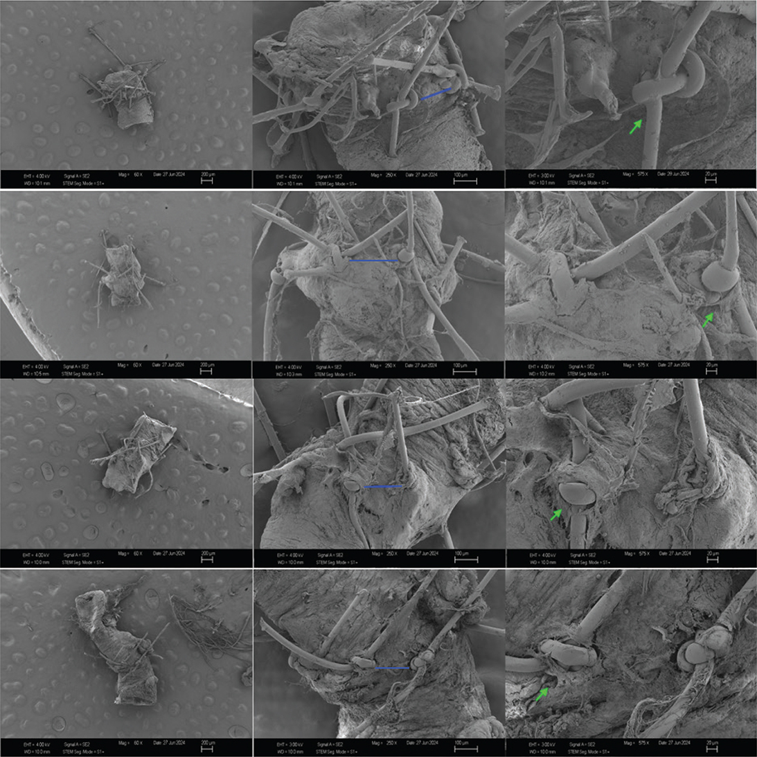
Figure 4. Representative SEM images of the four technique groups (IS-CK, IS-AK, OLS-CK, OLS-AK; top to bottom) at magnifications of 60×, 250× and 575× (left to right). Blue lines in the 250× images mark the intervals between adjacent sutures, the feature scored as suture spacing. Green arrows in the 575× images indicate the knots, the feature scored as knot quality. Image contrast has been enhanced to improve legibility.
Discussion
Shorter anastomosis time reduces ischaemic injury and improves functional outcomes [8]. Numerous alternative methods have been developed to shorten anastomosis time [9–11], but hand-sewn anastomosis remains the gold standard for small-calibre vessels, coupler-assisted methods being costlier and riskier at this calibre [4, 12, 13]. Sert et al. previously compared these four suture–knot combinations in a single operator [14]. Our study extends that work in three respects: anastomoses were performed by nine microsurgeons across three experience levels, separating the contributions of technique and experience; each technique was assessed before and after a standardised practice session within the same operators; and the anastomoses were examined by SEM. The novelty lies not in showing that one combination is faster, which is established, but in apportioning the time between technique, knot and operator. Our primary finding is that anastomosis time was most strongly associated with the knot-tying method rather than the suturing technique: conventional knotting was approximately 11 min faster than airborne knotting, whereas the two suturing techniques differed by approximately 0.3 and 1.4 min within conventional and airborne knotting, respectively. The small advantage of the open-loop suture is consistent in direction with studies of continuous and semi-continuous suturing [5, 8, 15–17] and may reflect fewer instrument exchanges and discrete steps, but its magnitude is small relative to the ischaemia times that determine outcome in operations such as the replantations described by Tamai [18] and the early reconstructions advocated by Godina [19].
Speed must be balanced against anastomotic quality, and the SEM findings show that the two are dissociated. The open-loop suture produced more regular suture spacing; the airborne knot, despite being slower, produced knots that lay flat and without crossing; neither influenced suture–tissue interaction. The airborne technique was developed to avoid retrieving the suture from surrounding tissues and thereby reduce manipulation of the adventitia, the most plausible explanation for the difference observed here [7]. Two cautions follow. Firstly, SEM demonstrates knot morphology, not mechanical behaviour: showing that airborne knots are more secure would require tensile or leakage testing, which we did not perform, and we have accordingly avoided claims of knot security. Secondly, every anastomosis was patent at 1 h irrespective of technique, so the morphological differences have no demonstrated functional correlate and should be read as hypothesis-generating. SEM is an established tool for assessing endothelial injury and repair in experimental microsurgery [20], and our findings accord with recent studies comparing conventional and robotic microsurgery [21].
Surgeon experience was the dominant determinant: 38 min separated the most from the least experienced surgeons, against approximately 11 min for the knot-tying method and 1 min for the suturing technique.
The airborne knot was initially challenging for all participants, but benefited most from a short, intensive practice session, improving by approximately 15% against 5–10% for the conventional-knot techniques; the significant technique × practice interaction confirms that this difference is itself reliable. Because the session gave each technique the same number of repetitions, the unequal improvement cannot be an artefact of unequal practice and is therefore a property of the techniques themselves. Techniques that appear inefficient at first may therefore be integrated effectively with dedicated training, which argues for structured practice before clinical application, consistent with systematic reviews of microsurgical training programmes and simulators [22–24].
For a microsurgeon early in training the airborne knot may be a worthwhile investment for difficult-access cases, whereas conventional knotting remains preferable whenever ischaemia time is limiting.
This study has several limitations. All anastomoses were performed on healthy femoral arteries of young, systemically well rats; clinical microsurgery frequently involves vessels compromised by avulsion or crush injury, irradiation, atherosclerosis, diabetes or previous surgery, in which wall fragility, intimal irregularity and spasm alter both the difficulty and the duration of anastomosis, so our findings may not translate to such vessels or to injuries of the type described by Urbaniak [25]. Follow-up extended only to 1 h and assessed patency alone; thrombosis, neointimal hyperplasia and vessel-wall healing were not examined, and no mechanical testing of knot strength was performed, so the SEM differences cannot be linked to function. Technique order was fixed and identical for all surgeons, so sequence position is confounded with technique; moreover all participants used IS-CK routinely, and the greater familiarity this confers may favour that technique independently of its intrinsic efficiency. Several features of the data argue against a sequence explanation – the alternating rather than monotonic pattern of times, the fact that a single knot-type contrast accounts for 99% of the between-technique variance, and the absence of a systematic advantage for the later-performed technique – but a counterbalanced design would have permitted a definitive test.
The qualitative assessment carries its own limitations. SEM was confined to the 12 specimens from the most experienced group, so although no specimen selection was applied – every post-practice anastomosis of that group was examined – the quality findings describe expert performance only; the reliability estimates rest on comparisons among only four techniques and are correspondingly imprecise, the assessors were not calibrated on the scale beforehand, and the negligible agreement for suture–tissue interaction cannot be distinguished from insufficient discriminative capacity of the scale. The raters were the same nine surgeons who performed the anastomoses; although images were randomised, coded and assessed blind to technique and operator, assessment by external microsurgeons would have been preferable. The assessor who timed the recordings was not blinded, although the techniques are immediately distinguishable on video and the outcome has operationally defined start and end points. Finally, no a priori power calculation was performed, the between-stratum comparison rests on three surgeons per stratum, and the experience strata were defined pragmatically rather than from a validated scheme. Finally, both femoral arteries of each animal were used, so every rat contributed two anastomoses – always by the same surgeon in the same session, and always the two techniques adjacent in the fixed order, so that one animal received both interrupted-suture techniques and the other both open-loop techniques. Each animal therefore contributed one conventional-knot and one airborne-knot anastomosis, and the knot-type contrast on which our principal conclusion rests is estimated entirely within animals, so that variation between animals cancels from it. The comparisons between suturing techniques and between practice sessions are made between animals, and animal-to-animal variation contributes to the error terms used for them, which are correspondingly conservative. The animal was not recorded as a separate identifier and could not be modelled explicitly, but for the principal finding this is immaterial.
Conclusion
Anastomosis time was principally associated with the knot-tying method rather than the suturing technique: conventional knotting was approximately 11 min faster than airborne knotting, while the two suturing techniques differed by approximately 1 min or less. All techniques yielded patent results, and there is a trade-off between speed and specific aspects of morphological quality: the open-loop technique gives more regular suture spacing, the airborne knot better-seated knots, though neither difference was linked to patency. Surgical experience remains the single most critical factor, outweighing technique by a factor of approximately three, and techniques with a steeper learning curve improve most with practice. These findings apply to elective, uncomplicated anastomoses.
Declarations
Data availability
The raw data supporting all analyses are provided as Supplementary Material: anastomosis times for all 72 anastomoses (Supplementary Data 1) and the SEM assessment scores (Supplementary Data 2).
Ethical approval
Ethical approval was obtained from the Çanakkale Onsekiz Mart University Animal Experiments Ethics Committee (07/07/2023, No: 2023/06-02).
References
[1] Gardiner MD, Nanchahal J. Strategies to ensure success of microvascular free tissue transfer. J Plast Reconstr Aesthet Surg. 2010;63(9):e665–e673. https://doi.org/10.1016/j.bjps.2010.06.011
[2] Chen SH, Tang YB, Chen HC. Survival of transferred ileum after ischemia time longer than 1 hour: a clinical result different from animal studies. J Am Coll Surg. 2013;217(2):300–305. https://doi.org/10.1016/j.jamcollsurg.2013.03.018
[3] Svaerdborg M, Birke-Sorensen H. Monitored extended secondary arterial ischemia in a free muscle transfer. J Reconstr Microsurg. 2012;28(2):119–124. https://doi.org/10.1055/s-0031-1289163
[4] Alghoul MS, Gordon CR, Yetman R, et al. From simple interrupted to complex spiral: a systematic review of various suture techniques for microvascular anastomoses. Microsurgery. 2011;31(1):72–80. https://doi.org/10.1002/micr.20813
[5] Lykoudis EG, Spyropoulou GA, Liadakis GN, et al. A new combined ‘interrupted-continuous’ microvascular anastomotic technique. Experimental study and clinical application. J Reconstr Microsurg. 2008;24(2):79–84. https://doi.org/10.1055/s-2008-1076094
[6] Chen YX, Chen LE, Seaber AV, et al. Comparison of continuous and interrupted suture techniques in microvascular anastomosis. J Hand Surg Am. 2001;26(3):530–539. https://doi.org/10.1053/jhsu.2001.22933
[7] Agko M, Ciudad P, Chen HC. ‘Airborne’ suture tying technique: simple steps to make it easy. Head Neck. 2017;39(12):2558–2561. https://doi.org/10.1002/hed.24923
[8] Wang J, Ma H, Zhang H, et al. Continuous locked suture technique for arterial anastomosis in rat small bowel transplantation. Microsurgery. 2007;27(2):112–117. https://doi.org/10.1002/micr.20315
[9] Mudigonda SK, Murugan S, Velavan K, et al. Non-suturing microvascular anastomosis in maxillofacial reconstruction – a comparative study. J Craniomaxillofac Surg. 2020;48(6):599–606. https://doi.org/10.1016/j.jcms.2020.04.005
[10] Maruccia M, Fatigato G, Elia R, et al. Microvascular coupler device versus hand-sewn venous anastomosis: a systematic review of the literature and data meta-analysis. Microsurgery. 2020;40(5):608–617. https://doi.org/10.1002/micr.30585
[11] Davis CR, Rappleye CT, Than PA, et al. Sutureless microsurgical anastomosis using an optimized thermoreversible intravascular poloxamer stent. Plast Reconstr Surg. 2016;137(2):546–556. https://doi.org/10.1097/01.prs.0000475774.37267.3f
[12] Broer PN, Weichman KE, Tanna N, et al. Venous coupler size in autologous breast reconstruction – does it matter? Microsurgery. 2013;33(7):514–518. https://doi.org/10.1002/micr.22169
[13] Geierlehner A, Rodi T, Mosahebi A, et al. Meta-analysis of venous anastomosis techniques in free flap reconstruction. J Plast Reconstr Aesthet Surg. 2020;73(3):409–420. https://doi.org/10.1016/j.bjps.2019.11.033
[14] Sert G, Aksoyler D, Kara M, et al. Comparison of total anastomosis time between four different combinations of suturing and knot tying techniques in microsurgical anastomosis. J Plast Surg Hand Surg. 2023;57(1–6):240–246. https://doi.org/10.1080/2000656X.2022.2052083
[15] Sapountzis S, Kiranantawat K, Lim SY, et al. A novel ‘continuous-interrupted’ method for microvascular anastomosis. Microsurgery. 2014;34(1):82–84. https://doi.org/10.1002/micr.22174
[16] Simsek T, Eroglu L, Engin MS, et al. End-to-end microvascular anastomosis in the rat carotid artery using continuous horizontal mattress sutures. J Reconstr Microsurg. 2006;22(8):631–640. https://doi.org/10.1055/s-2006-956237
[17] Mao K, Tang MY, South JR. A comparison of continuous with interrupted sutures in microvascular anastomosis. Microsurgery. 1986;7(4):158–160. https://doi.org/10.1002/micr.1920070406
[18] Tamai S. Twenty years’ experience of limb replantation – review of 293 upper extremity replants. J Hand Surg Am. 1982;7(6):549–556. https://doi.org/10.1016/S0363-5023(82)80100-7
[19] Godina M. Early microsurgical reconstruction of complex trauma of the extremities. Plast Reconstr Surg. 1986;78(3):285–292. https://doi.org/10.1097/00006534-198609000-00001
[20] Kirse DJ, Young PH. A scanning electron microscopic study of arterial endothelial repair under turbulent flow conditions. Microsurgery. 1992;13(1):26–30. https://doi.org/10.1002/micr.1920130107
[21] Cho J, Kim DG, Kim TH, et al. Further validating the robotic microsurgery platform through preclinical studies on rat femoral artery and vein. J Reconstr Microsurg. 2024. https://doi.org/10.1055/a-2460-4940
[22] Sapino G, Gonvers S, Cherubino M, et al. Synthetic simulators for microsurgery training: a systematic review. Plast Reconstr Surg Glob Open. 2024;12(7):e6004. https://doi.org/10.1097/GOX.0000000000006004
[23] Esanu V, Carciumaru TZ, Ilie-Ene A, et al. Validated microsurgical training programmes: a systematic review of the current literature. J Clin Med. 2025;14(21):7452. https://doi.org/10.3390/jcm14217452
[24] Lee DH, Shin YK, Son SH, et al. Learning curve of microsurgical anastomosis: training for resident education. J Am Acad Orthop Surg. 2025;33(17):977–985. https://doi.org/10.5435/JAAOS-D-24-00981
[25] Urbaniak JR, Evans JP, Bright DS. Microvascular management of ring avulsion injuries. J Hand Surg Am. 1981;6(1):25–30. https://doi.org/10.1016/S0363-5023(81)80006-8