ORIGINAL RESEARCH ARTICLE
Long-term efficacy of selective arterial embolisation of renal angiomyolipoma
Jesper Swärda,b , Karl Bohlinc, Olof Henriksonc, Sven Lundstama,d,e, Ralph Peekera,d and Anna Grenabo Bergdahla,d
aInstitute of Clinical Sciences, Sahlgrenska Academy, University of Gothenburg, Gothenburg, Sweden; bRegion Västra Götaland, NU-Hospital Group, Department of Urology, Uddevalla, Sweden; cRegion Västra Götaland, Sahlgrenska University Hospital, Department of Radiology, Gothenburg, Sweden; dRegion Västra Götaland, Sahlgrenska University Hospital, Department of Urology, Gothenburg, Sweden; eRegion Västra Götaland, Sahlgrenska University Hospital, Department of Oncology, Gothenburg, Sweden
ABSTRACT
Objective: To evaluate the long-term efficacy of selective arterial embolisation in renal angiomyolipoma (AML), with emphasis on tumour shrinkage, potential regrowth and the necessity of supplementary procedures.
Material and methods: A retrospective review of all 58 consecutive embolisations at two institutions, between 1999 and 2018, was performed. Clinical notes, laboratory data and imaging were reviewed.
Results: The overall complication rate was 6.8%, with no Clavien-Dindo grades III–V complications. Kidney function was unaffected by embolisation as measured by creatinine. Median radiological follow-up was 4.8 years (interquartile range [IQR]: 2.8–7.8), and median clinical follow-up was 7.5 years (IQR: 4.7–14.0). Decreasing AML size was observed in 96% of procedures. Maximal shrinkage (30% median diameter decrease; IQR: 15–44) was reached after median 2.2 years (IQR: 0.6–4.8). During follow-up, regrowth occurred in 38% of patients, and four bleeding episodes occurred in three patients with tuberous sclerosis. Growing size and/or rebleeding prompted a redo embolisation in 9% of spontaneous AML and 50% of tuberous sclerosis-associated AML.
Conclusions: Being a well-tolerated treatment with few complications, selective arterial embolisation renders a pronounced size-reduction in most patients with AML, and kidney function is preserved. Regrowth is common, and a radiological follow-up is necessary. Tuberous sclerosis is a risk factor for the need of reintervention.
KEYWORDS: Angiomyolipoma; bleeding; embolisation; rebleeding
Citation: Scandinavian Journal of Urology 2023, VOL. 58, 86–92. https://doi.org/10.2340/sju.v58.12318.
Copyright: © 2023 The Author(s). Published by MJS Publishing on behalf of Acta Chirurgica Scandinavica. This is an Open Access article distributed under the terms of the Creative Commons Attribution-NonCommercial 4.0 International License (https://creativecommons.org/licenses/by-nc/4.0/), allowing third parties to copy and redistribute the material in any medium or format and to remix, transform, and build upon the material for non-commercial purposes, provided proper attribution to the original work.
Received: 12 April 2023; Accepted: 7 September 2023; Published: 31 October 2023
CONTACT Jesper Swärd jesper.sward@vgregion.se Department of Urology, NU Hospital Group, 45153 Uddevalla, Sweden
Supplemental data for this article can be accessed online at https://doi.org/10.2340/sju.v58.12318
Competing interests and funding: The authors report no conflicts of interest.
Introduction
Angiomyolipoma (AML) is a benign renal tumour with a prevalence of 0.2% – 0.6% [1]. It is histologically composed of fatty tissue, dysmorphic blood vessels and smooth muscle in varying proportions. There are two major types of renal AML: one associated with tuberous sclerosis and one sporadic form. Renal AML associated with tuberous sclerosis is often multiple, bilateral, symptomatic and without a male or female predominance [2]. Sporadic renal AML is mainly unifocal, asymptomatic and with a female predominance [3].
The widespread use of abdominal imaging has increased the incidental detection of all renal masses, including AML. Albeit its benign nature, AML can be complicated by retroperitoneal bleeding, which can become life threatening or bleeding into the urinary collection system. It is the most common cause of spontaneous renal bleeding [4]. AML presents a challenge to the clinician to decide when to follow with active surveillance and when to intervene; for smaller lesions, most advocate active surveillance, whereas intervention is prompted in larger AML [2, 5, 6]. The size to trigger intervention remains undefined.
Treatment options have traditionally been surgical removal (radical/partial nephrectomy) but are in many centres increasingly replaced by selective angioembolisation. Embolisation is less invasive; however, reports on long-term outcome and radiological follow-up are scarce. The principal aim of the present study was to disclose the long-term efficacy of embolisation in renal AML, with emphasis on tumour shrinkage, regrowth and necessity of supplementary procedures.
Patients and methods
Study population
Patients who underwent selective arterial embolisation for AML at Sahlgrenska University Hospital and NU Hospital Group were included in this retrospective study. A shared institution radiology database was queried for patients who underwent the procedure between 1999 and 2018. The list was cross-referenced with the database for medical records at the two Departments of Urology, searching the diagnosis code D30.0 to ensure completeness. Patients without available cross-sectional imaging before embolisation were excluded. All AML were diagnosed with CT based on intra-tumoural fat without calcification or necrosis [7]. Fat was defined as areas with a density of less than -10 Hounsfield Units (HUs). Treatment decisions were based on multidisciplinary discussions involving urologists and radiologists. Since the patients were gathered over 20 years, multiple clinicians and interventionists were involved in both the treatment decisions and the embolisation procedures. Follow-up CT was mainly done in the first year after embolisation and thereafter individualised. One patient, with two AML, starting everolimus treatment was excluded from the assessment after the therapy was commenced. Continuous variables were reported as means (range) and medians (interquartile range [IQR]).
Embolisation
Embolisation was performed predominantly under general anaesthesia through the common femoral artery using 4–5 Fr angiographic catheters and coaxial microcatheters. Selective renal angiography was performed to assess AML circulation. Next, superselective catheterizations of AML feeding vessels were performed to spare as much renal parenchyma as possible. Various embolic agents were used alone or in combination, such as polyvinyl alcohol and ethanol combined (47%), polyvinyl alcohol only (12%), polyvinyl alcohol and coils (12%), coils only (10%), Onyx only (9%), polyvinyl alcohol, ethanol and coils combined (7%) and finally Onyx combined with coils (3%). Embolic material was injected under fluoroscopic guidance, and coils were used to occlude large aneurysmal formations that would have been unsuitable for particle embolisation alone. In a few cases, coils were used mainly to block AV shunts in the AML. In the interventions where polyvinyl alcohol and ethanol were used, no contrast was added when injecting this mixture. Polyvinyl alcohol and ethanol were given in small portions of 0.3–0.5 ml, and a small amount of contrast media was given under fluoroscopy afterwards to see the embolic effect. Usually, a couple of these portions were given until stasis was reached.
Imaging analysis
Complete technical success was defined as stasis of flow in arteries feeding the tumour and lack of opacification on post-embolisation angiogram. Partial success was defined as slight/insignificant tumour enhancement around residual feeders, as judged by a subsequent re-evaluation by an independent radiologist. Failure was defined as obvious enhancement in the tumour.
Two radiologists reviewed the last CT-scan before embolisation, the angiography and all the follow-up CT-scans until December 2021 using an AW Server 3.2 Ext. 3.4 workstation (GE Healthcare, Milwaukee, Wisconsin, USA). Time from embolisation to last CT was defined as radiological follow-up time. Radiological outcome was evaluated by measuring maximal tumour diameter before and after embolisation. Response was defined as decreasing diameters from embolisation to the smallest diameter measured (nadir). Procedural response was arbitrarily sub-divided in non-response (no decrease in tumour diameter), minor response (1% – 19% decrease) and major response (≥20% decrease). Long-term efficacy was evaluated radiologically and in tumours with regrowth, the smallest measured diameter before regrowth was defined as nadir. Three categories of efficacy were defined: no progress in size, minor progress (increase 1% – 19%) and major progress (increase ≥20%).
Clinical follow-up
Preoperative creatinine was defined as the latest test available before embolisation and postoperative creatinine as the latest test before discharge. Ninety-day complications were registered according to the Clavien-Dindo system. Clinical follow-up time was defined as time from embolisation to either end of study (December 2021), start av everolimus or death. Bleeding episodes, reembolisations and surgeries were registered.
Statistical analysis
Data were analysed using SPSS version 29. Kaplan–Meier estimates were used to analyse probabilities of bleeding/reintervention in tuberous sclerosis and sporadic AML patients, and differences were compared with log rank test. A binomial logistic regression was performed to analyse risk factors (tuberous sclerosis, gender, size; <6 vs. ≥6 cm) for regrowth and likelihood of needing a reintervention.
Ethical approval
Ethical approval was applied and granted by the regional board of ethics, application 238-17 with amendment granted 2020-01723.
Results
A total of 56 individual patients were identified. One patient was excluded due to missing radiology before embolisation. Another patient underwent angiography but no embolisation due to dissection in the artery feeding the lesion and was likewise excluded except in the registration of complications. A third patient was excluded due to an extremely infiltrative appearance making it impossible to differ tumour from normal kidney parenchyma. The final cohort consisted of 53 patients undergoing a total of 58 embolisations of individual AML. Median radiological follow-up was 4.8 years (2.8–7.8). Baseline demographics are outlined in Table 1.
Twenty-three patients were embolised within 6 months of diagnosis due to bleeding (n = 15) and size (n = 8). The remaining 30 patients were initially managed with active surveillance but growing size (n = 27) and rebleeding (n = 3) eventually prompted embolisation. Median time from diagnosis to embolisation for those on active surveillance was 3.3 years (1.1–6.8). Nine out of 30 patients on active surveillance had an initial bleeding episode at diagnosis that was managed conservatively, resulting in a total of 24 patients (45%) with a bleeding episode before embolisation, defined as either retroperitoneal bleeding (n = 22) or haematuria (n = 2). Mean and median size at first bleeding was 7.8 cm (3.1–23.9) and 6.5 cm (5.0–9.9), respectively. Three bleeding AML were smaller than 4 cm.
Embolisation
Thirty-one percent of the embolisations were carried out during 1999–2008 and 69% during 2009–2018. Most procedures were undertaken in general anaesthesia (75%). Fifteen percent were performed in epidural anaesthesia, and 10% in sedation only. Technical success differed between sporadic (93%) and tuberous sclerosis-associated AML (50%). The remaining procedures had partial success, and none was a failure. No patient was scheduled upfront for a second procedure.
Complications
The overall complication rate was 6.8% (4/59). There were no Clavien III–V complications. The aforementioned patient with dissection in an artery feeding the tumour was deemed a Clavien-Dindo grade II complication even if it eventually resulted in a self-embolisation of the AML without non-target embolisation. Another three patients had a complication within 90 days; one with a minor myocardial infarction managed conservatively, one had a new bleeding episode from a newly embolised lesion managed conservatively, and one had a pneumonia treated with antibiotics. All three complications were graded as Clavien-Dindo II. Postembolisation syndrome, characterised by pain, fever and nausea, was not routinely documented in the clinical records, why the rate of this well-known Clavien-Dindo I complication is unknown in this material. Kidney function was unaffected by embolisation as measured by creatinine (Table 2).
Response to treatment
Radiological follow-up was available in 55/58 procedures (95%). Procedural response was obtained in 53/55 procedures (96%). Two had unchanged or increased size after embolisation (non-responders). Patients with tuberous sclerosis had larger tumours, and fewer (20%) were major responders compared to patients with sporadic AML (71%) (Table 3). Appendix S1 and Figure 1a–b present minor responders and major responders, respectively, concerning patients with sporadic AML. In the responders, median diameter decrease (at nadir) was 30% (15–44). Nadir occurred after a median of 2.2 years (IQR: 0.6–4.8).
| SAE for renal AML1 | ||||||
| Sporadic, n = 45 | TSC associated, n = 10 | |||||
| Non-response | Minor response | Major response | Non-response | Minor response | Major response | |
| Number of tumours (%) | 2 (4) | 11 (24) | 32 (71) | 0 | 8 (80) | 2 (20) |
| Median tumour diameter at SAE, cm (IQR) | 6.7 | 8.6 (7.6–9.9) | 5.8 (5.0–8.0) | - | 11.3 (8.2–13.8) | 8.8 |
| Technical success | ||||||
| Complete | 2 | 8 | 32 | 0 | 5 | 1 |
| Partial | 0 | 3 | 0 | 0 | 3 | 1 |
| Failure | 0 | 0 | 0 | 0 | 0 | 0 |
| 1In total, 55 procedures. Missing radiology in one sporadic- and two TSC-patients. | ||||||
| AML: angiomyolipoma; TSC: tuberous sclerosis; SAE: selective arterial embolisation. | ||||||
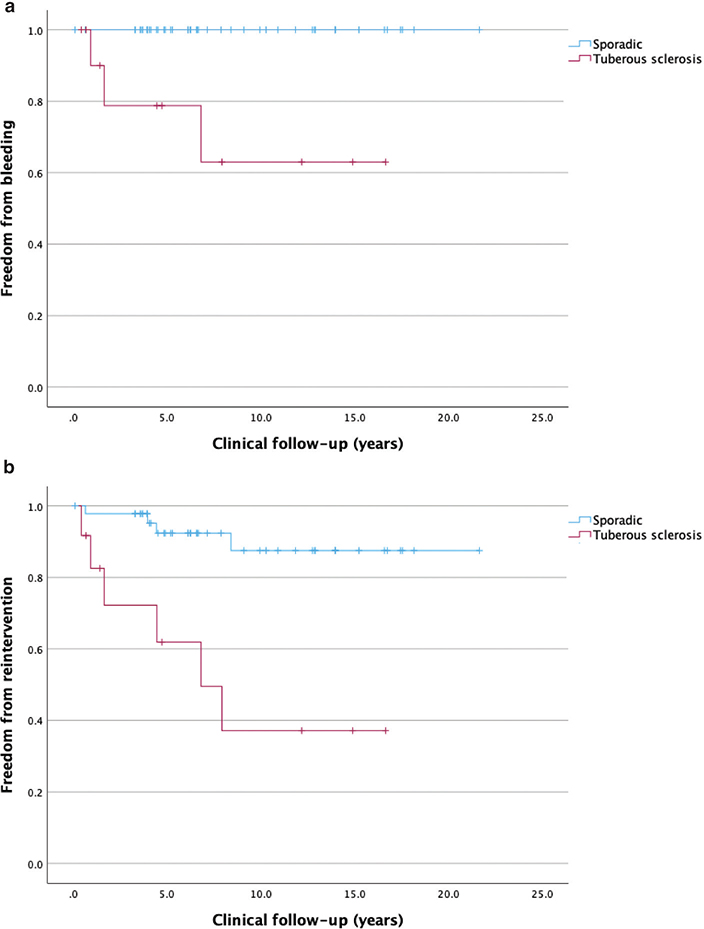
Figure 1. Kaplan–Meier curves showing freedom from (a) bleeding and (b) reintervention, with respect to time (years).
Progress during follow-up
In patients with procedural response, 20 (38%) regrew during follow-up: 13 (25%) with minor and seven (13%) with major progress. Median time from embolisation to nadir for sporadic AML with major progress was 0.5 years (0.5–2.3) (Table 4). Figure 2 in Appendix S1 presents patients with sporadic AML with major progress.
| SAE for renal AML1 | ||||||
| Sporadic, n = 43 | TSC associated, n = 10 | |||||
| No progress | Minor progress | Major progress | No progress | Minor progress | Major progress | |
| Number of tumours (%) | 26 (60%) | 12 (28%) | 5 (12%) | 7 (70%) | 1 (10%) | 2 (20%) |
| Median tumour diameter at SAE, cm (IQR) | 6.7 (5.6–9.7) | 5.6 (5.0–7.5) | 5.7 (5.3–7.7) | 12.2 (8.0–13.8) | 10.5 | 8.8 |
| Technical success | ||||||
| Complete | 24 | 11 | 5 | 3 | 1 | 1 |
| Partial | 2 | 1 | 0 | 4 | 0 | 1 |
| Median time to nadir, years (IQR) | 3.4 (1.2–5.0) | 2.8 (0.7–4.2) | 0.5 (0.5–2.3) | 0.5 (0.3–1.1) | 2.0 | 1.9 |
| Median time to first increase, years (IQR) | - | 4.2 (0.9–10.4) | 3.4 (1.5–6.4) | - | 4.3 | 5.0 |
| Median follow-up, years (IQR) | 4.1 (2.5–7.6) | 5.3 (3.1–8.2) | 9.7 (9.0–14.4) | 1.3 (0.5–3.3) | 6.9 | 6.4 |
| 153 procedures. Missing radiology in one sporadic- and two TSC-patients. The two sporadic AML without tumour shrinkage (non-response) in Table 3 are not included due to re-SAE. | ||||||
| AML: angiomyolipoma; TSC: tuberous sclerosis; SAE: selective arterial embolisation; IQR: interquartile range. | ||||||
Rebleeding and reintervention
Two patients had one new bleeding episode during follow-up, and another one had two. Two were retroperitoneal haemorrhages, and two were haematurias. All three were female tuberous sclerosis patients. All were successfully treated with a new embolisation. Details are shown in Appendix S1 and Table 1. Of the totally 10 reembolisations (17%) after median 4.2 years (1.1–6.2), three were due to bleeding, and seven were due to unchanged size after embolisation or progress during follow-up. Tuberous sclerosis was a significant risk factor for the need of reinterventions, OR 13.7 (P < 0.015). After a median clinical follow-up of 7.5 years (4.7–14.0), the need for a reintervention was significantly increased in tuberous sclerosis versus sporadic AML patients (P < 0.001) (Figure 1).
One patient developed a retroperitoneal abscess and pulmonal empyema after reembolisation that was managed with drainage and antibiotics resulting in a long hospital stay. It was classified as Clavien-Dindo IIIa. Another patient treated 6.9 years earlier with embolisation had a retroperitoneal rebleeding and was treated with emergency embolisation but died 3 days post-intervention in multiorgan failure.
One of the 10 patients with reinterventions needed a third procedure after 5.2 years due to bleeding. Two patients with AML sized 11.9 and 23.9 cm, respectively, had kidney surgery after reembolisation. One had two reembolisations without any decrease in AML size, and partial nephrectomy was performed. The other underwent one reembolisation but continuous abdominal pain eventually led to a nephrectomy.
Discussion
The present study of AML treated with selective arterial embolisation is one of the largest published series with long-term follow-up, reported so far [8–11]. Our study shows a high technical success rate (84%) and a low complication rate (6.8%). Thirty-eight percent of the AML grew during follow-up, but only 5% had rebleedings. Growing size and rebleeding prompted reembolisation in 17% of tumours.
Although relatively rare, most urologists probably come across a few cases of AML every year. The absence of prospective randomised studies makes clinical decision-making difficult, and today’s evidence relies on reported case-series. Since the first report on embolisation, by Moorehead in 1977, the use of embolisation has increased, but surgery is still the most reported method of treatment [12–14]. Other techniques like cryoablation and radiofrequency ablation show promising results, but reports are still scarce [15, 16].
The overall technical success rate in this study was 84%, and the rest were deemed partially successful, tallying with the reports by Wang et al. (100% success) and Lee et al. (89% success) [9, 11]. Herein, radiological success on follow-up imaging was seen in 96% of procedures; median tumour diameter shrinkage was 30% at nadir. Others have reported outcomes (in means) between 21% and 40% [9, 17]. It is obvious that embolisations do not eradicate the tumours, they only reduce their size.
In the present series, 38% of AML with initial response to embolisation grew during follow-up. This rate of regrowth indicates that radiological follow-up is to be recommended, especially in tuberous sclerosis patients. This is a drawback in comparison with surgery for AML, where the tumour is removed, and no radiological follow-up is necessary. However, some complicated central tumours are not accessible to nephron-sparing surgery. The seven patients with major progress were also the ones with the longest follow-up, whereas the 33 tumours with no progress had the shortest follow-up. A possible explanation might be that regrowth is a common occurrence after some time. Our study has, to our knowledge, the second longest radiological follow-up reported, only surpassed by Anis et al. with a mean follow-up of 10 years on a material of 55 embolisations. Their and our observation cast some doubt on the method’s durability over time in terms of size reduction.
Reintervention rate is another way of measuring durability. We report that 17% of treated tumours needed reintervention, that is, the freedom from reintervention at a median clinical follow-up of 7.5 years, was 83%. Anis reported a reintervention rate of 41% at 10 years, and Nozadze a 20% rate at a median of 4.6 years [8, 17]. Murray’s review encompassing 31 studies including 524 cases reported a reintervention rate of 20% at a mean follow-up of 3.3 years [18]. Due to lack of evidence-based guidelines, indications for the initial embolisation vary greatly. Probably do indications for retreatment vary even more, why comparisons of reintervention rates are difficult. Reintervention due to acute haemorrhage is a more comparable indication. We report a 5% rebleeding rate, all prompting a redo embolisation. Anis reported a 15% rebleeding rate, Lee 1% and Nozadze 0% [8, 11, 17]. It is difficult to draw any conclusions from four bleeding episodes in three patients other than noticing that they all were female and had tuberous sclerosis.
We observed a low complication rate of 6.8%, consistent with other recent series [10, 17, 19–21]. No Clavien-Dindo grade III–IV complication was observed after the primary embolisation, which supports that it is a safe procedure. However, redo embolisation seems to be associated with higher risks. Renal function was unaffected by embolisation in our material, in consistency with contemporary studies reporting this outcome [11, 17, 22].
Since Oesterling’s pioneering work in 1986, a size of 4 cm has been a widely accepted trigger for intervention, although intervention was recommended only in symptomatic cases over 4 cm [23]. This 4 cm rule has been questioned, and a higher size-limit has been proposed; we ourselves suggested 5 cm based on findings in a recent paper [6].
In the present study, 24 patients had acute bleedings before embolisation. Due to selection bias, conclusions regarding when to proceed with prophylactic intervention cannot be drawn from this study. However, the mean and median diameters at bleeding were fairly large, 7.8 cm and 6.5 cm, respectively. Only three were smaller than 4 cm. The four rebleedings after embolisation occurred in large AML measuring between 6.6 and 19.6 cm at time of bleeding. Although a somewhat higher threshold for active treatment than 4 cm can be considered, the observed death due to rebleeding 6.9 years after embolisation reminds us of the potential gravity of the disorder.
This study, as well as most others, has a mix of patients with sporadic and tuberous sclerosis-associated AML. We report 19% tuberous sclerosis, whereas others report between 7% and 70% tuberous sclerosis-associated AML [8–11, 24]. Patients with tuberous sclerosis are younger, have a higher ASA-score and larger AML. Their more complex disease is reflected in a 58% longer intervention time and a lower proportion of complete technical success (50% vs. 93%). Patients with tuberous sclerosis responded in a lesser degree to embolisation in terms of size reduction. There was no difference in progress after nadir compared to sporadic AML, but this can perhaps be explained by a shorter follow-up of the tuberous sclerosis patients. All three patients with bleedings after embolisation had tuberous sclerosis, indicating the disorder to be a risk factor for new bleeding episodes after embolisation. Growing size and rebleeding resulted in a redo embolisation in 9% of spontaneous AML and 50% of tuberous sclerosis-associated AML. Tuberous sclerosis was the only significant predictor of reintervention in binomial regression analysis.
Replacing expensive hospitalisations with less expensive outpatient care is very attractive in today’s healthcare. Nozadze et al. performed 89.4% of embolisations in local anaesthesia, resulting in a median of 0 days of hospitalisation [17]. In our study, 75% of procedures were performed under general anaesthesia, resulting in a median hospital-stay of two (sporadic) and four (tuberous sclerosis) days, respectively. Although patients in our study had a higher ASA-score and a higher proportion of acute embolisations, the study by Nozadze shows that outpatient care is possible, and that it might be a good strategy for us and others to adopt.
There are several limitations to the present study. First, due to the retrospective nature, data were not collected for predesigned research purposes and may be subject to bias. Second, the different case mix regarding sporadic versus tuberous sclerosis-associated AML, emergency embolisation versus elective embolisation, etc. make comparisons between groups more difficult, as well as comparisons with previously published studies. Third, follow-up differs between sporadic and tuberous sclerosis-associated AML, which also limits the analysis.
In conclusion, selective arterial embolisation renders a pronounced size-reduction in most patients. It is well-tolerated, with few and mild complications, and renal function is preserved. Regrowth is common why radiological follow-up is necessary. Tuberous sclerosis is a risk factor for reintervention.
Acknowledgements
We are thankful to The Research and Development Center at NU Hospital Group for supporting this study with grants.
ORCID
Jesper Swärd https://orcid.org/0000-0001-6035-6381
References
- [1] Fittschen, A., I. Wendlik, S. Oeztuerk, et al., Prevalence of sporadic renal angiomyolipoma: a retrospective analysis of 61,389 in- and out-patients. Abdominal Imaging, 2014. 39(5): p. 1009-1013. https://doi.org/10.1007/s00261-014-0129-6
- [2] Buj Pradilla, M.J., T. Martí Ballesté, R. Torra, et al., Recommendations for imaging-based diagnosis and management of renal angiomyolipoma associated with tuberous sclerosis complex. Clinical Kidney Journal, 2017. 10(6): p. 728-737. https://doi.org/10.1093/ckj/sfx094
- [3] Bernstein, J., T.O. Robbins and J.M. Kissane, The renal lesions of tuberous sclerosis. Semin Diagn Pathol, 1986. 3(2): p. 97-105.
- [4] Ahn, T., M.J. Roberts, A. Navaratnam, et al., Changing etiology and management patterns for spontaneous renal hemorrhage: a systematic review of contemporary series. Int Urol Nephrol, 2017. 49(11): p. 1897-1905. https://doi.org/10.1007/s11255-017-1694-8
- [5] Bhatt, J.R., P.O. Richard, N.S. Kim, et al., Natural History of Renal Angiomyolipoma (AML): Most Patients with Large AMLs >4cm Can Be Offered Active Surveillance as an Initial Management Strategy. Eur Urol, 2016. 70(1): p. 85-90. https://doi.org/10.1016/j.eururo.2016.01.048
- [6] Sward, J., O. Henrikson, D. Lyrdal, et al., Renal angiomyolipoma-patient characteristics and treatment with focus on active surveillance. Scand J Urol, 2020. 54(2): p. 141-146. https://doi.org/10.1080/21681805.2020.1716066
- [7] Jinzaki, M., S.G. Silverman, H. Akita, et al., Renal angiomyolipoma: a radiological classification and update on recent developments in diagnosis and management. Abdom Imaging, 2014. 39(3): p. 588-604. https://doi.org/10.1007/s00261-014-0083-3
- [8] Anis, O., U. Rimon, J. Ramon, et al., Selective Arterial Embolization for Large or Symptomatic Renal Angiomyolipoma: 10 Years of Follow-up. Urology, 2020. 135: p. 82-87. https://doi.org/10.1016/j.urology.2019.09.035
- [9] Wang, C., M. Yang, X. Tong, et al., Transarterial embolization for renal angiomyolipomas: A single centre experience in 79 patients. J Int Med Res, 2017. 45(2): p. 706-713. https://doi.org/10.1177/0300060516684251
- [10] Sheth, R.A., A.S. Feldman, E. Paul, et al., Sporadic versus Tuberous Sclerosis Complex-Associated Angiomyolipomas: Predictors for Long-Term Outcomes following Transcatheter Embolization. J Vasc Interv Radiol, 2016. 27(10): p. 1542-9. https://doi.org/10.1016/j.jvir.2016.05.029
- [11] Lee, S., H.S. Park, D. Hyun, et al., Radiologic and clinical results of transarterial ethanol embolization for renal angiomyolipoma. Eur Radiol, 2021. 31(9): p. 6568-6577. https://doi.org/10.1007/s00330-021-07831-y
- [12] Kuusk, T., F. Biancari, B. Lane, et al., Treatment of renal angiomyolipoma: pooled analysis of individual patient data. BMC Urol, 2015. 15: p. 123. https://doi.org/10.1186/s12894-015-0118-2
- [13] Fernandez-Pello, S., M. Hora, T. Kuusk, et al., Management of Sporadic Renal Angiomyolipomas: A Systematic Review of Available Evidence to Guide Recommendations from the European Association of Urology Renal Cell Carcinoma Guidelines Panel. Eur Urol Oncol, 2020. 3(1): p. 57-72. https://doi.org/10.1016/j.euo.2019.04.005
- [14] Moorhead, J.D., P. Fritzsche and H.L. Hadley, Management of hemorrhage secondary to renal angiomyolipoma with selective arterial embolization. J Urol, 1977. 117(1): p. 122-3. https://doi.org/10.1016/S0022-5347(17)58367-6
- [15] Makki, A., O. Graumann, S. Hoyer, et al., Cryoablation of Renal Angiomyolipoma: An Evaluation of Safety and Efficacy. J Endourol, 2017. 31(11): p. 1117-1122. https://doi.org/10.1089/end.2017.0376
- [16] Castle, S.M., V. Gorbatiy, O. Ekwenna, et al., Radiofrequency ablation (RFA) therapy for renal angiomyolipoma (AML): an alternative to angio-embolization and nephron-sparing surgery. BJU Int, 2012. 109(3): p. 384-7. https://doi.org/10.1111/j.1464-410X.2011.10376.x
- [17] Nozadze, G., S.B. Larsen, S. Heerwagen, et al., Selective arterial embolization of renal angiomyolipomas: A 10-year experience. BJUI Compass, 2022. 3(1): p. 86-92. https://doi.org/10.1002/bco2.107
- [18] Murray, T.E., F. Doyle and M. Lee, Transarterial Embolization of Angiomyolipoma: A Systematic Review. J Urol, 2015. 194(3): p. 635-9. https://doi.org/10.1016/j.juro.2015.04.081
- [19] Hocquelet, A., F. Cornelis, Y. Le Bras, et al., Long-term results of preventive embolization of renal angiomyolipomas: evaluation of predictive factors of volume decrease. Eur Radiol, 2014. 24(8): p. 1785-93. https://doi.org/10.1007/s00330-014-3244-4
- [20] Duan, X.H., M.F. Zhang, J.Z. Ren, et al., Urgent transcatheter arterial embolization for the treatment of ruptured renal angiomyolipoma with spontaneous hemorrhage. Acta Radiol, 2016. 57(11): p. 1360-1365. https://doi.org/10.1177/0284185115588125
- [21] Bardin, F., O. Chevallier, A. Bertaut, et al., Selective arterial embolization of symptomatic and asymptomatic renal angiomyolipomas: a retrospective study of safety, outcomes and tumor size reduction. Quant Imaging Med Surg, 2017. 7(1): p. 8-23. https://doi.org/10.21037/qims.2017.01.02
- [22] Lin, L., C. Wang, R. Pei, et al., Prophylactic selective arterial embolization for renal angiomyolipomas: efficacy and evaluation of predictive factors of significant shrinkage. Int Urol Nephrol, 2018. 50(10): p. 1765-1770. https://doi.org/10.1007/s11255-018-1953-3
- [23] Oesterling, J.E., E.K. Fishman, S.M. Goldman, et al., The management of renal angiomyolipoma. J Urol, 1986. 135(6): p. 1121-4. https://doi.org/10.1016/S0022-5347(17)46013-7
- [24] Chapman, D., M. Tyson and B. Buckley, Single-institution, retrospective review of elective and emergency embolization of renal angiomyolipoma. Can Urol Assoc J, 2021. 15(11): p. E598-E602. https://doi.org/10.5489/cuaj.7143