ORIGINAL RESEARCH ARTICLE
The Stockholm Spinal Cord Uro Study: Changing patterns of urological surgery in a regional prevalence group through 50 years – Outcomes and lessons learned
Elisabeth Farrellya,b , Lena Lindboc and Åke Seigera,c
aDepartment NVS, Division of Clinical Geriatrics, Karolinska Institute, Stockholm, Sweden; bDepartment of Diagnostics and Intervention, Urology and Andrology, Umeå University, Umeå, Sweden; cAleris Rehab Station, Frösundaviks allé, Stockholm, Sweden
ABSTRACT
Aims: To examine the number and types of urological surgical procedures carried out in a regional prevalence population of patients with traumatic spinal cord injury (SCI) during five decades, evaluate objective and patient-reported outcomes and to consider lessons learned for further improvement of surgical treatment in this patient group.
Methods: In a cross-sectional study of 412 patients with traumatic SCI, one-third had undergone urological surgery through a period of up to 50 years. Data on types of surgery, complications, follow-up and outcomes were collected in a retrospective review of patient files. S-creatinine, S-cystatin-C, renal ultrasound and a questionnaire regarding complications during the preceding year were assembled as part of a yearly follow-up. Descriptive statistics were calculated. Logistic regression was used to determine risk profiles for the incidence of urological surgery.
Results: A total of 137 patients had undergone 262 urological surgical interventions. The incidence was highest amongst persons with a cervical-thoracic neurological level of spinal cord lesion and during the first 2 years after SCI. Surgery for urinary stones constituted 29% of all procedures. One-fourth of the patients had undergone 47% of all procedures, notably urinary diversion and ensuing complications, implants and revisions, repeated stone interventions or bladder outlet procedures. After reconstructive surgery functional outcomes and patient-reported satisfaction were generally favourable, but long-term signs of renal complications were frequent.
Conclusions: Urological surgery after SCI involves imperative as well as reconstructive procedures, some of which are challenging and call for centralisation to devoted teams. Prospective studies of reconstructive urology are warranted, including more extensive patient-reported outcomes.
KEYWORDS: Spinal cord injury; neurogenic bladder; neurogenic lower urinary tract dysfunction; urological surgery; reconstructive urology; renal complications; follow-up programme; patient-reported outcomes
Citation: Scandinavian Journal of Urology 2024, VOL. 59, 173–180. https://doi.org/10.2340/sju.v59.40326.
Copyright: © 2024 The Author(s). Published by MJS Publishing on behalf of Acta Chirurgica Scandinavica. This is an Open Access article distributed under the terms of the Creative Commons Attribution 4.0 International License (http://creativecommons.org/licenses/by/4.0/), allowing third parties to copy and redistribute the material in any medium or format and to remix, transform, and build upon the material, with the condition of proper attribution to the original work.
Received: 9 March 2024; Accepted: 27 August 2024; Published: 24 October 2024
CONTACT Elisabeth Farrelly elisabeth.carlsson.farrelly@regionvasterbotten.se Department of Diagnostics and Intervention, Urology and Andrology, Umeå University, S-901 87 Umeå, Sweden
Disclosure statement and funding: The authors report no conflicts of interest. This study was supported by grants from the Norrbacka-Eugenia Foundation (45/2008) and Wellspect HealthCare (98/2007).
Introduction
The Stockholm Spinal Cord Uro Study [1] is a comprehensive survey of urinary tract function in a regional prevalence population of patients with chronic traumatic spinal cord injury (SCI). It was carried out as a cross-sectional study based on a yearly check-up visit at the Spinalis out-patient clinic, including a national programme for follow-up of SCI complications, a regional programme for follow-up of neurogenic lower urinary tract dysfunction (NLUTD) post-SCI and a study-specific questionnaire on patient-reported complications of the urinary tract during the preceding year. The basic characteristics of the patient group have been presented [1], as well as separate reports on urinary tract infections (UTIs) [2] and the urodynamic follow-up of bladder and renal function [3]. In this article, we report on the history and outcomes of urological surgery in the regional prevalence population.
Patients with SCI are at risk for multiple complications of the urinary tract, where surgical treatment may become necessary or desirable. Imperative urological procedures are carried out due to severe infections, urinary stones and to secure efficient bladder voiding. Reconstructive surgery is performed with the aim of improving quality of life and reducing the risk of renal complications, spasticity and autonomic dysreflexia [4]. Indications for imperative procedures are usually health issues of a sub-acute nature, whilst reconstructive urology is aimed at relieving long-term complications.
Surgical treatment is one part of the comprehensive management of urinary tract function post-SCI and is applied in combination with pharmaceutical treatment, catheterisation and other technical aids [5]. During recent years, international guidelines [6–10] have been developed and now offer recommendations on some of the more common surgical procedures for incontinence and neurogenic detrusor overactivity (NDO). In this report, we summarise the changing patterns of surgical treatment during five decades and the outcomes of the most prevalent procedures, and discuss the lessons learned for further development and research.
Material and methods
Participants
Participants in the Stockholm Spinal Cord Uro Study were 412 men and women, aged 18 years or older, with a post-traumatic SCI for at least 1 year who attended a yearly check-up visit at the regional outpatient centre – the Spinalis clinic. All individuals were living in the greater Stockholm area and registered at the Spinalis clinic, which oversees follow-up for a vast majority of the regional SCI population. Amongst them, 137 patients had undergone urological surgical interventions at any time during the period from the event of traumatic SCI until 2009.
Methods
In the Stockholm Spinal Cord Uro study, we applied two programmes for follow-up of chronic SCI: a national Swedish programme for follow-up of medical SCI complications [11] and a regional programme for NLUTD [1], complemented with a study-specific questionnaire regarding urinary tract complications during the preceding year [1]. Objective measurements and patient-reported data were collected, as previously reported [1]. Neurological levels and AIS grades of injury (American Spinal Injury Association [ASIA] Impairment Scale) were registered [12].
In the study-specific questionnaire and structured interview, a history of urological surgery was obtained as well as the current method of bladder management, pharmacological treatment, patient-reported frequency of UTI, other complications of the urinary tract, self-reported satisfaction with previous surgery and current method of voiding. Patient files were retrospectively reviewed for records of surgery, complications and re-operations, follow-up of renal function and patient-reported outcomes during previous years.
In the regional follow-up programme, signs of renal complications were elevated levels of S-creatinine and/or S-cystatin-C, reduced values of cystatin-C-related glomerular filtration, ultrasound verified dilatation of upper urinary tracts and/or pathology of renal parenchyma, as described in a previous article [1]. An important aim of the follow-up programme was to make it patient friendly and at the same time accurately informative for medical evaluation. S-cystatin-C was chosen as a screening tool as it has been shown to give a good representation of glomerular filtration with high sensitivity and specificity in patients with SCI [13]. In clinical cases of uncertainty, a nephrologist was consulted, and iohexol clearance would sometimes be used as a standard reference.
Data analyses
Data were analysed using SPSS for Windows 28 software (IBM SPSS Statistics for Windows, Version 25.0; IBM Corp., Armonk, NY) Descriptive statistics were calculated. Logistic regression was used to determine risk profiles for the incidence of urological surgery.
Results
Amongst the 412 patients in the total study population, 137 persons (surgery population) had undergone urological surgery with a total of 262 procedures through 1960–2009.
Patient characteristics of the total study population, the surgery sub-population and the non-surgery sub-population are presented in Table 1. Age and gender distribution were similar between the surgery and the non-surgery population. In the surgery population, SCI duration was longer by a mean 7.6 years, and the proportion of patients with an SCI classification C1–C8 AIS A-C was twice that of the non-surgery group. The proportion of patients with T1–S3 AIS A-C was also higher, whilst patients with an AIS D lesion at any level were found at 25% in the surgery group and 75% in the non-surgery group.
The number and types of urological surgical interventions, indications for procedures and time periods when the various types of surgery were conducted are summarised in Table 2 and Figure 1.
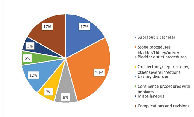
Figure 1. Types of urological surgical procedures and relative proportions.
Characteristics of SCI lesions for various types of surgery are illustrated in Figure 2.
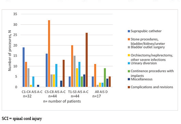
Figure 2. Spinal cord injury characteristics, number of patients, and number and types of urological surgical procedures.
SCI = spinal cord injury C1-C4 = cervical neurological level of spinal cord lesion C1, C2, C3, or C4. C5-C8 = cervical neurological level of spinal cord lesion C5, C6, C7, or C8. T1 - S3 = thoracic or sacral neurological level of spinal cord lesion at any level T1-T12, S1, S2, or S3.
American Spinal Injury Association (ASIA) Impairment Scale (AIS) grades: A Complete. No motor or sensory function is preserved in the sacral segments S4-S5. B Incomplete. Sensory function preserved but not motor function is preserved below the neurological level and includes the sacral segments S4-S5. C Incomplete. Motor function is preserved below the neurological level, and more than half of key muscles below the neurological level have a muscle grade less than 3. D Incomplete. Motor function is preserved below the neurological level and at least half of key muscles below the neurological level have a muscle grade of 3 or more
Length of follow-up, laboratory, radiological and patient-reported outcomes are presented in Table 3.
The gender ratio for all surgical procedures was 81% men/19% women. For urinary diversions and continence procedures with implants, the ratio was 57% men/43% women.
Primary surgery was performed at a median of 3 years after SCI with 50% occurring within 2 years. These procedures were almost exclusively sphincterotomies, insertion of a suprapubic catheter or bladder stone extractions. The latest surgical procedure was carried out at a median of 7 years post-SCI with a wide range of 1–50 years. More than half of all procedures after more than 10 years were stone procedures and revisions of urinary diversions or implants.
Indications for imperative surgery were symptomatic urinary stones, severe infections and to ensure efficient voiding by suprapubic catheter. These interventions constituted 53% of all urological operations.
The first surgery for bladder stones was conducted up to 45 years post-injury. Sixteen patients had recurring stone formation in the upper or lower urinary tracts during a median of 23.5 years. Amongst all patients who underwent stone surgery, 40% changed bladder management. Half of these changes were chronologically associated with stone surgery and included a transition from bladder reflex triggering to sphincterotomy, suprapubic catheter or urethral indwelling catheter. None of these patients had a recurrence of urinary stones.
Kidney infections resulting in nephrectomy were in all cases the result of chronic renal stone formation. Scrotal abscesses and gangrene were caused by epididymitis or pressure ulcers. Cystectomy/urethrectomy after urinary diversion was carried out due to chronic symptomatic infections.
Sphincterotomy was performed as a single procedure in 7 men and supplemented by repeat sphincterotomy, bladder neck incision or transurethral resection of the prostate (TUR-P) in 4 men. The patient-reported outcomes when all patients voided by condom catheter were retrieved from file records: independence was rated as good by all patients. On follow-up at a median of 26 years, 4 patients had switched to CIC, urinary diversion or suprapubic catheter. 50% had signs of renal complications.
After urinary diversion by suprapubic catheter or reconstructive diversion, the functional outcome was reported as ‘good’ or ‘very good’ by 80%. Enhanced independence and ease of voiding were stated by all patients. Five patients had undergone reconstructive hand surgery in preparation for a continent diversion with a catheterisable outlet. Renal complications and metabolic alterations after reconstructive diversions are displayed in Table 3. Revision surgery due to complications was relatively common (Table 2).
Follow-up of continence procedures with implants at a median of 8 years displayed that 6/9 patients (67%) had undergone one or two additional continence procedures. Six patients were continent. Urinary diversions and continence procedures were performed by a limited number of surgeons at one university hospital.
The incidence of surgical treatment and types of procedures were related to the neurological level, severity and duration of the SCI, as illustrated in Figure 2 and Table 2. The relative risk (RR) of urological surgery in the total study population was 0.5. Amongst patients with a cervical-thoracic neurological level of lesion and AIS grades A–C, the RR was 0.87, and for patients with an AIS D lesion, it was 0.13. The odds ratio (OR) was 6.75 for any kind of urological surgery amongst patients with a cervical-thoracic level of lesion and AIS grades A–C versus an AIS D lesion. There was no difference with age at the time of the SCI.
One-fourth of the patients in the surgery group had ≥3 procedures, most frequently urinary diversions with ensuing revisions, recurring urinary stones or multiple bladder outlet procedures. Amongst these patients, 59% had developed signs of renal complications after a median follow-up of 18 years.
The development of surgical procedures during the period 1960–2009 is illustrated by our data. The earliest bladder outlet procedure was a bladder neck resection in 1961, followed by sphincterotomies predominantly 1973–1994 and suprapubic catheters from the mid-1990s. Reconstructive urinary diversions were performed from 1972 with incontinent diversions during the entire period. Continent urinary reservoirs were carried out predominantly 1986–2001 and then generally replaced by bladder augmentation with/without a catheterisable outlet. One sacral anterior root stimulator was implanted in 1993 and one in 2001. Continence implants were used from 1992.
Treatment of bladder stones was carried out as open procedures in the early 1960s but from the mid-1960s as transurethral lithotripsy. Upper urinary tract stones were extracted via open surgery until the mid-1980s, and then extracorporeal shock wave lithotripsy (ESWL) and endoluminal methods took over. Nephrectomies and orchiectomies due to severe infections occurred sparsely during the 1970s–1990s but not after the year 2000.
Discussion
This study illustrates the trends and development in the surgical management of NLUTD after traumatic SCI in Sweden through 50 years and provides an insight into the objective and patient-reported outcomes. The types of surgery are in accordance with other reports in the international literature from corresponding periods of time [14–18]. During the 1960s, scientific and clinical awareness increased of the negative impact of SCI-related NDO on renal function. Consequently, bladder outlet surgery was popularised in SCI men. Sphincterotomy was established as an efficient way of alleviating high intravesical filling pressures and, with a condom catheter, securing social continence. The development of antimuscarinic medications during the 1980s and the introduction of CIC [19], however, encouraged many SCI men to aim for continence without the condom catheter, and sphincterotomy declined in frequency. The sacral anterior root stimulator was offered as a new treatment option in Sweden from 1990. Excellent results were reported by the very few patients in our study who had the stimulator implanted. Around 15 patients were screened for the implant, but interest remained low due to the dorsal rhizotomy involved in the procedure where reflex sexual function would be lost. During the late 1980s and the 1990s, continent urinary reservoirs were introduced and became popular as a way of eliminating the problem of NDO and providing continence and ease of voiding by catheterisation through an abdominal stoma. Long-term results, however, revealed that re-operations and metabolic complications were frequent, and renal dysfunction was not uncommon with time [15, 20]. Continent reservoirs were then largely replaced by bladder augmentation where the ureteral orifices in the bladder are kept intact, and the upper urinary tracts remain more protected. The augmented bladder with or without a catheterisable outlet is now seen as the reconstructive standard for SCI patients world-wide [5]. Incontinent urinary diversion has maintained its place as a useful option for patients who do not wish or are unable to perform self-catheterisation independently. During the last two decades, the increasing use of botulinum toxin has advanced the treatment of NDO and reduced the need for reconstructive urology [21].
The incidence of stone surgery increased through the study period. The introduction of follow-up programmes [1, 11] led to the discovery of more stones at an early stage, and serious infections could be avoided. Surgery evolved into less invasive methods, sparing renal function and reducing complications. There were no instances of nephrectomy due to stones with chronic infection after the year 2000.
Follow-up after reconstructive urinary diversion demonstrated good results for patient-reported satisfaction and independence of voiding. Outcomes on the rate of UTIs and incontinence were variable, whilst objective outcomes on long-term renal complications were discouraging. Other studies from Scandinavia including mixed populations with NLUTD and non-NLUTD have also reported significant improvements in health-related quality of life [22, 23]. Follow-up for up to 5 years revealed no signs of renal deterioration as evaluated by renal clearance studies [23]. International reviews [5, 24] state similar findings, but follow-up periods are limited. Our follow-up was more extensive with a median of 13 years and included evaluation by S-cystatin-C. These factors may account for a higher prevalence of renal impairment. Reconstructive urinary diversion was carried out by a limited number of experienced surgeons; however, a devoted team that brought together knowledge on the technical surgical aspects, the specifics of the neuro-urological pathophysiology after SCI and the importance of close follow-up evolved gradually from the late 1990s. This may account for a high frequency of re-operations during the earlier period.
Follow-up after sphincterotomy demonstrated that independence was achieved and appreciated by patients, although repeated procedures were necessary in some cases, and recurrent UTIs were common. The rate of long-term renal complications was high. In a larger study, Pan et al found a single procedure permanently successful in 32%, and a second procedure was needed in 36%. Recurrent UTIs, recurrent detrusor-sphincter dyssynergia and upper urinary tract dilatation were noted in two-thirds, but S-creatinine levels remained normal in all patients over a period of up to 20 years [25]. Our follow-up period was longer with a median of 19 years (range 3–50 years), and the evaluation of renal function included the more sensitive S-cystatin-C. The number of sphincterotomy patients in our study is small, including only those patients who were registered at the Spinalis clinic from 1991 onwards when the frequency of this operation was already in decline. The operations were executed by two surgeons. It is possible that more sphincterotomies were carried out earlier, but it is also likely that the procedure was not offered to all who could have benefitted as there was no organised follow-up of medical complications after SCI prior to 1991.
Patient-reported functional outcomes after urinary diversion as well as sphincterotomy seem to indicate a significant sense of positive trade-off despite the repeated surgeries and complications with some procedures.
Amongst patients who had ≥3 urological procedures, a majority had developed signs of renal complications. These patients had a higher neurological level and severity of injury and were a priori at greater risk for kidney impairment [1, 26]. Repeated surgery may have increased the strain on renal function. Urodynamic monitoring was not always carried out appropriately before and after surgery, and risk factors for renal function may have been missed [3]. SCI duration, patient age and cardiovascular or metabolic co-morbidity are additional factors that may have affected renal function during the follow-up period [1, 3].
Rates of UTI, urolithiasis and severe infections such as urosepsis co-vary with higher neurological level and severity of SCI and with different types of bladder management [3]. In a recent publication, urosepsis was fatal in 14% of community-acquired infections [27, 28] in the general population. Substantial reduction of these risks and complications should be the goal of urological surgery after SCI. There is a lack of infection-related outcomes as well as patient-reported outcomes in the literature. Prospective studies are highly warranted to gain a better understanding of which surgical interventions may provide the best risk reduction for severe infections and optimal functional improvements for persons with SCI [29].
We support that reconstructive urological treatment should be concentrated to specialised centres. SCI rehabilitation in Sweden has recently been concentrated to four national highly specialised centres, which may facilitate future coordination of research and method development in many aspects of rehabilitation, including urological surgery.
This study has several limitations. It is based on our original cross-sectional study [1], where inclusion took place in 2007–2010, and 75% of the regional prevalence population is covered. Surgery conducted until 2009 is included here with follow-up until 2021. Patients who lived in the area prior to 2007 or declined participation in the original study are not included. The Spinalis clinic was established in 1991 as the first regional follow-up centre in Sweden for persons with SCI. A comprehensive inventory of past medical history was taken for each patient who chose to register, and further medical details were subsequently registered at yearly check-up visits [14]. File records are nearly complete for urological surgery from 1991 onwards, but some interventions may have been missed if not reported by the patients. For the years antecedent to 1991, non-computerised file records of surgery were not always available. Since our original study in 2007–2010, the population in the Stockholm area has increased as well as the number of patients at the Spinalis clinic. Urological surgery has generally developed into more minimally invasive procedures, and robot-assisted laparoscopic surgery has become firmly established. This study is not fully representative of the regional patient population and the procedures of today but offers an understanding of the development of surgical treatment for persons with SCI through 50 years, including long-term outcomes and lessons learned.
Conclusions
One-third of the patients in the regional population had urological surgical interventions during a period of up to 50 years. Deterioration of renal function was common after repeated surgery. Therefore, follow-up in this patient group must be diligent and life-long. Centralisation of complex procedures to devoted teams is important. Prospective studies of urological surgical treatment after SCI are warranted with a focus on patient-reported outcomes as well as long-term renal safety.
Acknowledgements
This study was approved by the Regional Human Ethics Committee at Karolinska Institutet, Stockholm, Sweden (2007/35–31/3). The authors wish to thank the staff at the Spinalis clinic for performing interviews, database registration and physical examinations. We wish to acknowledge the late associate professor at the Karolinska Institute Hans Wijkström who was part of the original research team and sadly passed away during our joint collection of data for this study.
ORCID
Elisabeth Farrelly https://orcid.org/0000-0003-1245-9276
References
- [1] Farrelly E, Lindbo L, Wijkström H, et al. The Stockholm Spinal Cord Uro Study: 1. Basic characteristics and problem inventory. Scand J Urol. 2019; 53(6): 403–410. https://doi.org/10.1080/21681805.2019.1673812
- [2] Farrelly E, Lindbo L, Wijkström H, et al. The Stockholm Spinal Cord Uro Study: 2. Urinary tract infections in a regional prevalence group. Frequency, symptoms, and treatment strategies. Scand J Urol. 2020; 54(2): 155–161. https://doi.org/10.1080/21681805.2020.1734078
- [3] Farrelly E, Lindbo L, Seiger Å. The Stockholm Spinal Cord Uro Study: 3. Urodynamic characteristics in a regional prevalence group of persons with spinal cord injury and indications for improved follow-up. Scand J Urol. 2021; 55(5): 412–418. https://doi.org/10.1080/21681805.2021.1954994
- [4] Eldahan KC, Rabchevsky AG. Dysreflexia after spinal cord injury: systemic pathophysiology and methods of management. Auton Neurosci. 2018; 209: 59–70. https://doi.org/10.1016/j.autneu.2017.05.002
- [5] Wyndaele JJ, Birch B, Boraus A, et al. Surgical management of the neurogenic bladder after spinal cord injury. World J Urol. 2018; 36: 1569–1576. https://doi.org/10.1007/s00345-018-2294-7
- [6] European Association of Urology Guidelines. Edition presented at the EAU Annual Congress in Milan 2021. EAU Guidelines: Neuro-Urology | Uroweb. EAU Central Office, Arnhem, The Netherlands.
- [7] Elliott S, Gomez R, eds. Urologic management of the spinal cord injured patient. A joint SIU-ICUD International Consultation. SIU Academy; 2017. SIU Cental Office, Montreal, Canada. SIU-ICUD | Société Internationale d’Urologie (siu-urology.org)
- [8] Abrams P, Cardozo L, Wagg A, et al., eds. Incontinence 6th Edition. ICI-ICS. Bristol UK: International Continence Society; 2017. Neurological Urinary Incontinence. Surgical treatment of Incontinence. Chapter 10:6, 1142–1178.
- [9] Sekido N, Igawa Y, Kakizaki H, et al. Clinical guidelines for the diagnosis and treatment of lower urinary tract dysfunction in patients with spinal cord injury. Int J Urol. 2020; 27: 276–88. https://doi.org/10.1111/iju.14186
- [10] Ginsberg DA, Boone TB, Cameron AP, et al. The AUA/SUFU guideline on adult neurogenic lower urinary tract dysfunction: treatment and follow-up. J Urol. 2021; 206: 1106–1113. https://doi.org/10.1097/JU.0000000000002239
- [11] Levi R, Ertzgaard P. Quality indicators in spinal cord injury care: a Swedish collaborative project. The Swedish Spinal Cord Injury Council 1998. Scand J Rehabil Med Suppl. 1998; 38: 1–80.
- [12] Biering-Sørensen F, DeVivo MJ, Charlifue S, et al. International spinal cord injury core data set (version 2.0)-including standardization of reporting. Spinal Cord. 2017; 55(8): 759–764. https://doi.org/10.1038/sc.2017.59
- [13] Erlandsen EJ, Hansen RM, Randers E, et al. Estimating the glomerular filtration rate using serum cystatin C levels in patients with spinal cord injury. Spinal Cord. 2012; 50(10): 778–783. https://doi.org/10.1038/sc.2012.52
- [14] Noll F, Sauerwein D, Stöhrer M. Transurethral sphincterotomy in quadriplegic patients: long-term-follow-up. Neurourol Urodyn. 1995; 14(4): 351–358. https://doi.org/10.1002/nau.1930140409
- [15] Jonsson O, Olofsson G, Lindholm E, et al. Long-time experience with the Kock ileal reservoir for continent urinary diversion. Eur Urol. 2001; 40(6): 632–640. https://doi.org/10.1159/000049849
- [16] Johnson EU, Singh G. Long-term outcomes of urinary tract reconstruction in patients with neurogenic urinary tract dysfunction. Indian J Urol. 2013; 29(4): 328–337. https://doi.org/10.4103/0970-1591.120116
- [17] Singh G, Thomas DG. Artificial urinary sphincter in patients with neurogenic bladder dysfunction. Br J Urol. 1996; 77(2): 252–255. https://doi.org/10.1046/j.1464-410X.1996.85515.x
- [18] Amend B, Toomey P, Sievert KD. Artificial sphincter. Curr Opin Urol. 2013; 23(6): 520–527. https://doi.org/10.1097/01.MOU.0000434591.02823.d0
- [19] Lapides J, Diokno AC, Silber SJ, et al. Clean, intermittent self-catheterization in the treatment of urinary tract disease. Trans Am Assoc Genitourin Surg. 1971; 63: 92–96.
- [20] Perrouin-Verbe MA, Chartier-Kastler E, Even A, et al. Long-term complications of continent cutaneous urinary diversion in adult spinal cord injured patients. Neurourol Urodyn. 2016; 35(8): 1046–1050. https://doi.org/10.1002/nau.22879
- [21] Romo PGB, Smith CP, Cox A, et al. Non-surgical urologic management of neurogenic bladder after spinal cord injury. World J Urol. 2018; 36(10): 1555–1568. https://doi.org/10.1007/s00345-018-2419-z
- [22] Virring Brandt AS, Bjerggaard Jensen J, Buchard Brandt S, et al. Clam augmentation enterocystoplasty as management of urge urinary incontinence and reduced bladder capacity. Scand J Urol. 2019; 53: 417–423. https://doi.org/10.1080/21681805.2019.1692901
- [23] Schultz A, Boye B, Jonsson O, et al. Urostomy and health-related quality of life in patients with lower urinary tract dysfunction. Scand J Urol. 2015; 49: 2–7. https://doi.org/10.3109/21681805.2013.876095
- [24] Cody JD, Nabi G, Dublin N, et al. Urinary diversion and bladder reconstruction/replacement using intestinal segments for intractable incontinence or following cystectomy. Cochrane Database Syst Rev. 2012; 2: CD003306. https://doi.org/10.1002/14651858.CD003306.pub2
- [25] Pan D, Troy A, Rogerson J, et al. Long-term outcomes of external sphincterotomy in a spinal injured population. J Urol. 2009; 181(2): 705–709. https://doi.org/10.1016/j.juro.2008.10.004
- [26] Gao Y, Danforth T, Ginsberg DA. Urologic management and complications in spinal cord injury patients: A 40- to 50-year follow-up study. Urology. 2017; 104: 52–58. https://doi.org/10.1016/j.urology.2017.03.006
- [27] Holmbom M, Andersson M, Grabe M, et al. Community-onset urosepsis: incidence and risk factors for 30-day mortality – a retrospective cohort study. Scand J Urol. 2022; 56(5–6): 414–420. https://doi.org/10.1080/21681805.2022.2123039
- [28] Johansen TEB, Cai T. Facing urosepsis – the most deadly of all urological diseases. Scand J Urol. 2022; 56(5–6): 421–422. https://doi.org/10.1080/21681805.2022.2137230
- [29] Patel DP, Lenherr SM, Stoffel JT, et al. Study protocol: patient reported outcomes for bladder management strategies in spinal cord injury. BMC Urol. 2017; 17(1): 95. https://doi.org/10.1186/s12894-017-0286-3