ORIGINAL RESEARCH ARTICLE
Impact of emergency computed tomography on treatment and time to treatment for renal colic
Maria Uttera,b, Fredrik Altmarkb, Marcin Popiolekc, Andreas Forsvalla,b, Karl-Johan Lundströmd, Tomas Thiele,f and Magnus Wageniusa,b
1Department of Clinical Sciences, Division of Infection Medicine, Lund University, Lund Sweden; 2Department of Urology Helsingborg Hospital, Helsingborg, Sweden; 3Department of Urology, Faculty of Medicine and Health, Örebro University, Örebro, Sweden; 4Institution of Diagnostics and Intervention, Umeå University, Umeå, Sweden; 5Department of Urology, Karolinska University Hospital, Stockholm, Sweden; 6Department of Clinical Science, Intervention and Technology, Karolinska Institute, Stockholm, Sweden
ABSTRACT
Objective: The aim of this study was to evaluate the utilization and impact of emergency computed tomography (CT) on the management of renal colic, focusing on treatment decisions, time to treatment and the subsequent need for additional emergency department (ED) visits.
Materials and methods: Retrospective analysis of patient visits to the ED in Helsingborg with a diagnosis of urolithiasis (ICD codes N20-23) between July 1, 2019 and June 30, 2020.
Results: Out of 64,263 visits, 1.4% (880) visits were related to urolithiasis, involving 612 patients. Emergency CT (within 24 h) was performed in 43% of the cases, with an additional 9% undergoing CT at a subsequent emergency visit. Radiological confirmation of kidney or ureteral stone was found in 324 patients, of which 63% (204) required no treatment. Comparison between patients who underwent emergency CT and those who did not, revealed a significantly shorter time to treatment and closure in the emergency CT group. The median time to treatment was 28 days for those with an emergency CT and 59 days for those without (P < 0.001), acute surgery excluded. The median time to closure was 31 days for emergency CT compared to 37 days without emergency CT (P < 0.010), acute surgery excluded.
Conclusion: In this study, the use of emergency CT shortened the time to treatment and rendered the patient stone free earlier compared to deferred diagnostics, with a note of caution that emergency CT may have led to increased surgical treatments for stones that might otherwise have passed spontaneously.
ClinicalTrials.gov Identifier: NCT06535711
KEYWORDS Urolithiasis; acute renal colic; emergency medicine; urology; computed tomography; URS; ESWL; PCNLIntroduction
Citation: Scandinavian Journal of Urology 2025, VOL. 60, 29–35. https://doi.org/10.2340/sju.v60.42593.
Copyright: © 2025 The Author(s). Published by MJS Publishing on behalf of Acta Chirurgica Scandinavica. This is an Open Access article distributed under the terms of the Creative Commons Attribution 4.0 International License (http://creativecommons.org/licenses/by/4.0/), allowing third parties to copy and redistribute the material in any medium or format and to remix, transform, and build upon the material, with the condition of proper attribution to the original work.
Received: 10 August 2024; Accepted: 25 November 2024; Published: 5 February 2025
CONTACT Maria Utter maria.utter@med.lu.se Department of Urology Helsingborg Hospital, Charlotte Yhlens gata 10, Helsingborg 252 53, Sweden
Competing interests and funding: No potential conflict of interest was reported by the authors.
This work was supported by the Gorthon foundation.
Introduction
Kidney stone disease or urolithiasis is a common condition that frequently leads to emergency department (ED) visits due to the intense pain resulting from ureteral obstruction, known as renal colic. The incidence of diagnosed kidney stones in Sweden has surged from 16,654 cases in 2008 to 25,991 cases in 2019, representing a 56% increase [1]. Similar trends have been observed in other Western nations [2]. In Sweden, the prevalence of kidney stones is estimated at 10% in males and 3% in females [3] while recent data from Western nations suggest rates between 8% and 19% in males and 3%–5% in females [4]. Recurrence within 5 years occurs in approximately 25% of first-time stone formers [5].
In Sweden, patients presenting with suspected renal colic at EDs typically receive pain management. If this proves effective, patients are discharged with painkillers and scheduled for a computed tomography (CT) 3–4 weeks later, provided that there are no signs of infection or severe renal failure. This protocol aims to allow for spontaneous stone passage, with a follow-up CT confirming clearance. Notably, smaller stones have a higher likelihood of spontaneous passage [6]. One recent review on the subject reported that 64% of patients passed their stones spontaneously (49% of upper ureteral stones, 58% of mid-ureteral stones and 68% of distal ureteral stones). Among the smaller stones (<5 mm), almost 75% passed spontaneously [7]. However, for patients not passing the stone spontaneously, stone treatment might be delayed with this regime.
CT has replaced radiography as the preferred diagnostic imaging modality for kidney stones [8]. Although ultrasound is an alternative, its use for kidney stone diagnosis remains uncommon in Sweden due to lower sensitivity, particularly for ureteral stones [9]. Low-dose CT has become the standard in clinical practice, offering high diagnostic accuracy (sensitivity 93% and specificity 97%) [10] with minimal radiation exposure (< 3mSv).
Emerging evidence suggests that early treatment for kidney stones may enhance treatment outcomes [11]. Consequently, prompt identification of stone location and size is crucial. Some studies have reported superior outcomes with early surgical treatment, such as extracorporeal shock wave lithotripsy (ESWL) within 48 h [12]. This early treatment window is missed with deferred diagnostics.
In Europe, the debate on managing acute flank pain has primarily focused on diagnostic approaches rather than timing. Research on the optimal timing of diagnostic imaging remains limited, despite its potential impact on early treatment and differential diagnosis exclusion.
The prevalence of emergency CT utilization for renal colic cases in Sweden remains undocumented. Similarly, the timing and choice of treatment remains poorly understood. In addition, the focus on cancer care and standardized investigations has potentially diverted resources and operating room time away from benign conditions such as kidney stones.
The implementation of national guidelines on urolithiasis in Sweden in December 2022 aimed to standardize and improve the treatment of urinary stone disease [13]. This study seeks to evaluate the utilization and impact of emergency CT on the management of renal colic prior to guideline implementation. The primary aim of this study is to evaluate the utilization and impact of emergency CT on the management of renal colic, focusing on treatment decisions and time to treatment. The secondary aim is to evaluate the need for additional ED visits. Our study also aims to serve as a baseline reference for future evaluation of the implementation of emergency CT as recommended in the national guidelines on urolithiasis.
Materials and methods
This retrospective study encompassed all patients who received the diagnosis urolithiasis during their visits to the ED in Helsingborg from July 1, 2019 to June 30, 2020. Inclusion criteria comprised ICD codes N20, N22 and N23. The ED in Helsingborg is the only one providing emergency care for renal colic in the catchment area.
Data collection involved gathering information on patient demographics (age, sex), date of emergency visit, treatments, and medical closures, as well as the frequency of emergency visits and CT scans. Additional data included stone characteristics such as size, location, Hounsfield unit (HU), skin to stone distance (SSD), interventions such as stenting or nephrostomy, treatments, and follow-up details. Medical closure was defined as the date when the patient was deemed stone-free; for ureteroscopy (URS) cases this typically coincided with the day of surgery, while for other cases, it corresponded to the day of the follow-up CT.
Stone measurements followed guidelines from the Urological Radiographic Society in Sweden [14], with length and width measured in the coronary projection along with HU values where the ROI (region of interest) covered >2/3 of the stone. In the axial projection height and SSD were measured, with SSD representing the distance from the stone to the skin at the outer border of the psoas muscle. Treatment success was defined as no residual stone if ureteral stone and < 4 mm if kidney stone.
Emergency visits and CT scans count were limited to visits and scans related to renal colic (index stone) within the first year after the initial emergency visit. Any subsequent presentations with symptoms of another stone were reported as separate event. Emergency CT was defined as a CT conducted within 24 h of the first visit. The routine at the time was that CT should be performed in cases of therapy resistant pain, fever, solitary kidney or uncertain diagnosis. In addition, there was a local protocol in Helsingborg recommending emergency CT to rule out abdominal aortic aneurysm in patients > 50 years and no known urolithiasis. The deferred diagnostics were intended within 3–4 weeks.
The local ethics committee approved this study, 2021-06913-01, Göteborg.
Statistical analyses were performed using SPSS software, version 28. Statistical significance was set at P < 0.05. Employing the chi-squared test and Fisher’s exact test for categorical variables and Mann–Whitney U-test and T-test for unpaired comparisons. Continuous variables were reported as mean and standard deviation (SD) or median and interquartile range (IQR).
Results
A total of 64,263 patient visits to the ED were registered between July 1, 2019 and June 30, 2020. Among these, 1.4% (880) received the diagnosis kidney stone disease (code N20-23). After excluding 74 visits by 34 patients due to inaccurate diagnosis code or restricted access to medical charts, 806 patient visits corresponding to 612 unique patients were analyzed. The most common inaccurate diagnosis codes were side effects of stent or nephrostomy, isolated hydronephrosis or bladder stone. A flowchart of eligible patients throughout the study is given in Figure 1.
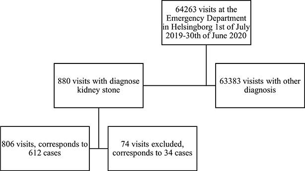
Figure 1. Flowchart of patients included.
Emergency CT was conducted in 43% (262) of patients with suspected renal colic and 57% (350) left the ED without a CT. Additionally, 9% (55) of patients underwent CT imaging at a subsequent emergency visit. In our analysis the group defined as the emergency CT group was confined to those with a CT (within 24 h) at their first visit. Characteristics regarding patients and stones are presented in Table 1.
Of the total of 612 patients, kidney stones were identified in 324 cases, (53%). In the emergency CT group, stones were found in 87.0% (228/262) of patients, compared to the deferred CT group where stones were found in 27% (96/350) on the follow-up CT. In the emergency CT group, 13% had no detectable stone on the emergency CT, but were still coded as having a kidney stone, with the majority of these cases being assessed as spontaneous stone passage.
Among the 324 patients with confirmed kidney stone 37% (120) underwent treatment. Of these 324 patients, 6% (19) underwent acute treatment within 3 days, all URS. In total URS was the most common treatment, with 21% (68) followed by ESWL in 16% (50) and percutaneous nephrolithotomy (PCNL) in 1% (2). In the group with emergency CT 72 patients underwent treatment compared to 48 patients among those without acute diagnostics, for details see Table 2, where further information regarding treatment and follow-up can be found.
| Variable | Total | Emergency CT < 24 h | No emergency CT < 24 h | P |
| Detected stones, number of patients (%) | 324 (53) | 228 (87) | 96 (27) | |
| Treatment (%) | <0.003 | |||
| • None | 492 (80) | 190 (72) | 302 (86) | |
| • URS | 68 (11) | 41 (16) | 27 (8) | |
| • ESWL | 50 (8) | 31 (12) | 19 (5) | |
| • PCNL | 2 (1) | 0 | 2 (1) | |
| Stent (%) | 0.6 | |||
| • None | 247 (76) | 170 (74) | 77 (80) | |
| • JJ | 62 (19) | 47 (21) | 15 (16) | |
| • Nephrostomy | 15 (5) | 11 (5) | 4 (4) | |
| Follow up (%) | <0.001 | |||
| • Completed | 442 (72) | 189 (72) | 253 (72) | |
| • No referral | 75 (12) | 15 (6) | 60 (17) | |
| • Patient absent | 36 (6) | 8 (3) | 28 (8) | |
| • No need (surgery, spontaneous passage) | 21 (4) | 16 (6) | 9 (3) | |
| • No stone on emergency CT | 37 (6) | 33 (13) | 0 | |
| Days to treatment, median (IQR) | <0.001 | |||
| • Total | 33 (8–60) | 22 (3–40) | 55 (30–91) | |
| • URS | 22 (2–53) | 4 (1–39) | 40 (10–79) | |
| • ESWL | 35 (23–72) | 26 (18–41) | 70 (39–115) | |
| • PCNL | 159* | 159 | ||
| Days to closure, median (IQR) | <0.001 | |||
| • Total | 34 (21–59) | 30 (7–54) | 37 (25–62) | |
| • No treatment | 34 (27–57) | 32 (26–50) | 47 (29–93) | |
| • URS | 34 (2–79) | 8 (1–52) | 53 (18–134) | |
| • ESWL | 112 (55–179) | 96 (41–174) | 113 (93–196) | |
| • PCNL | 225* | 225 | ||
| *Delay due to patient preference and Covid-19 pandemic. ESWL: extracorporeal shock wave lithotripsy; URS: ureteroscopy; IQR: interquartile range; PCNL: percutaneous nephrolithotomy. |
||||
Statistically significant differences were observed between the emergency CT and deferred CT groups regarding age, stone size, treatments, follow-up, and time to treatment and closure (see Tables 1 and 2). Patients in the emergency CT group were older, had smaller stones, more treatments, more follow-up, and shorter time to treatment and closure. These differences remained significant even after excluding acute surgery within 3 days, as presented in Table 3. The median time to treatment was 28 days for the emergency CT group compared to 59 days for the deferred group, the corresponding numbers for closure was 31 and 37, respectively. Thus, the emergency CT group had 31 days shorter time to treatment and 6 days shorter time to closure. In the deferred group stone treatment could not be planned until the follow-up CT was done and persisting stone with treatment indication was confirmed.
There was no statistically significant difference in the number of subsequent ED visits between patients who underwent emergency CT and those who did not.
A quarter of the stones were < 4 mm of which 5% (4/81) required treatment. Among stones sized 4–5 mm, 22% (26/116) required treatment. These results are visualized in a bar chart in Figure 2.
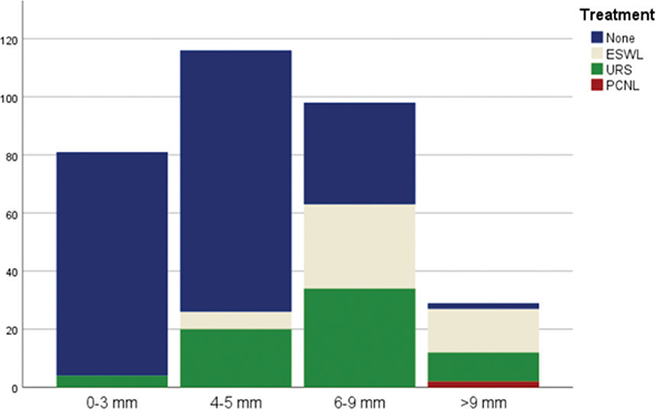
Figure 2. An overview of numbers of treatments and maximum size of the stone. Extracorporeal shock wave lithotripsy (ESWL). Ureteroscopy (URS). Percutaneous nephrolithotomy (PCNL).
Most patients, 72% (442) underwent a CT as a follow-up, while 19% (111) did not due to lack of referral or non-attendance, the remaining 9% was not in need of a follow-up. Overall, 19% lacked appropriate follow-up, with 9% in the emergency CT group and 26% in the deferred CT group, a statistically significant difference. Upon detailed examination of the group that lacked follow-up, we found that the majority 55% (60/111) were referred to primary care. Additionally, 32% (36/111) did not attend their follow-up CT, and 9% (10/111) did not reside in the region. Of the remaining few (5 cases), follow-up was not pursued for the following reasons: two patients had previously received a high radiation dose, two patients were not referred to primary care by mistake and one patient was pregnant and therefore opted to avoid radiation exposure.
In the emergency CT group, we have 1.8 CT scans/ patient (474/262) and in the deferred group 0.9 CT scans/patient (314/350).
One patient died during the follow-up period, with the kidney stone not being the cause of death.
Discussion
In this retrospective study we aimed to assess the use of emergency CT in renal colic and if it affected treatment, time to treatment and emergency visits. The group with an emergency CT were older, had smaller stones, more treatments, more follow-up and shorter time to treatment and closure.
During our study period, 43% of patients with suspected urolithiasis underwent emergency CT. This likely indicates cases where pain management alone was insufficient or where complex factors such as suspected infection, kidney failure, solitary kidney or uncertain clinical diagnosis necessitated further investigation. According to the routine at the time, all patients requiring hospitalization due to urolithiasis underwent an emergency CT. Patients in the deferred group were not hospitalized. The recent Swedish recommendations advocating emergency CT at the ED would have resulted in an additional 350 emergency scans over the year, a relatively small increase.
Of those who did not receive an emergency CT, 9% eventually underwent CT during emergency revisit. This raises questions about whether these additional visits could have been avoided with an emergency CT at their first visit.
Patients who underwent emergency CT tended to be older, had smaller stones, underwent more treatments, had more extensive follow-up, and experienced shorter time to treatment and closure. The issue whether emergency CT in fact shortens time to treatment or not has not been proven earlier. The shorter time to treatment could also just be a consequence of intractable pain leading to acute treatment in these cases, in our material 16% (19/120) of those who required treatment. In our material there was a statistically significant shorter time to treatment and to closure for the group with emergency CT when acute surgery (within 3 days) was removed. Thus, emergency CT shortened time to treatment with 33 days and to closure with 6 days. The deferred group’s delay of 33-days likely corresponds to the time between the emergency visit and the follow-up CT. Indicating that delaying CT will delay treatment to those who needs it.
Number of CT scans is higher in the emergency CT group as everyone in this group has an acute diagnostic imaging. Patients in the deferred group seems to be lost in the follow-up and a number of patient in this group have no diagnostics at all.
The difference in proportion of small stones between the groups is most likely attributed to the spontaneous passage of stone over time. Regarding treatments, we saw a higher rate of treatments in the group who had emergency CT. This suggests that we might be intervening unnecessarily in some cases, highlighting the need for judicious medical decisions. Lindqvist et al. also found a higher stone treatment rate for those with immediate imaging compared to deferred imaging [15]. Although it is a bit precarious to compare our two groups since they are not randomized and differ in several aspects.
The older aged patients in the emergency CT group emphasize the need to rule out serious differential diagnosis, as kidney stones are less likely to debut in older age [16]. Another factor contributing to the older age is that diagnostic imaging is recommended in Helsingborg to exclude abdominal aortic aneurysm in those without known kidney stone disease and age >50 years.
We observed a high rate of stones in the group with an emergency CT, 87% (228/262) of those with an emergency CT had a symptomatic stone. In the group with deferred CT, we found stone in 27% (96/350). The corresponding number from Lindquist et al. [15] was 70% and 39%, respectively, not entirely comparable as it was a randomized but smaller study. In the deferred group 56% (53/96) had a stone ≥6 mm and could likely have been scheduled for treatment approximately 3 weeks earlier if they had done an emergency CT.
The high success rate, 95%, of conservative management for small stones (<4 mm) emphasizes the importance of careful consideration before intervening. These figures are in line with the results reported by Jendeberg et al. in 2017 [6]. Among the slightly larger stones, (4–5 mm) 78% require no treatment, corresponding to figures from Yallapa et al. 2018 [7]. The presence of stones requiring PCNL in the deferred CT group aligns with the typical presentation of dull type of pain rather than acute renal colic for these stones.
Differences in time to closure between ureteroscopic (URS) and extracorporeal shock wave therapy (ESWL) may be attributed to factors such as stone location and modality. On the other hand, the accessibility to ESWL is probably better but apparently not enough to make up for the difference.
The second half of our period of observation coincided with the Covid-19 pandemic but despite this we saw the same number of visits concerning acute renal colic the first 6 months as the last 6 months and we conclude that Covid-19 19 had no or limited effect on this study. Several studies indicate lower admission rates during the Covid-19 pandemic, although comparing these rates is challenging due to the unique nature of Sweden´s lockdowns and recommendations [17–19].
One of the primary strengths of this study is the review that has been conducted, including the detailed examination of CT imaging with size measurements. In addition, all patients with a diagnosis of urolithiasis at the ED in Helsingborg were included in the study.
Several limitations should be acknowledged. This retrospective study has its uncertainties, such as the reasons for performing emergency versus deferred CT, which may involve different patient groups or unmeasured confounding factors. The local recommendation of emergency CT to exclude abdominal aneurysm could be seen as a limitation. Our study did not include cases of pyelonephritis or infected hydronephrosis due to stone-induced renal obstruction.
Our material reflects the management of acute kidney stones at a medium-sized regional hospital in Sweden and our results might be applicable to similar settings.
The implementation of the new Swedish guidelines may lead to increased CT utilization, raising concerns about overuse. However, it also presents an opportunity to improve care and reduce time to definitive treatment and find important differential diagnosis. It is a question of what to prioritize: minimizing radiation versus delaying treatment for those in need for treatment and for the patient to get a correct diagnosis. However, no information was available regarding the consequences of delayed treatment. We should, of course, aim for the lowest possible radiation dose, and an emergency CT does increase the radiation exposure. Nevertheless, it is important to remember that the CT performed is a low-dose CT with a radiation dose of < 3mSv. Randomized controlled trials are lacking regarding timing of diagnostics in renal colic and further research is needed.
In our study conducted between 2019 and 2020 almost half, 43%, of the patients received an emergency CT. This group with an emergency CT had significantly shorter time to treatment and were stone free earlier compared to the group with deferred diagnostics. We anticipate performing further evaluations of stone treatment following the implementation of the new Swedish guidelines where diagnostics is recommended.
Acknowledgment
The authors would like to thank their former colleague Adelina Mazhiqi for the data collection. They would also like to thank the Urology department in Helsingborg and Ängelholm.
Authors’ contribution
Data collection: AM, MU, MW. Analysis and interpretation: MU, FA, MP, AF, K-JL, TT, MW. Drafting the manuscript for important intellectual content: MU, FA, MP, AF, K-JL, TT, MW.
ORCID
Maria Utter  , https://orcid.org/0009-0002-4486-6810
References
- [1] Socialstyrelsen, Socialstyrelsen registerservice, statistik och data. https://sdb.socialstyrelsen.se/if_par/val.aspx.
- [2] Hesse A, Brändle E, Wilbert D, et al. Study on the prevalence and incidence of urolithiasis in Germany comparing the years 1979 vs. 2000. Eur Urol. 2003;44(6):709–713. https://doi.org/10.1016/S0302-2838(03)00415-9
- [3] Ljunghall S. Incidence and natural history of renal stone disease and its relationship to calcium metabolism. Eur Urol. 1978;4(6):424–430. https://doi.org/10.1159/000474013
- [4] Trinchieri A. Epidemiology of urolithiasis: an update. Clin Cases Miner Bone Metab. 2008;5(2):101–106. https://doi.org/10.1007/s12018-009-9025-y
- [5] Ferraro PM, Curhan GC, D’Addessi A, et al. Risk of recurrence of idiopathic calcium kidney stones: analysis of data from the literature. J Nephrol. 2017;30(2):227–233. https://doi.org/10.1007/s40620-016-0283-8
- [6] Jendeberg J, Geijer H, Alshamari M, et al. Size matters: the width and location of a ureteral stone accurately predict the chance of spontaneous passage. Eur Radiol. 2017;27(11):4775–4785. https://doi.org/10.1007/s00330-017-4852-6
- [7] Yallappa S, Amer T, Jones P,et al. Natural history of conservatively managed ureteral stones: analysis of 6600 patients. J Endourol. 2018;32(5):371–379. https://doi.org/10.1089/end.2017.0848
- [8] Türk C, Petřík A, Sarica K, et al. EAU guidelines on interventional treatment for urolithiasis. Eur Urol. 2016;69(3):475–482. https://doi.org/10.1016/j.eururo.2015.07.041
- [9] Ray AA, Ghiculete D, Pace KT, et al. Limitations to ultrasound in the detection and measurement of urinary tract calculi. Urology. 2010;76(2):295–300. https://doi.org/10.1016/j.urology.2009.12.015
- [10] Xiang H, Chan M, Brown V, et al. Systematic review and meta-analysis of the diagnostic accuracy of low-dose computed tomography of the kidneys, ureters and bladder for urolithiasis. J Med Imaging Radiat Oncol. 2017;61(5):582–590. https://doi.org/10.1111/1754-9485.12587
- [11] Arcaniolo D, De Sio M, Rassweiler J, et al. Emergent versus delayed lithotripsy for obstructing ureteral stones: a cumulative analysis of comparative studies. Urolithiasis. 2017;45(6):563–572. https://doi.org/10.1007/s00240-017-0960-7
- [12] Cornelius J, Zumbühl D, Afferi L, et al. Immediate shockwave lithotripsy vs delayed shockwave lithotripsy after urgent ureteral stenting in patients with ureteral or pyeloureteral urolithiasis: a matched-pair analysis. J Endourol. 2021;35(5):721–727. https://doi.org/10.1089/end.2020.0384
- [13] Nationellt vårdprogram för stensjukdom i övre urinvägar 2022. https://vardpersonal.1177.se/globalassets/nkk/nationell/media/dokument/kunskapsstod/vardprogram/nationellt-vardprogram-for-stensjukdom-ovre-urinvagarna.pdf
- [14] Svensk uroradiologisk förening, Mätning av urinvägskonkrement, Version 1.0/2021-05-10. 2021. https://slf.se/sfmr/surf/rekommendationer-och-riktlinjer/metodbocker/
- [15] Lindqvist K, Hellström M, Holmberg G, et al. Immediate versus deferred radiological investigation after acute renal colic: a prospective randomized study. Scand J Urol Nephrol. 2006;40(2):119–124. https://doi.org/10.1080/00365590600688203
- [16] Croppi E, Ferraro PM, Taddei L, et al. Prevalence of renal stones in an Italian urban population: a general practice-based study. Urol Res. 2012;40(5):517–522. https://doi.org/10.1007/s00240-012-0477-z
- [17] Byrne MHV, Georgiades F, Light A, et al. Impact of Covid-19-19 on the management and outcomes of ureteric stones in the UK: a multicentre retrospective study. BJU Int. 2023;131(1):82–89. https://doi.org/10.1111/bju.15882
- [18] Kaczmarek K, Kalembkiewicz J, Jankowska M, et al. Did the Covid-19-19 pandemic restrict access to emergency urological services: Assessment of reorganisation effectiveness for hospital treatment. Int J Environ Res Public Health. 2023;20(4). https://doi.org/10.3390/ijerph20043735
- [19] Nourian A, Uppaluri C, Chen M, et al. Comparison of management and outcomes of symptomatic urolithiasis during the Covid-19-19 pandemic to a comparative cohort. Urology. 2022;165:178–183. https://doi.org/10.1016/j.urology.2022.01.019