CASE REPORT
Unexpected Liver Metastasis Three Years after Nephrectomy for Renal Oncocytoma: A Case-report and Review of Literature
Pekka Lammia,b, Anna Junttilaa, Essi Ikonenc, Arto Salonend, Olli Lahtinenb,d, and Antti Kivivuoria,b
aDepartment of Gastrointestinal Surgery, Kuopio University Hospital, Kuopio, Finland; bUniversity of Eastern Finland, Kuopio, Finland; cDepartment of Clinical Pathology, Kuopio University Hospital, Kuopio, Finland; dDepartment of Urology, Kuopio University Hospital, Kuopio, Finland; eDepartment of Radiology, Kuopio University Hospital, Kuopio, Finland
KEYWORDS: Oncocytoma; nephrectomy; liver metastasis
Citation: Scandinavian Journal of Urology 2025, VOL. 60, 73–75. https://doi.org/10.2340/sju.v60.43205.
Copyright: © 2025 The Author(s). Published by MJS Publishing on behalf of Acta Chirurgica Scandinavica. This is an Open Access article distributed under the terms of the Creative Commons Attribution 4.0 International License (http://creativecommons.org/licenses/by/4.0/), allowing third parties to copy and redistribute the material in any medium or format and to remix, transform, and build upon the material, with the condition of proper attribution to the original work.
Received: 20 December 2024; Accepted: 17 February 2025; Published: 20 March 2025
CONTACT Antti Kivivuori Antti.Kivivuori@pshyvinvointialue.fi Department of Gastrointestinal Surgery, Kuopio University Hospital, Puijonlaaksontie 2, 70210 Kuopio, Finland
Supplemental data for this article can be accessed online at https://doi.org/10.2340/sju.v60.43205
Competing interests and funding: The authors declare no conflicts of interest.
Case
A 70-year-old male with a history of hypertension was referred to our centre in July 2021 due to the presence of four tumours in his left kidney. Primary computed tomography (CT) showed four solid sharply demarcated heterogeneously enhancing tumours. The largest one was measured to be 10 cm, and it had hypoattenuating central area resembling a stellate scar, which can be seen in oncocytomas and chromophobe renal cell carcinoma. However, multiple large renal tumours without distant metastases favoured the diagnosis of oncocytomas. The patient was referred to percutaneous biopsy, which was performed using 16-gauge needle. Histopathologic evaluation was suggestive for benign oncocytoma. After a thorough discussion with the patient, the decision was to undergo surgery despite the suggestive findings for benign oncocytoma. So, a nephrectomy was performed in August 2021. The final pathological analysis confirmed the diagnosis of oncocytoma. No additional follow-up imaging was scheduled because of known indolent nature of the disease.
In June 2024, 3 years after initial surgical treatment, the patient began experiencing abdominal discomfort and pain. A CT scan revealed two new tumours in the liver, measuring 6 and 7.5 cm in segments 5 and 8 (Figure 1), respectively. After CT findings, the patient underwent gadoxetate disodium (Primovist)-enhanced magnetic resonance imaging (MRI). The lesions were seen as well-circumscribed masses with intermediate T2 signal intensity predominantly. Central areas demonstrated typical T2 hyperintensity corresponding to loose matrix of the connecting tissue in the stellate scar seen in part of the oncocytomas. A biopsy of the liver lesions confirmed they were metastases from the previous renal oncocytoma. CT scan did not show any additional metastases. Liver metastases were technically operable, and a surgery was scheduled. Due to the small left liver lobe, two non-anatomical local liver resections were performed. The final pathological analysis confirmed the metastases originated from the renal oncocytoma, which both had been resected with clear margins.
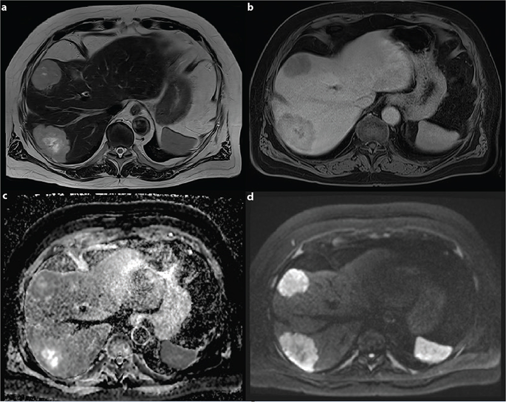
Figure 1. Image panel shows two liver masses in liver segments VII and VIII, proven to be renal oncocytoma metastasis in surgery. T2-weighted image (a) shows well-circumscribed masses with intermediate T2 signal intensity. T2 hyperintensity can be seen in the central parts corresponding to a stellate scar, which can be seen in oncocytomas. Masses demonstrate heterogenous enhancement with gadoxetate disodium (Primovist) in venous phase and delayed phase (b) with no visual enhancement in the scar. Diffusion restriction can be seen in the tumours except in the central scar areas in diffusion-weighted imaging (c, d).
Histopathologic evaluation of liver metastases
Both liver specimens contained sharply demarcated round tumours of 5 and 4 cm in diameter. In microscopy, the tumours consisted of cells with abundant eosinophilic cytoplasm. The cells formed sheets and small solid nests with loose intervening stroma. The nuclei had slight variation in size and shape. Mitotic activity was very low in both tumours, with no mitoses found in 50 high power fields. The findings were compared to the earlier renal samples, and the histology was similar. Immunohistochemistry had been done for the renal specimen and showed positivity for CD117 and negativity for vimentin. Cytokeratin 7 and alpha-methyl acyl-coenzymeA racemase (amacr) showed only few scattered faintly positive cells. The subsequent immunohistochemical profile of the larger liver tumour was similar, with positivity for CD117 (Figure S1). This confirmed the diagnosis of oncocytoma.
The patient recovered well from the open liver surgery and was discharged to home on the fifth postoperative day. Multidisciplinary team meeting was conclusive to restrain for any adjuvant oncologic treatment. Furthermore, intensive follow-up with CT scans is scheduled.
Discussion
Here, we present a rare case of metastatic renal oncocytoma, treated with an initial nephrectomy followed by radical liver resections with curative intent, 3 years after the original surgery.
Renal oncocytoma is an uncommon renal cortical neoplasm considered to be entirely benign. Differentiation from atypical renal cell carcinomas such as chromophobe renal cell carcinoma and eosinophilic renal cell carcinoma is essential. Although cross-sectional imaging can be suggestive for renal oncocytoma, a definitive diagnosis requires careful histopathologic examination with immunohistochemistry [1]. The histopathological diagnosis of oncocytoma requires immunohistochemistry in addition of light microscopy since the variants of renal cell carcinoma can mimic the morphology of oncocytoma [2]. The classic staining pattern for oncocytoma is positive for CD117 (c-kit) and negative for cytokeratin 7, alpha-methyl acyl-coenzymeA racemase (amacr) and vimentin. Scattered cytokeratin 7-positive cells may be seen. Chromophobic renal cell carcinoma has similar morphologic features and positivity for CD117 but has additional diffuse membrane positivity for cytokeratin 7 [3]. In cases of unclear morphology or unusual immunohistochemical findings, it is possible to use molecular profiling such as array-comparative genomic hybridization to confirm diagnosis [3].
If the diagnosis is confirmed with certainty, the treatment is usually conservative due to the tumour’s indolent nature. Surgery is generally indicated for cases with diagnostic uncertainty or progressive tumour’s identified during a follow-up [4, 5]. Only a few cases of metastatic renal oncocytoma have been reported in the literature.
Webster et al. recently published a report of a case with metastatic oncocytoma with extensive modern diagnostic workup. A preoperative sestamibi-SPECT scan demonstrated a right-sided renal tumour with eight liver metastases. The patient received neoadjuvant cabozanib for 6 months, which resulted reasonable tumour downstaging. After favourable neoadjuvant treatment, the patient underwent right nephrectomy and liver resections. Four months later, MRI showed five residual lesions, which were successfully treated with microwave ablations. After 30 months, the patient is disease free with only minor indeterminate stable hepatic lesions [6].
Oxley et al. presented a case of 70-year-old woman who underwent nephrectomy for renal oncocytoma and developed multiple liver metastases 9 years later. Treatment was palliative, resulting in death within 9 months [7]. Amin et al. reported a case of 48-year-old male who underwent nephrectomy for renal oncocytoma and later developed multiple bone metastases 1 year after surgery (8). No histology was obtained from metastases in either of these two cases [7, 8].
A few case reports of metastatic oncocytomas had been published earlier [9]. However, there is a major uncertainty of presented diagnosis in the case reports published before the use of immunohistochemistry.
Our case, along with a few earlier reports of metastatic renal oncocytoma, suggests that renal oncocytoma may have minor metastatic potential and should not be considered a completely indolent benign tumour. In a few reported cases, the treatment has ranged from expectant management to surgery with or without chemotherapy.
Conclusion
In conclusion, metastatic renal oncocytoma is an extremely rare condition. In suspected cases, histopathological differential diagnostics with immunohistochemistry is essential. However, in cases of true oligometastatic renal oncocytoma, patients may be treated with radical approach, including nephrectomy and surgical management of metastases.
Acknowledgement
This study was supported financially by the Department of Surgery at the University Hospital of Kuopio.
Authors’ contribution
PL and AK conceived and designed the study; PL and AK acquired the data; PL and AJ performed the experiment; PL and AK drafted the manuscript; all authors critically reviewed, edited and approved the manuscript; AK supervised the study and is the guarantor of the study.
References
- [1] Lobo JM, Clements MB, Bitner DP, et al. Does renal mass biopsy influence multidisciplinary treatment recommendations? Scand J Urol. 2020;54(1). https://doi.org/10.1080/21681805.2019.1703805
- [2] Amin MB, McKenney JK, Martignoni G, et al. Low grade oncocytic tumors of the kidney: a clinically relevant approach for the workup and accurate diagnosis. Mod Pathol. 2022;35. https://doi.org/10.1038/s41379-022-01108-5
- [3] International Agency for Research on Cancer. Urinary and male genital tumours. Vol. 8. WHO Classification of Tumours. 2022.
- [4] Miller BL, Mankowski Gettle L, Van Roo JR, et al. Comparative analysis of surgery, thermal ablation, and active surveillance for renal oncocytic neoplasms. Urology. 2018;112. https://doi.org/10.1016/j.urology.2017.09.016
- [5] Tzortzakakis A, Papathomas T, Gustafsson O, et al. 99mTc-Sestamibi SPECT/CT and histopathological features of oncocytic renal neoplasia. Scand J Urol. 2022;56(5–6). https://doi.org/10.1080/21681805.2022.2119273
- [6] Webster BR, Ricketts CJ, Vocke CD, et al. Molecular characterization of metastatic oncocytoma with exceptional response to treatment: a case report. JCO Precis Oncol. 2024;8:e2400188. https://doi.org/10.1200/PO.24.00188
- [7] Oxley JD, Sullivan J, Mitchelmore A, et al. Metastatic renal oncocytoma. J Clin Pathol. 2007;60(6). https://doi.org/10.1136/jcp.2006.044198
- [8] Amin R, Anthony P. Metastatic renal oncocytoma: a case report and review of the literature. Clin Oncol. 1999;11(4). https://doi.org/10.1053/clon.1999.9064
- [9] Perez-Ordonez B, Hamed G, Campbell S, et al. Renal oncocytoma: a clinicopathologic study of 70 cases. Am J Surg Pathol. 1997;21. https://doi.org/10.1097/00000478-199708000-00001