CASE REPORT
Inferior vena cava syndrome as the initial manifestation of metastatic prostate cancer: a rare case successfully treated with endovascular stenting
Dimitra Akrivoua , Andreas Forsvalla, Angelos Katevatisa, Tobias Kühmeb, Anna Eliassona and Magnus Wageniusa
aUrology Department, Helsingborg hospital, Helsingborg, Sweden; bVascular Surgery Department, Helsingborg Hospital, Helsingborg, Sweden
KEYWORDS: Inferior vena cava syndrome; prostate cancer; retroperitoneal metastasis; case report
Citation: Scandinavian Journal of Urology 2025, VOL. 60, 90–92. https://doi.org/10.2340/sju.v60.43708.
Copyright: © 2025 The Author(s). Published by MJS Publishing on behalf of Acta Chirurgica Scandinavica. This is an Open Access article distributed under the terms of the Creative Commons Attribution 4.0 International License (http://creativecommons.org/licenses/by/4.0/), allowing third parties to copy and redistribute the material in any medium or format and to remix, transform, and build upon the material, with the condition of proper attribution to the original work.
Received: 27 February 2025; Accepted: 17 April 2025; Published: 15 May 2025
CONTACT: Dimitra Akrivou dim.s.akrivou@gmail.com Urology Department, Helsingborg Hospital, Charlotte Yhlens gata 10, 251 87 Helsingborg, Sweden
Competing interests and funding: The authors report no conflicts of interest.
The research received no external funding.
Introduction
Inferior vena cava (IVC) syndrome is a rare clinical entity, occurring in various malignant and non-malignant medical conditions [1, 2]. The syndrome can result from congenital defects, extrinsic compression or intraluminal obstruction [1, 3], and the clinical presentation includes nonspecific symptoms, such as lower extremity oedema and pain and ascites [3, 4]. In cancer patients, IVC syndrome may be caused by external compression by intra-abdominal or retroperitoneal lymph-node (LN) masses [3]. Conservative management is frequently ineffective, making stent placement an attractive treatment option [4–7].
We report a man who developed IVC syndrome because of a retroperitoneal LN metastatic mass. The primary cancer site was in the prostate, and the condition was successfully treated with endovascular stent placement followed by prompt hormonal treatment. Given the rarity of the IVC syndrome, this case report may be of value for others.
Case report
A 70-year-old man with cardiovascular disease (myocardial infarction, hypertension, hyperlipidemia) presented at the emergency department with a 2-week history of right leg and scrotal oedema. After the initial physical examination, the oedema was suspected to be caused by a deep venous thrombosis (DVT). A DVT was confirmed with a Doppler ultrasound, and peroral anticoagulation therapy was started. The underlying cause was investigated with a contrast-enhanced computed tomography (CT) of the abdomen, which showed extensive metastasis in the lungs, liver, bone and retroperitoneal LNs. The primary tumour was suspected in the urinary system because of bladder wall thickening and tumour infiltrating the bladder, prostate and left ureteric orifice. The IVC was compressed by a paraaortic LN mass. There were no signs of thrombosis in the IVC, but there was thrombosis present in the right common iliac vein (Figure 1a).
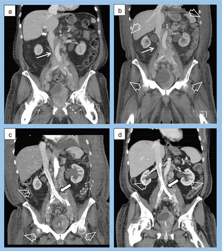
Figure 1. CT coronal views of the IVC thoughtout the course of events from diagnosis (a) up to 2-months follow-up after intervention (d). (a) First CT of the patient showing the narrowing of the IVC (white arrow), confirming the diagnosis. Visible tumour in the bladder and prostate of the patient (interrupted line arrow). (b) One-month follow-up CT after diagnosis, when the patient experienced worsening of the symptoms and was admitted to the hospital. Radiologically visible worsening of the narrowing (white arrow) and the subcutaneous oedema (open arrow). (c) First CT after stent-intervention, which was performed acutely due to hemodynamic instability. Visible intravascular stent in the IVC and common iliac veins (white arrow), hematoma in the left abdominal wall (interrupted line arrow) and subcutaneous oedema (open arrows). (d) Two-months follow-up after stent intervention. Visible intravascular stent in the IVC and common iliac veins (white arrow) and significantly decreased subcutaneous oedema (open arrows).
Further investigation included a cystoscopy, urine cytology, serum prostate-specific antigen (PSA) and digital rectal examination (DRE). The DRE raised suspicion of a locally advanced prostate cancer. Cystoscopy showed invasion of tumour from the bladder neck to the lateral bladder wall. The PSA value was 296 ng/ml (age-specific reference range < 5 ng/ml), so systematic prostate biopsies were obtained, according to the Swedish PCa guidelines [8]. Urine cytology: Paris 4. Both the bladder and prostate biopsies revealed a Gleason score 4 + 5 = 9 prostatic adenocarcinoma (immunohistochemical profile reported in Table 1), confirming the diagnosis of high-volume, de-novo (synchronous) metastatic prostate cancer. Treatment with luteinising-releasing hormone agonist and abiraterone was started.
At a 1-month follow-up appointment, physical examination revealed the progress of the leg and scrotal oedema, and further oedema was visible up to the umbilical level. A CT angiography was performed and showed no signs of tumour shrinking at any locations and progress of IVC compression (Figure 1b). Additionally, his PSA was 367 with testosterone < 0.4 nmol/L. The patient was admitted to the Department of Urology. After a multidisciplinary team discussion, he was offered an endovascular stent, combined with diuretics (furosemide), which he accepted. On the sixth hospital day, an IVC phlebography was performed via the right and left femoral vein, and two stents (BeYond Venous Self-Expanding, Bentley Innomed GmbH, Hechingen, Germany) were placed without complications. Directly after stenting, intermittent pneumatic compression (IPC) devices were applied on both legs. After these interventions, the urine production increased, and the patient’s general condition gradually improved. After 4 days, the patient became circulatory unstable and anaemic. An unprovoked hematoma was observed in the left abdominal wall and right arm. Routine blood samples showed impaired coagulation (INR 1,9) and hypoalbuminemia. An emergency CT angiography showed active bleeding, but a conservative approach was chosen (Figure 1c). Infusion of Hartmann’s solution was started; blood and fresh frozen plasma transfusions were given, as well as tranexamic acid treatment. The anticoagulant treatment was discontinued. The patient’s vital signs were closely monitored at the ward, and as they improved without vasopressors, there was no need for intensive care unit (ICU) transfer. The patient continued to respond well to conservative treatment, including full-dose low-molecular weight heparin, regained an adequate haemoglobin level and gradually returned to full mobilisation.
The cancer was considered de novo metastatic castration resistant and the hematomas a paramalignant phenomenon although post-stenting release of accumulated tinzaparin and apixaban used for DVT treatment was discussed as a possible contributing factor. One week after stent placement, the patient’s general condition had stabilised, and docetaxel therapy was started. The leg oedema decreased, but the patient’s weight remained 2.5 kg more than at hospital admission (93.3 kg) for 2 weeks after the stent placement, presumably a consequence of the extensive fluid resuscitation. On the third week, the weight was down to 80.8 kg.
At 1-month follow-up, the patient was feeling well without stent- or docetaxel-related complications. The leg and scrotal oedema were much less pronounced, and his weight was 78 kg. The coagulation tests were normal. At 2-month follow-up, the patient remained complication free and in better overall clinical condition. His weight was 74.8 kg, and the PSA value was 48 ng/ml. A CT scan showed reduced oedema, full resorption of the hematoma, but no radiological response of the metastases (Figure 1d).
Discussion
Retroperitoneal LN metastasis may disturb the flow in the IVC and in rare instances cause an IVC syndrome [1–3]. The incidence of this condition is unknown [1]. There are only a few case reports of an IVC syndrome in prostate cancer patients [9, 10].
With recent technical advances, the management of malignant IVC syndrome has evolved from radiotherapy to minimally invasive methods, particularly endovascular stent placement has been reported to have a high success rate [3–7].
To our knowledge, this is the first report of a patient in whom an IVC syndrome was the first manifestation of a de novo metastatic prostate cancer and who was successfully treated with endovascular stents.
The rarity of malignant IVC syndrome makes comparative clinical trials nearly impossible. It seems reasonable to refer the patient to a high-volume vascular centre, where multidisciplinary management can be planned by a team of vascular surgeons and oncologists with support from intensive care specialists. As demonstrated by this case, close observation for potential post-stenting complications is warranted.
Ethics approval and consent to participate
In Sweden, ethical approvement is not required for case reports. The patient has been informed and given his consent to this publication.
Acknowledgments
We would like to thank our colleague Jan Blond, Department of Vascular Surgery, Helsingborg Hospital, Helsingborg, for his contribution to the management of the patient and input to this case report.
Author contributions
DA, AE, AF, TK and MW – designed the case report study and participated in drafting the manuscript; DA – obtained patient’s consent; DA and AK– searched into the medical library (Pubmed) for references and collected and analyzed the data; AF, AK, TK and MW – conducted key revision points. All authors contributed to critical revision of the manuscript for important intellectual content. All authors gave final approval of the version to be published. All authors participated fully in the work, take public responsibility for appropriate portions of the content, and agree to be accountable for all aspects of the work in ensuring that questions related to the accuracy or completeness of any part of the work were appropriately investigated and resolved.
References
[1] Lawrensia S, Khan YS. Inferior vena cava syndrome. Treasure Island, FL: StatPearls Publishing; 2024 [updated 2023 May 20]. Available from: https://www.ncbi.nlm.nih.gov/books/NBK560885/ [cited November 7th 2024]
[2] Chaudhary RK, Nepal P, Kumar S, et al. Imaging of inferior vena cava normal variants, anomalies and pathologies, Part 2: acquired. SA J Radiol. 2023;27(1):2694. https://doi.org/10.4102/sajr.v27i1.2694
[3] Friedman T, Quencer KB, Kishore SA, et al. Malignant venous obstruction: superior vena cava syndrome and beyond. Semin Intervent Radiol. 2017;34(4):398–408. https://doi.org/10.1055/s-0037-1608863
[4] Morita S, Sugawara S, Suda T, et al. Stent placement for malignant inferior vena cava syndrome in a patient with recurrent colon cancer. Intern Med. 2020;59(22):2885–2890. https://doi.org/10.2169/internalmedicine.5033-20
[5] Kuetting D, Thomas D, Wilhelm K, et al. Endovascular management of malignant inferior vena cava syndromes. Cardiovasc Intervent Radiol. 2017;40(12):1873–1881. https://doi.org/10.1007/s00270-017-1740-z
[6] Epelboym Y, Stecker MS, Fan CM, et al. Treatment of malignant inferior vena cava obstruction with Gianturco-Rosch-Z stents: a single center 13-year experience. Clin Imaging. 2020;59(2):95–99. https://doi.org/10.1016/j.clinimag.2019.09.015
[7] Augustin AM, Lucius LJ, Thurner A, et al. Malignant obstruction of the inferior vena cava: clinical experience with the self-expanding Sinus-XL stent system. Abdom Radiol (NY). 2022;47(10):3604–3614. https://doi.org/10.1007/s00261-022-03587-1
[8] Stranne J. 2023/2024 update of the national prostate cancer guidelines in Sweden. Scand J Urol. 2024;59:210–211. https://doi.org/10.2340/sju.v59.42656
[9] Wei W, Gong C, Wei R, et al. Metastatic prostate cancer presenting as generalized lymphadenopathy and progressing with inferior vena cava syndrome: a case report and literature review. Oncol Lett. 2024;28(6):557. https://doi.org/10.3892/ol.2024.14690
[10] Makusha LP, Kulon M, Pucar D, et al. Inferior vena cava syndrome on skeletal scintigraphy secondary to metastatic prostate cancer. World J Nucl Med. 2020;19(3):324–326. https://doi.org/10.4103/wjnm.WJNM_53_19