ORIGINAL RESEARCH ARTICLE
Detection of prostate cancer by magnetic resonance imaging before biopsy – real-world evidence from a nationwide cohort study
Amalie H. Simonia, Anne G. Ordinga, Jakob N. Hedegaarda, Michael Borreb,c, Søren P. Johnsena and Henrik Møllera,d
aDanish Center for Health Services Research, Department of Clinical Medicine, Aalborg University, Gistrup, Denmark; bDepartment of Clinical Medicine, Aarhus University, Aarhus, Denmark; cDepartment of Urology, Aarhus University Hospital, Aarhus, Denmark; dThe Danish Healthcare Quality Institute, Aarhus N, Denmark
ABSTRACT
Objective: Magnetic resonance imaging (MRI) improves prostate cancer detection, but some tumours may be undetectable. This study assessed prostate cancer detection in biopsy-naïve men undergoing first-time prostate MRI.
Material and methods: In this nationwide Danish cohort (2018–2022), men aged ≥ 40 years undergoing first-time prostate MRI were classified as MRI-positive if biopsied within 30 days, and MRI-negative if not. This served as a proxy for the radiology report findings. One-year cumulative incidence of prostate cancer diagnosis in the Danish Prostate Cancer Registry, repeated MRI, biopsy and mortality was estimated by MRI-status (positive/negative) and prostate-specific antigen (PSA) level, using the Aalen–Johansen estimator. Occurrence of high-grade cancer was also assessed.
Results: Among 10,777 men, 40% were MRI-positive and 60% MRI-negative. The cumulative incidence of prostate cancer was higher in the MRI-positive than the MRI-negative men during 1-year follow-up (63% vs. 5% at PSA levels 2–3.9 µg/L), increasing with higher PSA levels (91% vs. 38% at PSA levels ≥ 20 µg/L). At 1 year, repeated MRI occurred in 21% of MRI-positive men and 9% of MRI-negative men; the subsequent 1-year biopsy incidence was 15% among MRI-negative men. Mortality was comparable between MRI-positive and MRI-negative men (1% vs. 1%). The risk of high-grade prostate cancer at the time of diagnosis was 80% in MRI-positive and 74% in the MRI-negative men.
Conclusions: Among biopsy-naïve men undergoing first-time prostate MRI, MRI-positive status strongly predicts prostate cancer, even in men with low PSA. In MRI-negative men with PSA < 4.0 µg/L, prostate cancer risk remains low within the 2-year follow-up.
KEYWORDS: Biopsy; multiparametric magnetic resonance imaging; population surveillance; prostatic neoplasms
Citation: Scandinavian Journal of Urology 2026, VOL. 61, 178–184. https://doi.org/10.2340/sju.v61.46161.
Copyright: © 2026 The Author(s). Published by MJS Publishing on behalf of Acta Chirurgica Scandinavica. This is an Open Access article distributed under the terms of the Creative Commons Attribution 4.0 International License (http://creativecommons.org/licenses/by/4.0/), allowing third parties to copy and redistribute the material in any medium or format and to remix, transform, and build upon the material, with the condition of proper attribution to the original work.
Received: 12 December 2025; Accepted: 12 May 2026; Published: 7 July 2026
CONTACT: Amalie H. Simoni asimoni@dcm.aau.dk Danish Center for Health Services Research, Department of Clinical Medicine, Aalborg University, Selma Lagerløfs Vej 249, 9260 Gistrup, Denmark
Competing interests and funding: The authors report no conflicts of interest.
This work was supported by the Danish Cancer Society, grant number R305-A18514.
Introduction
Prostate cancer is the second most common cancer among men worldwide [1], with over 4,100 new cases diagnosed in Denmark in 2024 [2]. Typically, prostate-specific antigen (PSA) testing serves as an entry point to the diagnostic pathway [1, 3]. Due to imaging improvements, prostate magnetic resonance imaging (MRI) has become a key diagnostic tool, providing detailed information on the lesion characteristics and enabling targeted biopsies [4, 5]. Prostate MRI has significantly improved sensitivity and specificity in prostate cancer detection compared to PSA testing alone [4, 5], and the European Association of Urology has since 2019 recommended a prostate MRI before biopsy [6]. Individual risk assessment still considers age, PSA level and prior negative biopsy history [3, 7].
Meta-analyses have demonstrated that MRI-informed targeted prostate biopsy outperforms systematic biopsy in men undergoing first-time biopsy [8], and performs well in detecting prostate cancer among men with PSA levels of 4–10 µg/L [9]. Other studies indicate that long-term adverse oncologic outcomes (recurrence, metastases and prostate cancer-specific mortality) are low in the MRI-negative population [10, 11], and omitting biopsy in MRI-negative men eliminated more than half of the actively treated low-risk prostate cancers [11, 12]. Still, some aggressive prostate cancers may not be detectable by prostate MRI [10, 13]. The proportion and characteristics of MRI-negative men who are cancer-free remain unknown [2, 14]. Hence, further research is recommended to characterize the size and clinical profile of the anticipated large population with MRI-negative findings who are not initially diagnosed with prostate cancer [2, 12].
We aimed to examine the prostate cancer detection in men undergoing a first-time prostate MRI, in a large real-world cohort of biopsy-naïve men.
Materials and methods
We used existing registries, with prospectively collected information from the Danish healthcare system, covering the entire nation (approximately 5.9 million people) [15]. All residents have access to tax-financed primary and secondary healthcare [16]. By linking these registries, we conducted a population-based cohort study of men undergoing a first-time prostate MRI. The study is registered at the institutional review board (Journal number: 2022-068-03232).
Data sources
The Danish Civil Registration System (CRS) holds information on sex, date of birth, migration and vital status for the entire population [17]. The Danish National Patient Registry (DNPR) has recorded all inpatient hospital contacts since 1977 and outpatient encounters since 1995, providing information on discharge diagnosis and secondary diagnoses according to the 10th revision of the International Classification of Diseases (ICD) since 1994 [18]. The Danish Prostate Cancer Database (DaProCa) has recorded information on patient, cancer and treatment characteristics for all men with prostate cancer since 2003 [19]. The Danish National Laboratory Registry has recorded clinical biochemical laboratory measurements in Denmark since 2013, based on the international Nomenclature for Properties and Units coding [20]. Statistics Denmark, the Danish Health Data Authority, the Danish Healthcare Quality Institute, and the Danish Prostate Cancer Registry provided the data for the study. According to Danish data protection rules, we cannot share individual-level data. Researchers who fulfil the requirements set by the data providers can apply to obtain similar data.
Study population
We included all men aged ≥ 40 years with a first-time prostate MRI (procedure code: UXMD92) in the DNPR during 2018–2022, with no data on previous prostate biopsies since 1996. Data on prostate biopsy procedures were collected from the DNPR (Procedure code: KTKE). The date of the first prostate MRI was defined as the index date. Men were excluded if they had a prior prostate cancer diagnosis, had no registration of a PSA measure at the time of the prostate MRI, or died within 30 days after the MRI. Men who were diagnosed with histopathologically verified prostate cancer within 30 days after the prostate MRI but had no registration for a diagnostic biopsy were excluded.
Men were classified as ‘MRI-positive’ if they underwent a biopsy within 30 days of the index, or ‘MRI-negative’ if they did not. This definition was used as a proxy for the prostate MRI result because radiology reports were not available. The definition represents the clinical diagnostic pathway of the prostate MRI-informed biopsy decision pathway.
Baseline variables
Information on age was obtained from the CRS and classified as 40–49, 50–59, 60–69, 70–79 and 80+ years. The PSA level measured most recently before the prostate MRI was collected from the Danish Laboratory Registry and classified as 0–1.9, 2–3.9, 4–9.9, 10–19.9 and ≥ 20 µg/L. The Charlson comorbidity index (CCI) score was used to summarize the comorbidity burden at index, using scores of 0, 1, 2, 3 and ≥ 4 points [21].
Follow-up
All men were followed from the index date of the first prostate MRI for 2 years, to a diagnosis of prostate cancer (in the DaProCa database, where the diagnosis date is defined as the first requisition date in the pathology registry with a relevant diagnosis), emigration, death, or 31 December 2023, whichever occurred first. Additionally, they were monitored for repeated prostate MRI within 1 year, and the MRI-negative men were followed for biopsies between 31 days and 1 year after the initial MRI. Information on incident prostate cancer and Gleason score at diagnosis was collected from the DaProCa database and categorized as low-grade (< 7) or intermediate-to-high-grade cancer (≥ 7).
Statistical analysis
Characteristics were summarized as frequencies and proportions of the categorical variables, separately for MRI-positive and MRI-negative men.
We calculated the cumulative incidence of a prostate cancer diagnosis among the biopsy-naïve men undergoing first-time prostate MRI within 30 days, 60 days, 1- and 2 years after the index date, accounting for the competing risk of death using the Aalen–Johansen estimator, according to MRI-positive or MRI-negative status and within strata of PSA level.
The cumulative incidences of a new prostate MRI, prostate biopsy at 1-year after the index date, were calculated, accounting for the competing risk of prostate cancer or death using the Aalen–Johansen estimator. The cumulative incidence of mortality 1 year after the index was also calculated according to MRI-status. The crude and adjusted risk of intermediate-to-high-grade prostate cancer (Gleason score ≥ 7) was calculated among the MRI-positive and -negative men. Adjustment was restricted to PSA level category and calendar year, which capture the primary confounding factors, while avoiding adjustment for variables that lie on the causal pathway of the MRI-informed biopsy decision. A 95% confidence interval (CI) was applied to all estimates. All data analysis was performed using STATA18.1 (StataCorp.2023. Statistical Software: Release18. College Station, TX: StataCorp LLC).
Results
The study included 10,777 biopsy-naïve men with a first-time prostate MRI, without prior prostate cancer. A flowchart of the cohort construction is shown in Figure 1. Of these, 40% (N = 4,314) were MRI-positive, whereas 60% (N = 6,463) were MRI-negative, defined by the presence or absence of biopsy within 30 days, respectively.

Figure 1. Flowchart of cohort construction: men with first-time prostate MRI and no prior biopsy or prostate cancer. N: number of unique men; MRI: magnetic resonance imaging; PSA: prostate-specific antigen.
Baseline characteristics of the study population are presented in Table 1. Use of prostate MRI increased extensively from 2018 to 2022. Among MRI-positive men, a higher proportion were aged 70 years or older and had an elevated PSA level compared to MRI-negative men. The comorbidity levels were comparable between the two groups.
The cumulative incidence of prostate cancer at 30-, 60 days, 1- and 2 years following prostate MRI, accounting for the competing risk of death, stratified by MRI result (positive or negative MRI-informed biopsy decision) and PSA level, is presented in Figure 2. The cumulative incidence of cancer was highly dependent on the MRI result and baseline PSA level.
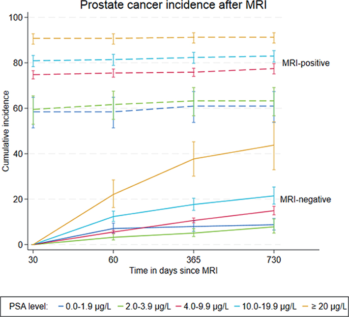
Figure 2. The cumulative incidence of prostate cancer, according to positive and negative magnetic resonance imaging and prostate-specific antigen level. The competing risk of death was accounted for using the Aalen–Johansen estimator. MRI: magnetic resonance imaging; PSA: prostate-specific antigen.
Most MRI-positive men received a prostate cancer diagnosis within 30 days; only few were diagnosed later during follow-up (Figure 2). The prostate cancer incidence rose with increasing PSA levels. The 1-year cumulative incidence of prostate cancer was 61% (95% CI: 54–67) at 0–1.9 µg/L, 63% (95% CI: 57–69) at 2–3.9 µg/L, 76% (95% CI: 74–78) at 4–9.9 µg/L, 82% (95% CI: 80–85) at 10–19.9 µg/L and 91% (95% CI: 89–93) at PSA levels ≥ 20 µg/L.
Among MRI-negative men, prostate cancer incidence was especially low among those with PSA levels < 4 µg/L but rose during follow-up according to the higher PSA levels (Figure 2).
The 1-year cumulative incidence of prostate cancer was 8% (95% CI: 6–10) at 0–1.9 µg/L, 5% (95% CI: 4–7) at 2–3.9 µg/L, 11% (95% CI: 10–12) at 4–9.9 µg/L, 18% (95% CI: 15–20) at 10–19.9 µg/L and 38% (95% CI: 30–45) at PSA levels ≥ 20 µg/L. Across all PSA levels, MRI-positive men had a consistently higher cumulative incidence of prostate cancer over the 2-year follow-up period compared to the MRI-negative men.
Within 1 year of the initial prostate MRI, the cumulative incidence of a repeated MRI, accounting for the competing risk of prostate cancer or death, was 9% (95% CI: 9–10) among the MRI-negative men and 21% (95% CI: 18–23) among the MRI-positive men. The cumulative incidence of a biopsy, accounting for the competing risk of prostate cancer or death, was 15% (95% CI: 14–16) among the MRI-negative men.
Among the men diagnosed with prostate cancer, the crude risk of intermediate-to-high-grade cancer (Gleason score ≥ 7) was 74% (95% CI: 71–77) in MRI-negative and 80% (95% CI: 79–82) in MRI-positive men. After adjusting for PSA level and calendar year, the corresponding risks were 76% (95% CI: 73–79) and 80% (95% CI: 79–81), respectively. The cumulative incidence of mortality within 1 year after the prostate MRI was 1% (95% CI: 1–1) for the MRI-negative and 1% (95% CI: 1–2) for the MRI-positive men.
Discussion
In this nationwide cohort study, 40% of biopsy-naïve men undergoing prostate MRI in Denmark (2018–2022) underwent biopsy within 30 days and were thus classified as MRI-positive. Prostate cancer incidence was lower in the MRI-negative men and rose with PSA levels in both groups.
Prostate cancer detection in MRI-positive men
When prostate MRI was followed by a biopsy within 30 days, the detection rate of prostate cancer was between 60% and 95%, depending on the PSA level. The further detection after the 30-day timepoint was very limited, despite the evidence of further clinical investigation with prostate MRI and biopsy in many of the men, where prostate cancer was not detected in the initial biopsy. This shows that the combination of prostate MRI and subsequent biopsy can be considered conclusive regarding the presence or absence of prostate cancer, and that the continued clinical surveillance after MRI and a negative biopsy may be redundant. This aligns with previous studies, which reported only a small number of recurrences, metastases, cases of incurable prostate cancer, or prostate cancer-specific mortality among men with MRI-negative findings [10, 11]. The investigation with prostate MRI and biopsy left many men with no evidence of prostate cancer in the biopsy. Two potential explanations may account for this observation. Firstly, the basic assumption that a prostate MRI with immediate biopsy was indicative of a positive MRI result, with a high suspicion of prostate cancer, may have been incorrect in some of these cases. We speculate that in the early years of prostate MRI use, the clinical decision in men with suspected prostate cancer may sometimes have been to perform prostate MRI followed by biopsy, regardless of the MRI result. Secondly, the observed pattern may indicate that a positive MRI result may arise in some men where cancer is not subsequently detectable by biopsy. With no recorded information on the radiological findings, we cannot estimate the relative importance of the two possible mechanisms. Other studies have indicated that false-positive MRI results may arise [22, 23].
Regarding the predictive value of the PSA measurement, it is not surprising that there is a high rate of detection of prostate cancer in men with high PSA (≥ 20 µg/L), prostate MRI and biopsy. Importantly, emerging evidence suggests that the likelihood of clinically significant disease varies markedly according to MRI suspicion level [24]. On the other hand, the detection of prostate cancer in 60% of the men with low PSA (0–1.9 µg/L or 2–3.9 µg/L) is indicative of false-negative PSA results. This occurs when the PSA level is low despite the presence of prostate cancer. This can happen because not all prostate cancers produce elevated PSA levels, leading to approximately 15% of cases being missed by the PSA test [25, 26].
Prostate cancer detection in MRI-negative men
After the gradual introduction of an MRI-first strategy in prostate cancer detection, it has become clinical practice to pursue rapidly with a tissue biopsy in men with MRI findings suggestive of prostate cancer. We therefore consider that when the prostate MRI was not followed by a biopsy within the following 30 days, this indicated a negative MRI result. The rate of detection of prostate cancer in these supposedly ‘MRI-negative’ men was highly dependent on the pre-MRI PSA measure and reached about 40% in men with the highest PSA levels, indicating the benefit of combining information from the prostate MRI and PSA test in the diagnostic pathway of prostate cancer [11].
Among the men diagnosed with prostate cancer, the risk of intermediate-to-high-grade disease was marginally lower in MRI-negative cases compared to MRI-positive ones. This observation may highlight the limitations of prostate MRI in detecting certain tumours that lack characteristic imaging features [7, 10, 13]. Alternatively, some MRI-negative patients may experience a delayed diagnosis of initial low-grade disease, allowing time for disease progression to a more advanced grade by the time of detection. These findings emphasize the need for a comprehensive diagnostic approach that integrates prostate MRI results with clinical assessments and histopathological data to minimize the risk of underestimating cancer severity [6, 10, 13]. Given that the cumulative 1-year mortality was very low and nearly identical between MRI-positive and MRI-negative men, and considering the limited follow-up period, these small differences are unlikely to represent cancer-specific mortality or to indicate any clinically meaningful divergence in outcomes.
Possible implications for early detection and organized screening
Focusing on men with very low PSA measurements, we are interested in the large contrast between cancer detection in men with negative or positive MRI results, varying from less than 10% at the 2-year timepoint in MRI-negative men to 60% in MRI-positive men. There is much debate about organized screening for prostate cancer [11, 27, 28], and we, as many others [27, 29, 30], remain unconvinced about the usefulness of such screening and concerned about the possible overdiagnosis and overtreatment that occur with the detection of slow-growing tumours, known from autopsy studies to be highly prevalent in the male population [29, 30]. However, as the ‘MRI-first’ strategy (i.e. MRI before biopsy) is introduced in prostate cancer diagnostics, prognostication and treatment choice, an ‘MRI-first’ strategy may be considered if organized screening were to be developed. The data suggest that the combination of MRI-negative imaging and low PSA largely rules out prostate cancer and makes tissue sampling unnecessary, whereas an MRI-positive finding may usefully be followed by a targeted biopsy, regardless of the PSA measure [27, 31].
Strengths and limitations
A key strength of this study is its nationwide cohort design within a universal, tax-funded healthcare system, which enables individual-level linkage across registries containing prospectively collected data [17–20]. The mandatory reporting and minimal financial or insurance-related barriers further enhance data completeness and reduce information and selection bias [17–20]. In 2018–2022, prostate MRI use in men with prostate cancer in Denmark rose to 52%, and to 80% among those treated or monitored [2]. This enabled the build-up of the present large cohort of men undergoing their first MRI, with no prior biopsy or prostate cancer diagnosis [2].
By incorporating the prostate imaging-reporting and data system (PI-RADS), prostate MRI supports biopsy-informed decision-making regarding a subsequent prostate biopsy [4, 6]. No data on the PI-RADS score from the prostate MRI were available in the present study. However, findings from a recent Swedish population-based study demonstrate that the proportion of reports including PI-RADS scores increased markedly over time, from 38% in 2015–2016 to 83% in 2022–2023 [24]. This improvement appears to have occurred in parallel with the increasing implementation and clinical use of prostate MRI, reflecting a broader maturation and standardization of MRI-based prostate cancer diagnostics. Consequently, upcoming data could provide improved opportunities to evaluate MRI-informed and PI-RADS-guided clinical decision-making in future studies. Hence, we cannot rule out that some biopsies were delayed beyond 30 days in some men with an actual positive MRI result. The standard timeframe in a Danish prostate cancer pathway is 36 calendar days from suspicion of prostate cancer until all examinations are completed and results are available, and the 30-day time window between prostate MRI and biopsy was based on specific advice from Danish consultant urologists. Still, this erroneous classification of the MRI result may have contributed to the emergence of prostate cancer in the period from 30 days to 60 days after the prostate MRI. However, this cannot explain the continued detection of prostate cancer beyond the 60-day timepoint, and most certainly not beyond the 360-day timepoint. This finding suggests the presence of false-negative MRI results, particularly among men with elevated PSA levels. This aligns with previous studies, which have reported approximately 15% false-negative results in MRI for prostate cancer diagnostics [5, 7, 13]. Still, our findings should be interpreted as reflecting the MRI‑informed biopsy decision process rather than the diagnostic performance of the prostate MRI results. Furthermore, biopsy decisions in clinical practice may be influenced by factors beyond MRI findings, which may lead to additional misclassification. Information on clinical T stage and detailed biopsy characteristics (including biopsy approach and number of positive cores) was not available in a valid form for the study period, which limited our ability to describe these aspects. We report Gleason scores for all detected cancers, providing a clinically meaningful measure of tumour grade and aggressiveness relevant to the focus on cancer detection after prostate MRI. Because several age-specific strata contained very small cell counts, cumulative incidence analyses stratified by PSA level and age could not be reported due to microdata confidentiality restrictions; this limits our ability to provide age-specific risk estimates across diagnostic pathways in the present study. The absence of sensitivity analyses limits our ability to assess the robustness of the findings under alternative assumptions and definitions.
Importantly, our findings should be interpreted as reflecting clinical decision-making processes following prostate MRI, rather than the intrinsic diagnostic performance of the imaging. Biopsy decisions are influenced by multiple clinical factors in addition to MRI results, and therefore, the observed patterns of prostate cancer detection cannot be directly translated into measures of prostate MRI accuracy.
Conclusion
Among biopsy-naïve men undergoing first-time prostate MRI, MRI-positive status, defined by MRI‑informed biopsy decision, was strongly associated with a high incidence of prostate cancer, including clinically significant cases, even in individuals with PSA levels below 4.0 µg/L. Conversely, MRI-negative men, especially with PSA levels under this threshold, exhibited a low risk of prostate cancer during 2 years of follow-up. These findings suggest that an MRI-negative status combined with a low PSA level indicates a lower likelihood of prostate cancer. Still, the relatively limited follow-up duration and absence of central MRI results restrict the ability to draw firm conclusions regarding the long-term necessity of continued surveillance. Overall, the results support the role of prostate MRI as a reliable triage tool in the detection and diagnostic pathway for prostate cancer. Clinically, this may justify deferring biopsy in MRI-negative men with low PSA, thereby reducing unnecessary procedures, anxiety and potential complications. Adopting MRI-based risk stratification could lead to more cost-effective use of healthcare resources, minimize overdiagnosis and overtreatment and align with emerging European guidelines [6] that advocate personalized biopsy decisions based on prostate MRI findings and PSA density.
Acknowledgement
Information on incident prostate cancer and Gleason score at diagnosis was collected from the Danish Prostate Cancer Database.
Data sharing
According to Danish data protection rules, we cannot share individual-level data. Researchers who fulfil the requirements set by the data providers can apply to obtain similar data.
Ethics and consent
The study was registered at the institutional review board at Aalborg University (record no. 2022-068-03232). According to Danish law, register-based studies do not require ethical approval.
References
[1] Bergengren O, Pekala KR, Matsoukas K, et al. 2022 update on prostate cancer epidemiology and risk factors – a systematic review. Eur Urol. 2023;84:191–206. https://doi.org/10.1016/j.eururo.2023.04.021
[2] Borre M, Elversang J, Hansen S, et al. The Danish Prostate Cancer Database annual report 2024. Aarhus: the Danish Healthcare Quality Institute; 2025.
[3] Jahnen M, Hausler T, Meissner VH, et al. Predicting clinically significant prostate cancer following suspicious mpMRI: analyses from a high-volume center. World J Urol. 2024;42(1):290. https://doi.org/10.1007/s00345-024-04991-6
[4] Nakai H, Takahashi H, LeGout JD, et al. Estimated diagnostic performance of prostate MRI performed with clinical suspicion of prostate cancer. Insights Imaging. 2024;15(1):271. https://doi.org/10.1186/s13244-024-01845-y
[5] Wimper Y, Fütterer JJ, Bomers JGR. MR imaging in real time guiding of therapies in prostate cancer. Life. 2022. Feb 17;12(2):302 https://doi.org/10.3390/life12020302
[6] Mottet N, van den Bergh RCN, Briers E, et al. EAU-EANM-ESTRO-ESUR-SIOG guidelines on prostate cancer – 2020 update. Part 1: Screening, diagnosis, and local treatment with curative intent. Eur Urol. 2021;79: 243–262. https://doi.org/10.1016/j.eururo.2020.09.042
[7] Wysock JS, Mendhiratta N, Zattoni F, et al. Predictive value of negative 3T multiparametric magnetic resonance imaging of the prostate on 12-core biopsy results. BJU Int. 2016;118(4):515–520. https://doi.org/10.1111/bju.13427
[8] Goldberg H, Ahmad AE, Chandrasekar T, et al. Comparison of magnetic resonance imaging and transrectal ultrasound informed prostate biopsy for prostate cancer diagnosis in biopsy naïve men: a systematic review and meta-analysis. J Urol. 2020 Jun;203(6):1085–1092. https://doi.org/10.1097/JU.0000000000000595
[9] Guo E, Xu L, Zhang D, et al. Diagnostic performance of MRI in detecting prostate cancer in patients with prostate-specific antigen levels of 4–10 ng/mL: a systematic review and meta-analysis. Insights Imaging. 2024;15(1): 147. https://doi.org/10.1186/s13244-024-01699-4
[10] Wibmer AG, Lefkowitz RA, Lakhman Y, et al. MRI-detectability of clinically significant prostate cancer relates to oncologic outcomes after prostatectomy. Clin Genitourin Cancer. 2022;20(4):319–325. https://doi.org/10.1016/j.clgc.2022.04.001
[11] Hugosson J, Godtman RA, Wallstrom J, et al. Results after four years of screening for prostate cancer with PSA and MRI. N Engl J Med. 2024;391(12):1083–1095. https://doi.org/10.1056/NEJMoa2406050
[12] Padhani AR, Godtman RA, Schoots IG. Key learning on the promise and limitations of MRI in prostate cancer screening. Eur Radiol. 2024 Sep;34(9): 6168–6174. https://doi.org/10.1007/s00330-024-10626-6
[13] Diez NG, de Pablos-Rodríguez P, Sánchez-Mateos Manzaneque D, et al. Can we rely on magnetic resonance imaging for prostate cancer detection and surgical planning? Comprehensive analysis of a large cohort of patients undergoing transperineal mapped biopsies. World J Urol. 2024;42(1):548. https://doi.org/10.1007/s00345-024-05233-5
[14] Nguyen-Nielsen M, Høyer S, Friis S, et al. The Danish prostate cancer database. Clin Epidemiol. 2016 Oct;25:8:649–653. https://doi.org/10.2147/CLEP.S100256
[15] Statistics Denmark. StatBank Denmark [Population] [Internet]. 2023 [cited 2025 Sep 10]. Available from: https://www.statbank.dk/statbank5a/default.asp?w=2048
[16] Frank L. Epidemiology. When an entire country is a cohort. Science. 2000;287(5462):2398–2399. https://doi.org/10.1126/science.287.5462.2398
[17] Schmidt M, Pedersen L, Sorensen HT. The Danish Civil Registration System as a tool in epidemiology. Eur J Epidemiol. 2014;29(8): 541–549. https://doi.org/10.1007/s10654-014-9930-3
[18] Schmidt M, Schmidt SA, Sandegaard JL, et al. The Danish National Patient Registry: a review of content, data quality, and research potential. Clin Epidemiol. 2015;7:449–490. https://doi.org/10.2147/CLEP.S91125
[19] Dansk Prostata Cancer Database (DaProCa) – RKKP [Internet]. [cited 2023 May 16]. Available from: https://www.rkkp.dk/kvalitetsdatabaser/databaser/dansk-prostata-cancer-database/
[20] Grann AF, Erichsen R, Nielsen AG, et al. Existing data sources for clinical epidemiology: The clinical laboratory information system (LABKA) research database at Aarhus University, Denmark. Clin Epidemiol. 2011;3:133–138. https://doi.org/10.2147/CLEP.S17901
[21] Charlson ME, Pompei P, Ales KL, et al. A new method of classifying prognostic comorbidity in longitudinal studies: development and validation. J Chronic Dis. 1987;40(5):373–383. https://doi.org/10.1016/0021-9681(87)90171-8
[22] Arafa MA, Rabah DM, Khan F, et al. False-positive magnetic resonance imaging prostate cancer correlates and clinical implications. Urol Ann. 2023;15(1):54–59. https://doi.org/10.4103/ua.ua_22_22
[23] Cheng Y, Fan B, Fu Y, et al. Prediction of false-positive PI-RADS 5 lesions on prostate multiparametric MRI: development and internal validation of a clinical-radiological characteristics based nomogram. BMC Urol. 2024;24(1):76. https://doi.org/10.1186/s12894-024-01465-0
[24] Ventimiglia E, Gedeborg R, Westerberg M, et al. Nationwide population-based longitudinal data on magnetic resonance imaging of the prostate and subsequent prostate biopsy results. Scand J Urol. 2026;61:64–71. https://doi.org/10.2340/sju.v61.45540
[25] Carter HB. Prostate cancers in men with low PSA levels – must we find them? N Engl J Med. 2004;350(22):2292–2294. https://doi.org/10.1056/nejme048003
[26] Bociański J, Sklinda K, Walecki J, et al. A case of high-grade prostate cancer with low PSA levels. Urol Case Rep. 2025 Aug 16;62:103161. https://doi.org/10.1016/j.eucr.2025.103161
[27] Obiora D, Orikogbo O, Davies BJ, et al. Controversies in prostate cancer screening. Urol Oncol. 2025 Jan;43(1):49–53.
[28] Oh CU, Kang H. The effectiveness and harms of PSA-based prostate cancer screening: a systematic review. Healthcare. 2025. Jun 9;13(12):1381
[29] Mahase E. Is the UK really ready to roll out prostate cancer screening? BMJ. 2023 May 17;381:p1062. https://doi.org/10.1136/bmj.p1062
[30] Vickers A, O’Brien F, Montorsi F, et al. Current policies on early detection of prostate cancer create overdiagnosis and inequity with minimal benefit. BMJ. 2023 May 17;381:e071082. https://doi.org/10.1136/bmj-2022-071082
[31] Fazekas T, Shim SR, Basile G, et al. Magnetic resonance imaging in prostate cancer screening. JAMA Oncol. 2024;10(6):745. https://doi.org/10.1001/jamaoncol.2024.0734