ORIGINAL RESEARCH ARTICLE
Paediatric stone treatment in the Nordic countries – a multicentre study from the Scandinavian Collaboration Group for Urinary Stones
Øyvind Ulvika,b, Line Vigrestadc, Dan Helgød, Stein Øverbye, Kristian Stritesky Larssenf, Pertti Nurmineng, Jóhann Páll Ingimarssonh, John Åkerlundi,j, Rita Ferritok and Tomas Thiell
aHelse Bergen HF, Department of Urology, Haukeland University Hospital, Bergen, Norway; bDepartment of Clinical Medicine (K1), University of Bergen, Bergen, Norway; cDepartment of Urology, St Olavs University Hospital HF, Trondheim, Norway; dDepartment of Urology, Akershus University Hospital HF, Lørenskog, Norway; eDepartment of Urology, Vestfold Hospital Trust HF, Tønsberg, Norway; fDepartment of Urology, Oslo University Hospital Aker HF, Oslo, Norway; gDepartment of Urology, Turku University Hospital, Turku, Finland; hDepartment of Urology, Landspítali – The National University Hospital of Iceland, Reykjavik, Iceland; iInstitute of Clinical Sciences, Sahlgrenska Academy, University of Gothenburg, Gothenburg, Sweden; jDepartment of Urology, Region Västra Götaland, Sahlgrenska University Hospital, Gothenburg, Sweden; kDepartment of Clinical Science, Intervention and Technology, Karolinska Institutet, Stockholm, Sweden; lDepartment of Urology, Karolinska University Hospital, Stockholm, Sweden
ABSTRACT
Objective: To explore differences and similarities in paediatric stone treatment between hospitals in the Nordic countries.
Material and methods: A retrospective review of the medical records was performed for children receiving stone treatment in nine Nordic hospitals between January 2014 and December 2023. Variables of interest included diagnostic imaging, treatment modalities, complication rates and stone free rates (SFR).
Results: Five hundred and sixty two treatments among 319 children were included; 117 girls and 202 boys with a median age of 10 years (interquartile range 5 – 14). Preoperative diagnostic imaging with computed tomography was performed in 442 cases (79%), and 120 patients (21%) were diagnosed with ultrasound, MRI or X-ray. In 150 cases (27%), stones treated were located in the ureter only and in 412 cases (73%) in the renal pelvis ± ureter. Ureteroscopic stone treatment (URS) was performed in 248 (44%), shock wave lithotripsy (SWL) in 242 (43%) and percutaneous lithotripsy (PCNL) in 72 cases (13%). The distribution of treatment modalities varied considerably between hospitals. URS treatments increased, and SWL procedures declined throughout the study period. A follow-up consultation was carried out in 505 cases (90%). Overall SFR after each session across treatment modalities and location was 49%. The SFRs after URS was 68%, SWL 30% and PCNL 61% and significantly higher after URS compared to SWL, p < 0.001. Postoperative complications leading to readmission occurred in 10% of patients.
Conclusion: The study reveals differences in preferred treatment modalities and results between the hospitals. Standardising diagnostics, treatment and follow-up could improve outcomes for children with kidney stone disease.
KEYWORDS: Paediatric stone treatment; urolithiasis; intervention; ureteroscopy; percutaneous nephrolithotomy; extracorporeal shock wave lithotripsy
Citation: Scandinavian Journal of Urology 2026, VOL. 61, 170–177. https://doi.org/10.2340/sju.v61.46270.
Copyright: © 2026 The Author(s). Published by MJS Publishing on behalf of Acta Chirurgica Scandinavica. This is an Open Access article distributed under the terms of the Creative Commons Attribution 4.0 International License (http://creativecommons.org/licenses/by/4.0/), allowing third parties to copy and redistribute the material in any medium or format and to remix, transform, and build upon the material, with the condition of proper attribution to the original work.
Received: 15 February 2026; Accepted: 27 May 2026; Published: 12 June 2026
CONTACT: Øyvind Ulvik doc.ulvik@online.no Helse Bergen HF, Department of Urology, Haukeland University Hospital, Jonas Lies vei 65, 5021 Bergen, Norway
Competing interests and funding: The authors report no conflicts of interest.
None.
Introduction
Kidney stone disease (KSD) is a common urological complaint worldwide, and the prevalence is rising [1]. Despite the increasing incidence in the paediatric population, especially among adolescent females, urolithiasis in children is rare [2]. Paediatric stone treatment differs from adult treatment in several aspects. In addition to diagnostics, where there is a focus on reducing radiation exposure, the challenges are also related to the treatment. Endourological equipment is rarely customised for children, and anaesthesia may require special competence.
European Association of Urology (EAU) recommends shockwave lithotripsy (SWL) as the first-line treatment option for ureteral stones in children [3]. However, the guidelines recognise the increasing role of ureteroscopy (URS) in this special population, particularly in renal stones when unfavourable factors for SWL are present [3]. Indications for percutaneous nephrolithotomy (PCNL) include stones larger ≥ 2 cm. This is identical to the guidelines for adults although the kidneys are smaller in children [3]. However, smaller stones resistant to SWL and URS may also be treated with PCNL, and stone-free rates (SFRs) are comparable to PCNL in adults [3, 4].
Most reports on stone treatment in children originate from a relatively small pool of nations with large population sizes [5–8]. Outcomes from smaller nations remain under-reported, and to date only one publication on URS stone treatment in children in a Nordic setting exists [9].
Investigating how paediatric KSD is treated in the Scandinavian countries and exploring the treatment results may narrow the knowledge gap in this special population.
Aims of the study
The primary aim of the study is to explore how paediatric stone disease is treated in the Nordic countries. Furthermore, to point out similarities and differences in practice patterns within these countries and also, to explore any similarities and differences regarding preoperative diagnostics, performance of the stone treatment procedures and follow-up.
Secondary aims are to investigate SFRs following different treatment modalities, intraoperative and postoperative complication rates and to compare results across treatment modalities.
Materials and methods
Study population and setting
The study originates from the Scandinavian Collaboration Group for Urinary Stones (SCGUS). A retrospective review of the medical records for children <18 years treated for KSD in nine Nordic stone centres in the period January 1st, 2014 – December 31st, 2023 was performed. Patients eligible for participation were recruited from Finland, Iceland, Norway and Sweden. Children treated for ureteral and/or renal stones with URS, SWL, PCNL or endoscopic combined intrarenal surgery (ECIRS) were included. Draining procedures without stone treatment were not registered, and patients treated conservatively were excluded.
The eight stone centres in Iceland, Norway and Sweden represent an approximate coverage of the paediatric population of 100, 90 and 50% in the three countries, respectively. The limited number of patients recruited from Finland is clearly not representable for the whole country.
Variables of interest included preoperative characteristics such as comorbidity, preoperative imaging modality and stone status; perioperative characteristics including treatment modality and relevant details from the surgical procedures, and postoperative status with length of stay, follow-up, SFRs and complications. No uniform definition of stone-free status was decided, and SFRs depended on the radiology report, which usually reported the zero-fragment status. The American Society of Anaesthesiologists (ASA) score was used to assess the patients’ overall physical status.
Ureteroscopy, shock wave lithotripsy and percutaneous approach
URS was performed under general anaesthesia using semirigid and/or flexible endoscopes. The size of the endoscope used varied from 4.5F to 9.8F. The decision to use an access sheath (UAS) or a safety guide wire (SGW) was left to the surgeon. Holmium:Yttrium-Aluminium-Garnet lasers and Thulium Fiber lasers were used for stone fragmentation and dusting. Fragments were retrieved with forceps, baskets or left in situ for spontaneous passage. Whether a fragmentation and retrieval- or a dusting strategy was chosen depended on the surgeon in each case, as well as post-endoscopic drainage with a JJ-stent.
SWL was performed in sedation or under general anaesthesia. X-ray or ultrasound (US) was used for stone identification and focusing shock waves. Shock wave frequency was decided by the operator in each case, typically 60/min.
PCNL and ECIRS were performed under general anaesthesia using nephroscopes with tract sizes 12F–30F depending on the surgeon’s preference. Percutaneous access was made by the surgeon or by a radiologist. The stones were fragmented using laser, ShockPulse (Olympus America, USA) or Swiss Lithoclast Master (EMS, Switzerland). Post-endoscopic drainage with a nephrostomy tube and/or JJ-stent was left to the surgeon’s discretion.
Statistics
The results are reported as absolute numbers and as medians with corresponding interquartile ranges (IQR). Continuous data were skewed and therefore compared using Mann-Whitney U test. Associations between categorical variables were assessed using exact chi-squared tests or Fisher´s exact tests. Statistical analyses were conducted using IBM SPSS Statistics 31.0.2.0 (IBM, Armonk, NY). A p-value of < 0.05 was considered statistically significant.
Ethics and approvals
Ethical approvals were collected from the health authorities in Finland (T431/2025), Iceland (VSN-24-098), Norway (REC-729556) and Sweden (2025-04759-01). In accordance with the ethical approvals, eligible patients and their parents received written information about the study prior to inclusion. Moreover, participation was voluntary, and all patients and parents had the opportunity to opt out of inclusion.
Results
Between January 1st, 2014 and December 31st, 2023, a total of 319 children underwent 562 treatments for KSD at the nine participating hospitals. There were 117 girls and 202 boys with a median age of 10 years (IQR 5–14). In total, 300 children (54%) had ASA-score 1. Details regarding patient characteristics are provided in Table 1. Stone size was similar for renal stones in the URS (9 mm) and SWL groups (9 mm), p = 0.8. However, more stones were located in the lower calyx in children treated with URS (63%) compared to those treated with SWL (50%), p = 0.018. Similar sizes were also registered for ureteral stones treated with URS (6 mm) and SWL (8 mm), p = 0.1. Also, the location in the ureter differed among modalities with more stones located in the lower part in the URS group (74%) compared to the SWL group (51%), p = 0.005.
| Characteristic | Total | URS | SWL | PCNL |
| Treatment side, n (%)a | ||||
| Right | 302 (54%) | 134 (54%) | 135 (57%) | 33 (46%) |
| Left | 255 (46%) | 113 (46%) | 103 (43%) | 39 (54%) |
| Earlier treatment of the same stone, n (%) | ||||
| Yes | 226 (40%) | 95 (38%) | 105 (43%) | 26 (36%) |
| No | 336 (60%) | 153 (62%) | 137 (57%) | 46 (64%) |
| Anatomy upper urinary tract, n (%) | ||||
| Normal | 494 (88%) | 212 (84%) | 225 (93%) | 57 (79%) |
| Single kidney | 6 (1%) | 2 (1%) | 4 (2%) | - |
| Stenosis/stricture | 40 (7%) | 29 (12%) | 4 (2%) | 7 (10%) |
| Pelvic kidney | 3 (1%) | - | 1 (1%) | 2 (3%) |
| Horseshoe kidney | 3 (1%) | 2 (1%) | - | 1 (1%) |
| Duplex ureters/pelvises | 4 (1%) | 2 (1%) | 2 (1%) | - |
| Atrophic kidney | 7(1%) | 1 (1%) | 1 (1%) | 5 (7%) |
| Renal stone characteristicsb | ||||
| Largest diam in mm, median (IQR) | 10 (7–15) | 9 (6–13) | 9 (6–12) | 18 (14–25) |
| Volume in mm3, median (IQR)c | 300 (120–1320) | 225 (53–531) | 216 (110–1038) | 1850 (1001–3740) |
| Number of stones, n (%) | ||||
| 1 stone | 227 (68%) | 76 (62%) | 122 (72%) | 29 (69%) |
| 2 stones | 65 (19%) | 30 (25%) | 26 (15%) | 9 (21%) |
| ≥3 stones | 42 (13%) | 16 (13%) | 22 (13%) | 3 (10%) |
| Location, n (%)d | ||||
| Lower calyx | 241 (58%) | 99 (63%) | 98 (50%) | 44 (68%) |
| Renal pelvis, upper and mid calyx | 177 (42%) | 58 (37%) | 98 (50%) | 21 (32%) |
| Stone density (HU), median (IQR) | 881 (642–1200) | 847 (570–1100) | 873 (660–1200) | 1000 (730–1275) |
| Ureteral stone characteristicsb | ||||
| Largest diam in mm, median (IQR) | 7 (5–10) | 6 (4–9) | 8 (5–10) | 10 (9–13) |
| Volume in mm3, median (IQR)c | 144 (75–441) | 144 (59–416) | 126 (84–450) | 273 (230–397) |
| Number of stones, n (%) | ||||
| 1 stone | 135 (80%) | 85 (78%) | 44 (88%) | 6 (55%) |
| 2 stones | 26 (15%) | 18 (17%) | 5 (10%) | 3 (27%) |
| ≥3 stones | 9 (5%) | 6 (5%) | 1 (2%) | 2 (18%) |
| Location, n (%)e | ||||
| Upper | 61 (34%) | 30 (26%) | 26 (49%) | 5 (46%) |
| Lower | 118 (66%) | 85 (74%) | 27 (51%) | 6 (54%) |
| Stone density (HU), median (IQR) | 731 (527–1040) | 700 (464–1000) | 800 (645–1070) | 640 (600–905) |
| Preoperative bacteriuria, n (%)f | 118 (22%) | 57 (24%) | 36 (16%) | 25 (35%) |
| Preoperative obstruction, n (%) | 212 (38%) | 99 (40%) | 82 (34%) | 31 (43%) |
| Pre-stented before surgery, n (%) | 123 (22%) | 51 (21%) | 60 (25%) | 12 (17%) |
| aInformation on the treatment side was missing for five patients. bComplete information on stone characteristics was missing for several patients. cStone volume (mm3) was measured using the ellipsoid formula: volume = 4/3πr3. dSome patients had stones in multiple locations. If one or more stones were located in the lower calyx, this was registered as a lower calyx stone even though the rest of the stones may be located in other locations (renal pelvis, upper or middle calyx). eIf multiple stones were located at different levels of the ureter, the most proximal location was registered. fBacteriuria was defined as ≥3+ leukocytes or positive nitrite on urine dipstick, positive urine culture or external catheter. If bacteriuria was present, the patient was treated with appropriate antibiotics prior to stone treatment. |
||||
Preoperative imaging with a computed tomography (CT) scan was performed in 442 cases (79%), while the remaining 120 (21%) were diagnosed with US, magnetic resonance imaging (MRI) or X-ray.
In total, 248 patients (44%) were treated with URS, 242 (43%) with SWL and 72 (13%) with PCNL. Of the latter, 25 procedures (35%) were performed as ECIRS with simultaneous retrograde and antegrade access. Figure 1 illustrates the number of different treatments across the recruiting hospitals, while Figure 2 presents trends in the number of procedures per year across treatment modalities. Details of the surgical procedures are listed in Table 2.
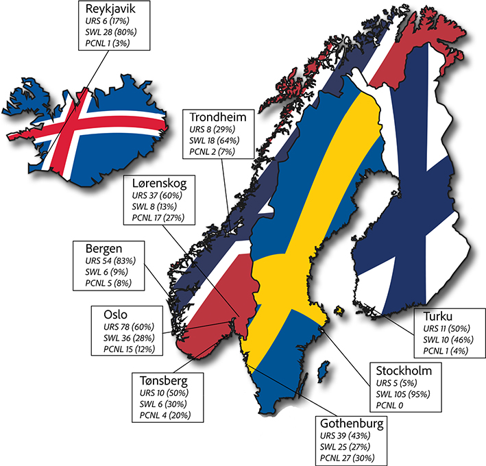
Figure 1. Distribution of treatments across recruiting hospitals.
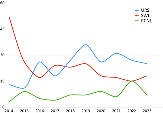
Figure 2. Numbers of procedures per year.
| Characteristic | URS | SWL | PCNL |
| Number of procedures, n (%) | 248 (44%) | 242 (43%) | 72 (13%) |
| Surgical experience, n (%) | |||
| Resident | 8 (3%) | NA | - |
| Urologist | 26 (11%) | NA | 2 (3%) |
| Endourologist | 214 (86%) | NA | 70 (97%) |
| Anaesthesia, n (%) | |||
| General anaesthesia | 248 (100%) | 169 (70%) | 72 (100%) |
| Sedation | - | 73 (30%) | - |
| Antibiotic prophylaxis, n (%) | |||
| Yes | 231 (93%) | 78 (32%) | 71 (99%) |
| No | 17 (7%) | 164 (68%) | 1 (1%) |
| Location of the treated stone, n (%) | |||
| Renal pelvis (± ureter) | 153 (62%) | 194 (80%) | 65 (90%) |
| Ureter only | 95 (38%) | 48 (20%) | 7 (10%) |
| Postoperative drainage, n (%) | |||
| Yes | 197 (79%) | 69 (29%) | 71 (99%) |
| No | 51 (21%)a | 173 (71%) | 1 (1%) |
| Drainage specified, n (%) | |||
| JJ-stent | 192 (97%) | 60 (87%) | 17 (24%) |
| Nephrostomy tube | 2 (1%) | 9 (13%) | 41 (58%) |
| JJ-stent and nephrostomy tube | 3 (2%) | - | 13 (18%) |
| Operative time, median minutes (IQR) | 64 (43–86) | NA | 127 (98–163) |
| Success determined by surgeon, n (%)b | |||
| Success | 193 (79%) | 116 (49%) | 56 (78%) |
| Partly successful | - | 103 (44%) | - |
| Unsuccessful | 52 (21%) | 17 (7%) | 16 (22%) |
| Stone free assessed at follow-up, n (%)c | |||
| Yes | 127 (68%) | 69 (30%) | 38 (61%) |
| No | 61 (32%) | 160 (70%) | 24 (39%) |
| Need for auxiliary treatment, n (%) | |||
| Yes | 33 (54%) | 106 (66%) | 8 (33%) |
| No | 28 (46%) | 54 (34%) | 16 (67%) |
| aPost-endoscopic stent placement was unsuccessful in one child undergoing URS. bTreatment success was not assessed by all surgeons. cImaging at follow-up assessing stone-free status was registered in 479 cases (85%). |
|||
A semirigid-, flexible- and a combination of the two types of endoscopes were used in 66 (27%), 87 (35%) and 95 (38%) URS procedures, respectively. A 7.95F ureteroscope was most frequently used (90 cases, 39%) throughout the study period. An SGW was used in 144 cases (58%) and an UAS in 81 (33%). An 11/13F UAS was most frequently used. Passage through the orifice and intramural ureter was uncomplicated in 219 cases (88%). In 23 cases (9%), dilatation was necessary, and passage was unsuccessful in 6 (3%). Fragmentation and retrieval were performed in 181 children (73%), while a dusting strategy leaving residuals for spontaneous passage was done in 40 (16%). In 27 cases (11%), no strategy was specified.
X-ray for stone identification and focus of shock waves was used in 222 (92%) SWL procedures, US in 14 (6%) and a combination of both X-ray and US in six cases (2%). A shock frequency of 60/min was most common (126 cases, 56%).
Percutaneous access in PCNL was made by a radiologist in 51 (71%) or a urologist in 21 (29%) procedures. A single tract approach was used in 64 cases (89%), while two tracts were necessary in 8 cases (11%). A 24F nephroscope was most commonly used (22 children, 31%) throughout the study period.
A total of 11 procedures (2%) were interrupted because of intraoperative complications: 6 URS (2%), 2 SWL (1%) and 3 PCNL (4%). Table 3 lists intraoperative complications across the treatment modalities.
| Characteristic | URS | SWL | PCNL |
| Complications leading to interruption, n (%)a | 6 (2%) | 2 (1%) | 3 (4%) |
| Bleeding impairing vision, n (%) | 4 (2%) | - | 2 (3%) |
| Perforation, n (%)b | 2 (1%) | - | 1 (1%) |
| Mucosal abrasion, n (%) | 1 (0.5%) | - | - |
| Other, n (%)c | - | 2 (1%) | - |
| Minor events not interrupting surgery, n (%) | 13 (5%) | 3 (1%) | 5 (7%) |
| Bleeding impairing vision, n (%) | 7 (3%) | - | 2 (3%) |
| Perforation, n (%) | 3 (1%) | - | 3 (4%) |
| Mucosal abrasion, n (%) | 6 (2%) | - | - |
| Other, n (%)d | - | 3 (1%) | - |
| aSome patients had more than one adverse event. There were no ureteral avulsions. bPerforation was assessed endoscopically or as contrast leakage on a retrograde pyelogram. cAdverse events related to the SWL procedure, including pain and pneumothorax. dMinor adverse events related to the SWL procedure, including pain and inability to identify the stone. |
|||
Early postoperative complications occurring before hospital discharge occurred in 27 children after PCNL (38%), 29 after URS (12%) and 35 after SWL (14%). In total, 83 (91%) of the early complications were classified as Clavien-Dindo grade 1 or 2. Median hospital stay was 1 day (IQR 1–2).
In total, 505 children (90%) had a follow-up consultation after stone treatment. The timing for follow-up varied considerably with a median time to follow-up of 5 weeks (IQR: 3 – 12). In addition, the imaging modality used to assess treatment success differed across hospitals. While US and X-ray were the preferred imaging modalities at follow-up in Norway, CT was most common in Finland, Iceland and Sweden. Overall, CT was the most frequent imaging modality used at follow-up (46%). The remaining patients were assessed by US (24%), X-ray (21%), US and X-ray (4%) or MRI (0.2%). In 26 patients (5%), there was no information about the imaging modality at follow-up. The proportion of CT at follow-up did not differ between treatment modalities: URS 43%, SWL 52% and PCNL 56%, p = 0.071.
Overall SFR across treatment modalities was 49%. The SFRs for the different treatment modalities, irrespective of stone location, were 68, 30 and 61% for URS, SWL and PCNL, respectively. Pre-stenting status did not influence the SFR in any of the three treatment modalities. Despite more stones being located in the lower calyx in the URS group, treatment of renal stones was associated with significantly higher SFR after URS compared with SWL, 59% vs 28%, p < 0.001. Similarly, the SFR for ureteral stones was significantly higher after URS (79%) compared to SWL (39%), p < 0.001, although slightly more stones were located in the lower part in those treated with URS.
Data regarding postoperative complications leading to readmission are presented in Table 4. In total, 54 children (10%) needed readmission in the first 3 months after stone treatment: 27 children (11%) in the URS group, 22 (9%) in the SWL group and 5 (7%) in the PCNL group.
| Characteristic | URS | SWL | PCNL |
| Complications leading to readmission, n (%)a | 27 (11%) | 22 (9%) | 5 (7%) |
| Infection, n (%) | 16 (6%) | 12 (5%) | 2 (3%) |
| Pain/obstruction, n (%) | 13 (5%) | 7 (3%) | 1 (1%) |
| Stent bother, n (%) | 5 (2%) | 3 (1%) | - |
| Micturition difficulties, n (%) | 2 (1%) | 2 (1%) | - |
| Other causes, n (%) | 2 (1%) | 6 (2%) | 3 (4%) |
| aSome patients had more than one reason for readmission. | |||
Discussion
In the present SCGUS study, paediatric stone treatment performed in nine Nordic centres during a 10-year period has been reviewed. The study represents the largest series of paediatric stone treatments in a Nordic setting, with a total of 562 procedures registered in 319 children. The results reveal significant differences in the preferred treatment modality, diagnostic investigation and follow-up routines across the hospitals. In addition, there were significant differences in success rates between the treatment modalities.
EAU Guidelines emphasise the importance of reducing radiation exposure in children, and US is considered the primary imaging technique in this special population [3]. However, low-dose CT protocols can reduce radiation exposure and improve diagnostic accuracy in children and is considered an alternative if US cannot provide the required information [3, 10]. In addition, ultra-low-dose CT with an effective radiation dose of 0.5mSv, equivalent to plain abdominal X-ray, has shown sensitivity and specificity of 97 and 95%, respectively [11]. In comparison, US in the paediatric population may have a sensitivity for detecting stones as low as 67% and specificity of 97% [12]. Despite superior sensitivity and specificity, CT may require sedation or general anaesthesia in the youngest children, and US is therefore still considered the preferred modality among these [3]. In the present study, non-contrast CT was the most frequent modality used for pretreatment diagnostic imaging (79%) and follow-up (46%). The frequent use of CT in the Nordic countries is probably a result of both institutional practices and availability.
The study revealed significant diversity in the preferred treatment modality among the participating hospitals. EAU Guidelines highlight SWL as the treatment of choice for both ureteral and renal stones in children although URS is presented as a feasible alternative [3]. As illustrated in Figure 1, SWL was the preferred treatment modality in the hospitals in Stockholm (95%), Reykjavik (80%) and Trondheim (64%), while URS was the predominant modality for paediatric stone treatment in Bergen (83%), Oslo (60%) and Lørenskog (60%). The preference for a specific treatment modality may be explained by special competence rather than strict adherence to current guidelines. Furthermore, the preference for a given treatment modality in children may coincide with the preferred stone treatment in adults at each hospital. Similar to reports in adults, we found increasing numbers of URS procedures in recent years and a decrease in SWL treatments in the current cohort [13, 14].
The present study illustrates important differences in practice patterns between the treatment modalities. While almost all children received antibiotic prophylaxis before endourological treatment, the corresponding number in the SWL group was 32%. This is in accordance with the EAU Guidelines [3]. Furthermore, all endourological procedures were performed under general anaesthesia while 30% of the SWL procedures were done in sedation only. Sedation and light analgesics are well documented for SWL in the adult population but is rarely described in paediatric treatment [15, 16]. However, although sedation may be feasible for the oldest children, the majority still require general anaesthesia for SWL, as demonstrated in the present study.
URS was the most frequent treatment modality among the Nordic children. However, lack of customised paediatric endoscopic equipment is an obvious barrier to this treatment. Although a paediatric semirigid ureteroscope 4.5F exists, its usability is limited due to the small working channel. In fact, the Croes study examining paediatric stone treatment showed that ureteroscopes sized 7–9F were most frequently used [17]. Furthermore, Kahraman and colleagues have demonstrated that a flexible ureteroscope 7.95F with a 4.9F tapered tip may improve results in paediatric stone treatment [18]. In the present study, a 7.95F ureteroscope identical to the scope in Kahraman’s series was most frequently used throughout the 10-year period. Uncomplicated passage through the orifice and intramural ureter was registered in 219 cases (88%). This confirms the feasibility of using a regular-sized ureteroscope in this population. No systematic changes in scope size were observed during the study period. Modern and miniaturised ureteroscopes may improve outcomes in the paediatric population in the future.
Pre-stenting has been shown to facilitate endoscopic access, improve SFR and reduce the risk of complications [19, 20]. In this study, 21% of children had an indwelling JJ-stent at the time of URS. Interestingly, pre-stenting status did not influence SFRs. Moreover, in a recent review and meta-analysis, Irshid et al. recommend primary URS without pre-stenting despite a potentially lower SFR [21]. In contrast to the modest pre-stenting rate, 79% of the children received a JJ-stent as an exit strategy after URS. The number seems high as post-endoscopic stenting is considered redundant following uncomplicated URS, also in children [3].
As expected, the largest stones were treated with PCNL. Specific paediatric equipment was not available, and PCNL with 24F tract size was used in most cases. Furthermore, as in the adult population, percutaneous access was most frequently made by a radiologist [22]. However, in 29% of procedures, the urologists made the access themselves.
Early postoperative complications were registered in 16% of children, and 91% of these were classified as Clavien-Dindo grade 1 or 2. Postoperative complications were most frequent after PCNL. The complication rates in the present study are comparable to those reported in other publications after paediatric stone treatment [17, 23, 24].
EAU Guidelines provide no specific recommendations on follow-up in children [3]. Follow-up after stone treatment was registered in 505 cases (90%), and CT was the most frequent imaging modality, followed by US. The SFR was significantly higher after URS (68%) compared to SWL (30%), p < 0.001. Interestingly, the urologists’ predictions of stone free status at the end of the procedure were overly optimistic compared to the image-based assessment at follow-up. The optimistic misperception of successful treatment among urologists has been documented earlier [25]. The SFRs reported in the present study are based on the radiology report at follow-up. No specific definition of stone free status was used in this study although zero fragment status was most commonly reported. Furthermore, several different imaging modalities were used for stone free assessment at follow-up. Thus, the SFRs may be even lower than reported. Owing to its noninvasive nature, SFR after SWL is often reported after multiple treatments [26, 27]. It should be noted that SFR in the present study is reported after a single session only, which may explain the discrepancy in SFR compared to the existing literature [27]. Arguably, the non-invasive nature of SWL is challenged by the frequent requirement of repeated procedures performed under general anaesthesia in the paediatric population. In contrast to the adult population, a strategy of repeated SWL procedures to achieve stone-free status may therefore not be considered as appropriate and uncomplicated in children. Furthermore, including staged procedures when determining stone-free status makes comparisons between treatment modalities difficult. As a consequence, other publications also report SFRs after a single procedure comparing SWL and URS in paediatric populations [7].
This study has several limitations, including a heterogenous cohort. The retrospective design may have contributed to rough registration of data, such as intraoperative adverse events and surgeons’ predictions of stone-free status. However, in most cases, a complete medical record enabled us to assess both complications and the urologists’ predictions of stone-free status. The lack of a uniform definition of stone-free status and various follow-up regimes regarding timing and imaging modalities are obvious limitations that affect the interpretation of the results. Thus, caution is warranted while interpreting the comparison of SFRs between treatment modalities. Similar limitations are reported in other large paediatric studies [17]. On the other hand, the limitations may be balanced by the strength of the multicentre design. The study also reveals current practice in paediatric stone treatment in nine Nordic centres. The reported practice may differ from other hospitals.
Conclusion
In this study, current practice in paediatric stone treatment in the Nordic countries is presented. There was an increasing trend for URS throughout the study period and a decline in SWL procedures. Although the overall treatment success is acceptable and complication rates are low, the study reveals differences in preferred treatment modality and follow-up regimes between the hospitals. Standardising diagnostics, treatment and follow-up could potentially improve the outcome for patients in this special population.
Authors’ contribution
Study concept and design: ØU
Data collection: ØU, LV, DH, SØ, KSL, PN, JPI, JÅ and TT
Analysis and interpretation: ØU
Drafting the manuscript: ØU
Critical revision of the manuscript: LV, DH, SØ, KSL, PN, JPI, JÅ and TT.
Supervision: ØU
References
[1] Lang J, Narendrula A, El-Zawahry A, et al. Global trends in incidence and burden of urolithiasis from 1990 to 2019: an analysis of global burden of disease study data. Eur Urol Open Sci. 2022;35:37–46. https://doi.org/10.1016/j.euros.2021.10.008
[2] Dwyer ME, Krambeck AE, Bergstralh EJ, et al. Temporal trends in incidence of kidney stones among children: a 25-year population based study. J Urol. 2012;188(1):247–252. https://doi.org/10.1016/j.juro.2012.03.021
[3] Skolarikos A, Jung H, Neisius A, et al. EAU guidelines on urolithiasis [Internet]. Edn. presented at the EAU Annual Congress Madrid 2025: EAU Guidelines Office, Arnhem, the Netherlands; 2025 [cited 2026 Feb 1] Available from: http://uroweb.org/guidelines/compilations-of-all-guidelines
[4] Soltani MH, Karimi A, Salimi M, et al. Feasibility, safety, and effectiveness of adult-sized instruments in pediatric percutaneous nephrolithotomy: a systematic review and meta-analysis. J Pediatr Urol. 2022;18(3):303–310. https://doi.org/10.1016/j.jpurol.2022.04.014
[5] Tejwani R, Wang HH, Wolf S, et al. Outcomes of shock wave lithotripsy and ureteroscopy for treatment of pediatric urolithiasis. J Urol. 2016;196(1):196–201. https://doi.org/10.1016/j.juro.2016.02.2975
[6] Sen H, Seckiner I, Bayrak O, et al. Treatment alternatives for urinary system stone disease in preschool aged children: results of 616 cases. J Pediatr Urol. 2015;11(1):34.e1–e5. https://doi.org/10.1016/j.jpurol.2014.11.010
[7] Marchetti KA, Lee T, Raja N, et al. Extracorporeal shock wave lithotripsy versus ureteroscopy for management of pediatric nephrolithiasis in upper urinary tract stones: multi-institutional outcomes of efficacy and morbidity. J Pediatr Urol. 2019;15(5):516.e1–e8. https://doi.org/10.1016/j.jpurol.2019.06.006
[8] Ellison JS, Chu DI, Nelson CP, et al. Percutaneous nephrolithotomy vs ureteroscopy for kidney stones in children. JAMA Netw Open. 2025;8(6):e2516749. https://doi.org/10.1001/jamanetworkopen.2025.16749
[9] Juliebø-Jones P, Æsøy M, Gjengstø P, et al. Ureteroscopy for stone disease in the paediatric population: lessons learned and outcomes in a Nordic setting. Ther Adv Urol. 2022;14:17562872221118727. https://doi.org/10.1177/17562872221118727
[10] Tasian GE, Copelovitch L. Evaluation and medical management of kidney stones in children. J Urol. 2014;192(5):1329–1336. https://doi.org/10.1016/j.juro.2014.04.108
[11] Kluner C, Hein PA, Gralla O, et al. Does ultra-low-dose CT with a radiation dose equivalent to that of KUB suffice to detect renal and ureteral calculi? J Comput Assist Tomogr. 2006;30(1):44–50. https://doi.org/10.1097/01.rct.0000191685.58838.ef
[12] Roberson NP, Dillman JR, O’Hara SM, et al. Comparison of ultrasound versus computed tomography for the detection of kidney stones in the pediatric population: a clinical effectiveness study. Pediatr Radiol. 2018;48(7):962–972. https://doi.org/10.1007/s00247-018-4099-7
[13] Juliebo-Jones P, Gjengsto P, Æsøy MS, et al. National trends in hospital encounters, outpatient consultations and surgeries for urolithiasis in Norway. Scand J Urol. 2025;60:244–249. https://doi.org/10.2340/sju.v60.45214
[14] Geraghty RM, Jones P, Somani BK. Worldwide trends of urinary stone disease treatment over the last two decades: a systematic review. J Endourol. 2017;31(6):547–556. https://doi.org/10.1089/end.2016.0895
[15] Bach C, Zaman F, Kachrilas S, et al. Drugs for pain management in shock wave lithotripsy. Pain Res Treat. 2011;2011:259426. https://doi.org/10.1155/2011/259426
[16] Browne C, Redmond E, Kelly T, et al. Strategies to maximise patient comfort during extracorporeal shockwave lithotripsy – a randomised controlled trial. Surgeon. 2021;19(4):207–211. https://doi.org/10.1016/j.surge.2020.07.002
[17] Guven S, Basiri A, Varshney AK, et al. Examining pediatric cases from the clinical research office of the endourological society ureteroscopy global study. Urology. 2017;101:31–37. https://doi.org/10.1016/j.urology.2016.11.020
[18] Kahraman O, Dogan HS, Asci A, et al. Factors associated with the stone-free status after retrograde intrarenal surgery in children. Int J Clin Pract. 2021;75(10):e14667. https://doi.org/10.1111/ijcp.14667
[19] Gokce MI, Telli O, Akinci A, et al. Effect of prestenting on success and complication rates of ureterorenoscopy in pediatric population. J Endourol. 2016;30(8):850–855. https://doi.org/10.1089/end.2016.0201
[20] Ishii H, Griffin S, Somani BK. Ureteroscopy for stone disease in the paediatric population: a systematic review. BJU Int. 2015;115(6):867–873. https://doi.org/10.1111/bju.12927
[21] Bani Irshid BA, Mohamed T, Deameh MG, et al. The effect of preoperative ureteral stenting on the outcomes and complications of pediatric ureteroscopy: a systematic review and meta-analysis. J Pediatr Urol. 2025;21(4):844–854. https://doi.org/10.1016/j.jpurol.2025.04.012
[22] Armitage JN, Withington J, Fowler S, et al. Percutaneous nephrolithotomy access by urologist or interventional radiologist: practice and outcomes in the UK. BJU Int. 2017;119(6):913–918. https://doi.org/10.1111/bju.13817
[23] Chatzikrachtis N, Tzelves L, Geraghty R, et al. Complication rate after pediatric shock wave lithotripsy according to Clavien-Dindo grading system: results from a systematic review and meta-analysis of the existing literature. World J Urol. 2023;41(3):829–835. https://doi.org/10.1007/s00345-022-04267-x
[24] Mosquera L, Pietropaolo A, Madarriaga YQ, et al. Is flexible ureteroscopy and laser lithotripsy the new gold standard for pediatric lower pole stones? Outcomes from two large European Tertiary Pediatric Endourology Centers. J Endourol. 2021;35(10):1479–1482. https://doi.org/10.1089/end.2020.1123
[25] Ulvik Ø, Harneshaug J, Gjengstø P. What do we mean by ‘stone free’, and how accurate are urologists in predicting stone-free status following ureteroscopy? J Endourol. 2021;35(7):961–966. https://doi.org/10.1089/end.2020.0933
[26] Lu P, Wang Z, Song R, et al. The clinical efficacy of extracorporeal shock wave lithotripsy in pediatric urolithiasis: a systematic review and meta-analysis. Urolithiasis. 2015;43(3):199–206. https://doi.org/10.1007/s00240-015-0757-5
[27] Kaygisiz O, Cicek MC, Mert A, et al. Which frequency is better for pediatric shock wave lithotripsy? Intermediate or low: a prospective randomized study. World J Urol. 2021;39(10):3963–3969. https://doi.org/10.1007/s00345-021-03696-4