Original Research Article
Urosymphyseal fistula after pelvic radiotherapy in a tertial referral centre – a rare entity with significant comorbidity requiring multidisciplinary management
Johan Brändstedta,b, Johan Abrahamssona,b, Gediminas Baseckasa,b, Johannes Bobjera,b, Axel Gerdtssona,b, Adalsteinn Gunnlaugssonc, Petter Kollbergd, Marie-Louise Lydrupe,f, Martin Nyberga,b, Daniel Wengerf,g, Anne Sörenbya,b, Johan Thama,h, Åsa Warnolfa,b and Fredrik Liedberga,b
aDepartment of Translational Medicine, Lund University, Lund, Sweden; bDepartment of Urology, Skåne University Hospital, Malmö, Sweden; cDepartment of Haematology, Oncology and Radiation Physics, Skåne University Hospital, Lund, Sweden; dDepartment of Surgical and Perioperative Sciences, Urology and Andrology, Umeå University, Umeå, Sweden; eDepartment of Surgery, Skåne University Hospital, Malmö, Sweden; fDepartment of Clinical Sciences Malmö, Lund University, Lund, Sweden; gDepartment of Orthopaedics, Skåne University Hospital, Malmö, Sweden; hDepartment of Clinical Infection Medicine, Skåne University Hospital, Malmö, Sweden
ABSTRACT
Objective: To report population-based clinical presentation and outcomes in patients with urosymphyseal fistula (USF) after pelvic radiotherapy (RT).
Patients and methods: A retrospective chart review was performed in 33 consecutive patients diagnosed with suspicion of USF in a tertial referral center from 2014–2022 to ascertain information about diagnostic delay, clinical presentation, precipitating causes, treatments received and outcomes during the median 22 months follow-up. Out of 33 consecutive patients with suspicion of USF, one female with vesicovaginal fistula, one patient developing RT-associated bladder angiosarcoma, four patients with short follow-up (<3 months), and three patients that during chart review not were considered to have a USF were excluded.
Results: In all, 24 males with a median age of 77 years were diagnosed with USF. Local pain was the predominating symptom in 17/24 (71%) patients. Endourologic manipulations preceded the diagnosis of USF in 16 patients. Five patients had a diagnostic delay of more than 3 months. At diagnosis, 20/24 patients had radiological signs of osteomyelitis, and five had a concomitant rectourethral fistula. Due to comorbidity, five patients were not amenable to any other interventions than urinary catheter or suprapubic tube in conjunction with long-term antibiotics, of which three died from infections related to the USF. Out of the remaining 19 patients receiving some form of urinary diversion, five had recurrent osteomyelitis, of which four did not undergo cystectomy in conjunction with surgery for the USF.
Conclusions: Urethral endourologic interventions in patients previously subjected to pelvic RT should be performed cautiously.
KEYWORDS: Urosymphyseal fistula; osteomyelitis; radiotherapy; urinary diversion
Citation: Scandinavian Journal of Urology 2023, VOL. 58, 4–10. https://doi.org/10.2340/sju.v58.5765.
Copyright: © 2023 The Author(s). Published by MJS Publishing on behalf of Acta Chirurgica Scandinavica. This is an Open Access article distributed under the terms of the Creative Commons Attribution-NonCommercial 4.0 International License (https://creativecommons.org/licenses/by-nc/4.0/), allowing third parties to copy and redistribute the material in any medium or format and to remix, transform, and build upon the material for non-commercial purposes, provided proper attribution to the original work.
Received: 11 December 2022; Accepted: 9 March 2023; Published: April 24, 2023
CONTACT Fredrik Liedberg fredrik.liedberg@med.lu.se Department of Urology, Skåne University Hospital, Malmö, Sweden; Department of Translational Medicine, Lund University, Jan Waldenströms gata 7, SE-205 02 Malmö, Sweden
Competing interests and funding: None of the authors have any conflicts of interest.
Introduction
Urosymphyseal fistula (USF) is a fistulation from the urinary tract to the symphysis that after previous radiotherapy (RT) might result in osteomyelitis. USF in an irradiated tissue usually fails conservative treatment. Nonetheless, heterogenous treatment options from conservative management [1] to extensive surgery with pubectomy and urinary reconstruction as a treatment standard [2] have been proposed. Nonetheless, there is a knowledge gap in the current literature on the optimal management of USF. The mode of presentation is also heterogenous, but local pain and symptoms related to infection with osteomyelitis, abscesses, and/or cutaneous fistulation are the most frequently reported symptoms in the limited published literature on this rare entity [2–7]. The fact that conventional imaging usually fails to diagnose USF might in part explain why USF probably is underdiagnosed, yet MRI offers the advantage to reveal the extent of osteomyelitis and soft tissue involvement [8]. Although USF can occur after primary RT only, a transurethral intervention such as clean intermittent catheterization (CIC) or endourologic manipulations frequently underly the fistulation from the urinary tract to the symphysis [4], creating prerequisites for osteomyelitis in the irradiated bone.
The aim of the current study is to report clinical presentation, treatment patterns, and outcomes in patients diagnosed with USF after RT in a tertial referral center in southern Sweden.
Patients and methods
Since 2014, 33 patients previously treated with pelvic RT were identified and with a suspicion of USF at the Department of Urology, Skåne University Hospital, Malmö in Sweden, a tertial urological referral center for the Southern Healthcare Region in Sweden (with a population of 1,900,000 inhabitants). Patients were consecutively identified by the clinicians working in the cystectomy unit performing all cystectomies in the catchment area; however, no formal search was possible in hospital registries as the condition is lacking an ICD code or a combination of codes that can identify patients with USF. The absence of valid registration of patients with USF in hospital registries is related to the novelty of USF as a diagnostic entity, as USF was reported for the first time only 10 years ago [5]. One female with a concomitant vesicovaginal fistula and one patient who developed an angiosarcoma in the irradiated field were excluded. When reviewing the patient charts, three additional patients were excluded as they were not considered to have had a USF. Four additional patients were excluded based on too short follow-up to be reported (<3 months after diagnosis) (Figure 1). Thus, 24 patients remained in the study cohort. All the included patients were men. All but one patient with muscle-invasive bladder cancer had received their RT for prostate cancer.
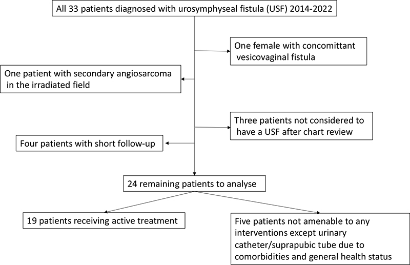
Figure 1. CONSORT-diagram describing the study population.
Outcome measures
During a chart review, the clinical pictures and diagnostic procedures were ascertained. Retrospective assessment of diagnostic delay between first symptom until USF was diagnosed was also performed. Clinical presentation, precipitating causes, and treatments were also assessed. Treatment associated 90-day complications were categorized according to the Clavien-Dindo classification [9]. Comorbidity by American Society of Anesthesiologists (ASA)-score was assessed during chart review or during preoperative assessment by anaesthesiologist among patients treated with surgical interventions under general anaesthesia. Findings in urinary cultures and cultures from tissue and/or bone were compared, and USF-status at end of follow-up was also considered an outcome measure.
Statistics
Descriptive statistics with proportions were applied to demonstrate the characteristics of the study population.
Ethical permission
The study was approved by the Research Ethics Board of Lund University, Sweden (Dnr 2018/224).
Results
Clinical characteristics
The median time from the date of USF-diagnosis to the end of follow-up or death (nine patients) was 22 (Inter Quartile Range (IQR) 6–40) months. Descriptive patient characteristics are provided in Table 1. Median age at diagnosis among the 24 patients with USF was 77 (IQR 71–81) years, and 17/24 (71%) had severe comorbidities corresponding to ASA-score 3 and 4. Local pain was by far the dominating initial symptom (17/24 (71%) patients).
| Total number of patients, n | 24 | |
| Median age at diagnosis, yrs (IQR) | 77 (71-81) | |
| ASA score | 2 | 7 (29%) |
| 3 | 12 (50%) | |
| 4 | 5 (31%) | |
| Diabetes, n | 2 (8%) | |
| Smoking, n | 2 (8%) | |
| Median preoperative S-albumin, g/L (IQR) * | 24 (22-28) | |
| Androgen deprivation, n | Bicalutamide | 5 (31%) |
| GnRH agonist | 2 (8%) | |
| Radiotherapy, n | Primary | 20 (83%) |
| Salvage | 4 (17%) | |
| Predominant symptom present before and at diagnosis of USF, n | Pain | 17 (71%) |
| Infection and abscess | 2 (8%) | |
| Rectal bleeding | 2 (8%) | |
| Urinary fistulae | 3 (13%) | |
| Precipitating intervention causing USF, n | Transurethral surgical procedure/dilatation | 14 (58%) |
| CIC | 2 (8%) | |
| Suprapubic tube | 3 (13%) | |
| Rectal biopsies | 1 (4%) | |
| None | 4 (17%) | |
| Diagnostic modality | CT | 24 (100%) |
| MRI | 19 (79%) | |
| Median time from first symptom to diagnosis, days (IQR) | 15 (9-59) | |
| IQR: Inter Quartile Range; USF: urosymphyseal fistula; CIC: clean intermittent catheterization; n: numbers; GnRH: Gonadotropin Releasing Hormone agonist; CT: Computed Tomography; MRI: Magnetic Resonance Imaging.. | ||
| *Nine missing values for preoperative S-albumin, the remaining categories are complete. | ||
A cystoscopy was performed in all patients, and in five patients with concurrent rectourethral fistula a sigmoidoscopy was also performed as part of the preoperative workup. An MRI was performed to evaluate the extent of the osteomyelitis and fistulation. Indirect signs of USF were considered diagnostic, and fistulographies were not routinely performed. Urine cultures were obtained in all patients. Prior to urinary diversion a CT-urography was performed to assess the morphology of the upper urinary tracts. Nutritional assessment including calorie intake and serum albumin levels was evaluated in all patients prior to planning active treatment. After assessing each patient and informal contacts with specialists in infectious diseases and orthopaedics at the discretion of the treating urologist, individual treatment plans were decided for each patient.
The possible precipitating interventions causing USF were in 14 patients a transurethral surgical procedure/dilatation; three patients received a suprapubic tube; two patients practiced CIC; and one patient was subjected to rectal biopsies as part of clinical workup for proctitis. Four patients lacked an obvious underlying cause.
A USF with radiological signs of osteomyelitis was present in 20 patients; the remaining four had solely radiological signs of USF with or without abscess at diagnosis. The majority of patients (19/24 (79%) patients) were diagnosed by MRI in addition to CT, but four patients with radiological signs of osteomyelitis in the pubic bone and one with USF without osteomyelitis on CT were not examined with MRI. Five patients had a concurrent rectourethral fistula. The median time from first symptom to diagnosis was 15 (IQR 9–59) days, but five patients had symptoms for more than 3 months until the USF was diagnosed.
Eight patients were diagnosed in 2014–2017, whereas the remaining 16 were diagnosed in 2018–2022. The RT preceding the diagnosis of USF was received in median 5 (IQR 2–7) years previously, with treatment details described in Table 2. Four patients were treated with RT as salvage therapy after radical prostatectomy. Two patients received salvage cryotherapy for a local recurrence after external beam radiation with curative intent.
Surgical treatment
In five individuals, severe comorbidities and deteriorated general health status when diagnosed with USF precluded any extensive interventions except urinary catheter or suprapubic tube. Ten patients were treated with some form of supravesical urinary diversion without cystectomy (Table 3). Three patients received an ileal conduit. One patient had bilateral ureteric ligatures, and two patients were subsequently operated on in a staged second procedure with end-to-end ureteroureteroanastomosis with unilateral nephrostomy as permanent urinary diversion [10]. Nine patients tolerated cystectomy, and four of these with concomitant rectourethral fistula underwent a pelvic exenteration with intersphincteric resection of the rectum en bloc with the urinary bladder and the fistula (Table 3). Several sequential bone cultures were obtained at surgery except in one patient where biopsies were obtained percutaneously. The objective during surgery was to debride the superior and inferior rami of the symphysis, remove infected juxta-articular bone sequesters (Figure 2), resect the USF, and simultaneously obtaining fractionated bone cultures and to perform a urinary diversion. If feasible without exacerbation of the local infection, antibiotics were withdrawn 2 days prior to surgery to improve the possibility to obtain representative fractionated bone cultures. When available, an omental flap was mobilized and applied to cover the infected bone (Figure 3). Postoperatively, prolonged antibiotics were prescribed for several months to treat the osteomyelitis.
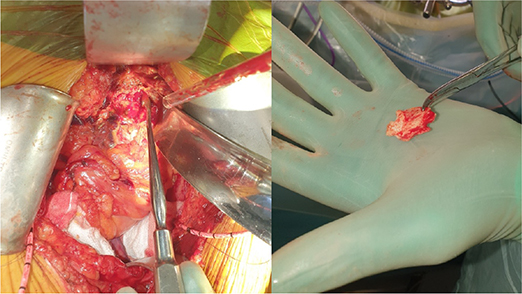
Figure 2. The symphysis and pubic bone are debrided and an infected juxta-articular bone sequester was removed.
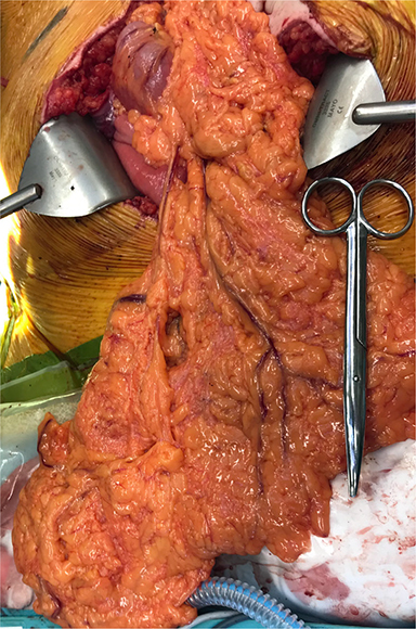
Figure 3. A generous omental flap has been mobilized in patient number 13, enabling coverage of the symphysis and filling out the pelvic cavity after the exenteration.
Complications
Eight out of the 19 patients (42%) receiving some form of active treatment suffered from complications within 90 days of the intervention. Five complications were related to infections (Clavien 2), and two patients had Clavien 3 complications. One of these latter two patients was diagnosed with a postoperative enterourethral fistula that later was subjected to surgery with uneventful recovery, and one patient with insufficiency in the end-to-end ureteroureteroanastomosis received a temporary contralateral nephrostomy tube, after which the anastomotic leakage healed spontaneously. The patient subjected to bilateral ureteric ligatures without cystectomy died 58 days after surgery due to fatal epistaxis (Clavien 5), not obviously related to the previous surgical intervention.
The osteomyelitis was treated with long-term antibiotics (months) based on the tissue biopsy cultures, where the duration was individualized according to clinical follow-up including c-reactive protein levels and radiology.
Urine and bone cultures
The most common findings in the bone cultures were enterococci, anaerobic and candida species. Bone cultures in two patients were negative despite radiologic signs of osteomyelitis, albeit preoperatively administered antibiotics can explain the lack of bacterial growth. In 11 patients, there was a discrepancy between the preoperative urine cultures and tissue biopsy culture findings, and polymicrobial growth occurred in eight patients either in the tissue biopsy cultures (Table 4).
| Patient number | Preoperative urine culture | Intraoperative bone culture |
| 1 | Enterococcus faecium | NP |
| 2 | Mixed flora | NP |
| 3 | Enterococcus faecium and Candida albicans | Negative* |
| 4 | NP | NP |
| 5 | Betastreptococcus group G and mixed grampositive flora | NP (Staphylococcus aureus and Enterococcus faecalis from abscess) |
| 6 | Staphylococcus aureus and Enterococcus faecalis | Enteroccus faecium, Klebsiella oxytoca, E. Coli, and Streptococcus anginosus |
| 7 | Aerococcus urinae | Aerococcus urinae, staphylococcus epidermidis and hominis |
| 8 | Proteus mirabilis | Proteus mirabilis and bacteroides fragilis |
| 9 | Staphylococcus aureus | NP |
| 10 | Staphylococcus aureus and E. Coli | NP |
| 11 | Staphylococcus aureus | NP (Staphylococcus aureus and E. Coli from abscess) |
| 12 | Enterococcus faecalis | NP |
| 13 | E. Coli and bacteroides fragilis | Pseudomonas aeruginosa, clostridium difficile, and Enterococcus faecium |
| 14 | Proteus mirabilis and proteus vulgaris | Candida albicans and streptococcus mitis |
| 15 | E.Coli and Enterococcus faecalis | NP |
| 16 | Enterococcus faecalis | Negative* |
| 17 | Streptococcus agalactiae and mixed anaerobe flora | Unspecified pevotella (bacteroides) species |
| 18 | Enterococcus faecalis | NP |
| 19 | Streptococcus agalactiae | E. Coli, haemophilus parainfluenzae, parabacteroides species, Enterococcus faecium, Enterococcus gallinarum, and Enterobacter cloacae |
| 20 | Enterococcus faecalis | NP |
| 21 | E. Coli and yeast species | Enterococcus faecalis |
| 22 | Negative | Enterococcus faecium |
| 23 | Enterococcus faecium and faecalis | Candida albicans |
| 24 | Enterococcus faecium | NP |
| NP: not performed. *Obtained during ongoing treatment with parenteral antibiotics. | ||
Outcomes
Out of the five patients not receiving any active treatment except long-term antibiotics and urinary catheter or suprapubic tube, three died during follow-up with infection caused by the USF as the primary cause of death. One of these five patients has been hospitalized on 20 occasions during the last 2 years for infections and catheter-related problems. The remaining patient is alive and asymptomatic 12 months after being diagnosed with USF, with regular changes of the urinary catheter over a guidewire.
Among the 19 patients receiving active treatment, five had a recurrent osteomyelitis and/or pelvic infection, of which four did not have a cystectomy at the time of surgery. All five patients with recurring osteomyelitis or pelvic infection subsequently died with infection caused by the USF as the primary cause of death. Thus, 1/9 (11%) patients having a cystectomy in conjunction with the surgery recurred compared to 4/10 (40%) when a cystectomy was not performed related to comorbidity.
Eight out of the 23 patients treated for prostate cancer had a PSA-recurrence at the end of follow-up.
Discussion
In our series, USF and osteomyelitis occurred primarily in elderly patients with significant comorbidity, and pain was the most common presenting symptom. For patients not amenable to active treatment, USF was a fatal condition. Three out of the five patients who due to comorbidity were treated with a conservative strategy in the current cohort died as a direct consequence of the USF due to infection and osteomyelitis. A multidisciplinary approach including orthopaedic, colorectal, and infectious diseases expertise likely improves the diagnostic workup and treatment of patients with USF. That is, knowledge about how to optimally obtain fractionated bone cultures and how to perform adequate extirpation of infected bone sequesters is useful. Also experience in rectal cancer surgery when fistulation involves the rectum and adequate knowledge to interpret obtained cultures (colonisation vs infection) and optimal choice of and duration of long-term antibiotic treatment and monitoring are of value. In our experience, refraining from cystectomy in conjunction with surgery might be associated with an increased risk of recurrent infection, since 4 out of 10 of these patients relapsed and subsequently died due to their recalcitrant infection.
Five patients included in our series were subjected to diagnostic delay beyond 3 months. This is likely due to a combination of the uncommon nature of USF and that the first published description of the diagnostic entity was only 10 years ago [5], explaining a limited awareness of the diagnosis. Our finding that local pain was the predominant symptom at diagnosis is in line with what has been reported previously, including chronic opioid use in 40% of the patients at diagnosis [7]. The pathogenesis of pain in patients with USF is likely related to the successive development of osteomyelitis in the pubic bone due to bacteriuria [11], facilitated by prior radiation that increases the susceptibility of the pubic bone to microbes [12]. The fact that 79% in the current series and between 76% and 100% of patients reported in the current literature [2–4] had a history of urethral manipulation preceding the diagnosis of USF, suggests that the main risk factor to develop USF with osteomyelitis is related to such interventions in patients previously treated with radiation therapy.
MRI is a cornerstone in diagnosing USF [8], including visualization of the osteomyelitis [13]. For patients with contraindication to MRI, FDG-PET-CT can to our most recent clinical experience be diagnostic showing increased FDG-uptake in the infected pubic bone. Still, the accumulation of patients diagnosed with USF during later years in this study and other series [7] suggests that clinical awareness about this entity still needs to be increased.
The optimal surgical treatment for USF remains to be determined. In one series with younger patients, 7/15 were subjected to reconstruction without cystectomy and urinary diversion and later artificial sphincter implantation in five [3]. Intriguingly, implantation of an artificial sphincter has been described to unmask subclinical USF in three patients after continence was restored [14]. A more radical approach was described in one recent study where all 25 patients were subjected to a formal pubectomy and five of them additionally received a VRAM (Vertical Rectus Abdominis Musculocutaneous) flap to fill out the defect in the symphysis [2]. Whether to use omental flaps or VRAM flaps in general during oncoplastic surgery and when operating USF in particular is also unknown [15], as well as if it is reasonable to refrain from an omental flap if not easily accessible through the lower midline incision used for the extirpative parts of the USF repair. Such extensive treatment strategies reported for patients with USF [2, 3, 7, 11] are in contrast to the heterogenic, and in some patients staged individualized, treatment options applied in the elderly patients in the current series. The fact that the median age was 77 years and the median preoperative S-albumin was 24 g/L in the current series suggests that our population-based and consecutive series included a broader category of patients, compared to the three largest recent publications where the median age was 69–71 years [2, 7, 11]. This is further underlined by the fact that one in five patients was not amenable to any active surgical treatment in the present study, whereas in other published series all included patients received surgical treatment. There might thus be a publication bias with underrepresentation of elderly and frail patients that are neither diagnosed nor treated for their USF.
The current study is limited by the retrospective design and the fact that additional patients with USF in the Southern Healthcare Region might be undiagnosed or not referred if conservative measures enough, that is, selection bias is possible. On the other hand, this study is to our knowledge the first population-based series published including description of outcomes also in patients receiving conservative and submaximal interventions. Other limitations are that only males were included, as the entity also has been described in females [16], and that the exact radiation doses were not possible to retrieve since many patients were treated with RT several years ago, when dose plans were not accessible. Additionally, more modern RT might deliver a lower dose to the symphysis, nonetheless more patients were diagnosed with USF during later years in the current series. The interpretation of the cultures obtained in the current study is also hampered by that the clinical scenarios did not allow to abstain from antibiotics for optimally 2 weeks prior to obtaining bone cultures. Thus, some of the findings in the cultures might be only colonization.
The knowledge about USF with osteomyelitis after radiation needs to be disseminated to avoid or minimize endourologic measures and rectal biopsies in these patients, creating the nidus for fistula and formation. Additionally, clinicians should be educated to perform an MRI in individuals with clinical suspicion of USF. To create multidisciplinary algorithms to treat patients with USF, as has been described for rectourethral fistula repair [17], would probably also be beneficial. Our data also suggest that reducing the extent of surgery by leaving the urinary bladder in situ may increases the risk of suboptimal outcomes. Based on cultures in our and other series [2] reporting yeast in bone cultures in some patients, empiric broad spectrum anti-fungal coverage can be considered despite that yeast in preoperative urine cultures is not always captured.
Conclusion
Urologists should avoid performing endourologic interventions in patients previously subjected to pelvic RT, as USF frequently is preceded by such measures. Pain is the predominant symptom of USF, and once a diagnosis is established by an MRI, a multidisciplinary and individually tailored treatment is suggested also in elderly patients not amenable to the most aggressive surgical approach.
Acknowledgements
This work was supported by the Swedish Cancer Society (CAN 2020/0709), Swedish Research Council (2021-00859), Lund Medical Faculty (ALF), Skåne University Hospital Research Funds, The Cancer Research Fund at Malmö General Hospital, Skåne County Council’s Research and Development Foundation (REGSKANE-622351), Gösta Jönsson Research Foundation, the Foundation of Urological Research (Ove and Carin Carlsson bladder cancer donation), and Hillevi Fries Research Foundation. The funding sources had no role in the study design, data analyses, interpretation of the results, or writing of the manuscript.
References
- [1] Peters G, Winston J, Nuwayhid F, Pointon O. Conservative management of a prostate-symphyseal fistula with osteomyelitis: a case report and management of a rare complication. Cureus. 2022;14:e24032. https://doi.org/10.7759/cureus.24032
- [2] Andrews JR, Hebert KJ, Boswell TC, et al. Pubectomy and urinary reconstruction provides definitive treatment of urosymphyseal fistula following prostate cancer treatment. BJU Int. 2021;128:460–7. https://doi.org/10.1111/bju.15333
- [3] Bugeja S, Andrich DE, Mundy AR. Fistulation into the pubic symphysis after treatment of prostate cancer: an important and surgically correctable complication. J Urol. 2016;195:391–8. https://doi.org/10.1016/j.juro.2015.08.074
- [4] Shapiro DD, Goodspeed DC, Bushman W. Urosymphyseal fistulas resulting from endoscopic treatment of radiation-induced posterior urethral strictures. Urology. 2018;114:207–11. https://doi.org/10.1016/j.urology.2017.12.020
- [5] Matsushita K, Ginsburg L, Mian BM, De E, Chughtai BI, Bernstein M, et al. Pubovesical fistula: a rare complication after treatment of prostate cancer. Urology. 2012;80:446–51. https://doi.org/10.1016/j.urology.2012.04.036
- [6] Lavien G, Chery G, Zaid UB, Peterson AC. Pubic bone resection provides objective pain control in the prostate cancer survivor with pubic bone osteomyelitis with an associated urinary tract to pubic symphysis fistula. Urology. 2017;100:234–9. https://doi.org/10.1016/j.urology.2016.08.035
- [7] Anele UA, Wood HM, Angermeier KW. Management of urosymphyseal fistula and pelvic osteomyelitis: a comprehensive institutional experience and improvements in pain control. Eur Urol Focus. 2022;8(4):1110–16. https://doi.org/10.1016/j.euf.2021.08.008
- [8] Sexton SJ, Lavien G, Said N, et al. Magnetic resonance imaging features of pubic symphysis urinary fistula with pubic bone osteomyelitis in the treated prostate cancer patient. Abdom Radiol (NY). 2019;44:1453–60. https://doi.org/10.1007/s00261-018-1827-2
- [9] Dindo D, Demartines N, Clavien PA. Classification of surgical complications: a new proposal with evaluation in a cohort of 6336 patients and results of a survey. Ann Surg. 2004;240:205–13. https://doi.org/10.1097/01.sla.0000133083.54934.ae
- [10] Jancke G, Baseckas G, Brändstedt J, et al. End-to-end ureteroureteroanastomosis with unilateral nephrostomy: revival of a forgotten technique suitable for a modern context? Scand J Urol. 2019;53:435–8. https://doi.org/10.1080/21681805.2019.1624389
- [11] Kahokehr AA, Boysen WR, Schild MH, et al. Urinary pubic symphysis fistula leads to histopathologic osteomyelitis in prostate cancer survivors. Urology. 2021;148:297–301. https://doi.org/10.1016/j.urology.2020.07.038
- [12] Tibbs MK. Wound healing following radiation therapy: a review. Radiother Oncol. 1997;42:99–106. https://doi.org/10.1016/S0167-8140(96)01880-4
- [13] Llewellyn A, Jones-Diette J, Kraft J, et al. Imaging tests for the detection of osteomyelitis: a systematic review. Health Technol Assess. 2019;23:1–128. https://doi.org/10.3310/hta23610
- [14] Kahokehr AA, Peterson AC. Unmasking of urinary-pubic symphysis fistula after implantation of artificial urinary sphincter in prostate cancer survivors-user beware. Urology. 2018;114:202–6. https://doi.org/10.1016/j.urology.2017.11.047
- [15] Chaudhry A, Oliver JD, Vyas KS, et al. Comparison of outcomes in oncoplastic pelvic reconstruction with VRAM versus omental flaps: a large cohort analysis. J Reconstr Microsurg. 2019;35:425–9. https://doi.org/10.1055/s-0038-1677524
- [16] Haveman I, van Altena AM, Verschuren CP, Aarts JW. Multidisciplinary management of patients with pubic osteomyelitis, a rare but serious complication after surgery and radiation therapy for advanced gynaecological cancer. BMJ Case Rep. 2021;14(1):e236289. https://doi.org/10.1136/bcr-2020-236289
- [17] Hayden JP, Boysen WR, Kowalik U, et al. Outcomes of an algorithmic, multidisciplinary approach to rectourethral fistula repair: a pre- and post-intervention quasi-experimental study. Dis Colon Rectum. 2023;66(4):598–608. https://doi.org/10.1097/DCR.0000000000002467