ORIGINAL RESEARCH ARTICLE
Primary mucosal melanomas of the urogenital tract: a clinical, pathological, and genetic nationwide survey of Danish patients 1990–2019
Victoria K. Znaidera, Lauge H. Mikkelsena,b, Christian Fuglesang S. Jensenc, Jens Sønksenc and Steffen Heegaarda,b
aDepartment of Pathology, Rigshospitalet, Copenhagen, Denmark; bDepartment of Ophthalmology, Rigshospitalet, Copenhagen, Denmark; cDepartment of Urology, Copenhagen University Hospital – Herlev and Gentofte, Herlev, Denmark
ABSTRACT
Purpose: To describe the epidemiologic, clinical, histopathological, and genetic features of primary mucosal melanoma of the urinary tract in a national Danish cohort with cases included from the year 1990 to 2019.
Material and methods: Patients of the Danish cohort were found using national databases. Only primary tumours were included in the cohort. Appropriate formalin-fixed paraffin-embedded blocks underwent next-generation sequencing.
Results: Eight cases of primary urinary bladder melanomas and 18 cases of primary urethral melanomas were included. Bladder melanomas had an incidence of 0.05 cases/million/year. Mean age at diagnosis was 67 years. The most frequent primary treatment was cystectomy. Adjuvant treatment was given in three cases and consisted of chemotherapy or radiotherapy. Mutations were found in the NF1, KRAS, ATRX, TP53, RAC1, and BRAF genes. Urethral melanomas were found to have an incidence of 0.12 cases/million/year. Average age at diagnosis was 77 years. The most frequent treatment was excision of the tumour. Adjuvant treatment was given in nine cases and most frequently consisted of radiotherapy. Mutations were found in the NF1, TERT PROMOTOR, NRAS, ATRX, TP53, ATM, TSC2, and CREBBP genes. The 5-year survival of patients with bladder melanoma was 12.5% and 22.2% for patients with urethral melanoma.
Conclusion: Our study highlights the rarity of urinary tract melanomas and their poor prognosis. The most widely used treatment for urogenital mucosal melanoma remains surgical while adjuvant therapy strategies are evolving. Next-generation sequencing showed mutational patterns with no location-specific patterns. The most frequent mutations were in the NF1, ATRX, NRAS, and TP53 genes.
KEYWORDS: Mucosal melanoma; urinary tract; pathology; genetics
Citation: Scandinavian Journal of Urology 2023, VOL. 58, 52–59. https://doi.org/10.2340/sju.v58.8489.
Copyright: © 2023 The Author(s). Published by MJS Publishing on behalf of Acta Chirurgica Scandinavica. This is an Open Access article distributed under the terms of the Creative Commons Attribution-NonCommercial 4.0 International License (https://creativecommons.org/licenses/by-nc/4.0/), allowing third parties to copy and redistribute the material in any medium or format and to remix, transform, and build upon the material for non-commercial purposes, provided proper attribution to the original work.
Received: 13 September 2022; Accepted: 4 June 2023; Published: 27 August 2023
CONTACT Steffen Heegaard sthe@sund.ku.dk Department of Pathology, Rigshospitalet, Teilumbygningen, Frederik V’s Vej 11 1st floor, DK-2100 Copenhagen, Denmark
Supplemental data for this article can be accessed online at https://doi.org/10.2340/sju.v58.8489
Introduction
Primary melanoma of the mucous membranes is a very rare but aggressive disease that represents 0.03% of all cancers and 1.3% of all melanomas [1].
The most common mucosal sites are head and neck (55.4%), anal/rectal (23.8%), female genital tract (18%), and urinary tract (2.8%) [1]. The risk factors for developing mucosal melanomas are still unknown.
Melanocytes derived from the neural crest migrate during embryogenesis to their final destination in different microenvironments including the mucous membranes where they can give rise to malignant melanoma [2, 3]. Mucosal melanomas have several genetic changes in intracellular signalling cascades, and these may constitute the pathogenetic mechanism of some mucosal melanomas. Genetically, mucosal melanomas have aberrations of the MAPK pathway, including NRAS, BRAF, NF1, and KIT [4]. However, mucosal melanomas are genetically heterogeneous compared with cutaneous melanoma [5].
Primary mucosal melanoma of the urethra accounts for less than 1% of all melanomas and about 4% of urethral cancers [6], whereas primary mucosal melanoma of the bladder is exceedingly rare. The prognosis for mucosal melanoma is poor and several factors have been proposed to explain this including advanced stages of disease at the time of diagnosis [7, 8]. Preferred treatment is surgical removal with generous free margins; however, there are no clear guidelines on management. The median survival rate of urethral melanoma after diagnosis is 25.6 months [9]. The overall survival rate for bladder melanoma after diagnosis is 28.5% [10].
The aim of this study was to describe the epidemiology, symptomatology, pathology, mutational patterns, and treatment of primary mucosal melanoma of the urinary tract in a national Danish cohort.
Material and methods
Patient and tissue samples
Patients diagnosed with primary melanoma of the urethra or bladder were identified by searching the digital archives of the Danish Registry of Pathology (Patobank). Patients with prior history of cutaneous or uveal melanoma were not included, since urogenital melanoma was considered secondary mucosal melanomas in these cases.
Medical records and pathology reports were retrieved from the local hospital departments and the following information was registered: age, gender, symptoms, clinical findings, size, macroscopic appearance, and microscopic morphology of the tumour and treatment (Tables 1 and 2).
Information on time of death for all patients was retrieved from the Danish Cause of Death Registry. The investigation adhered to the Declaration of Helsinki and approval was obtained from the Danish Data Protection Agency along with the local ethics committee (H-6-2014-060).
The standardised mortality rate (SMR) was calculated based on the survival (person years at risk) for the study population from the date of diagnosis until the date of death or end of follow-up. The expected number of deaths was calculated using data from the entire Danish population in the year 2019 available at Statistics Denmark databank (www.dst.dk).
Histopathology
Archived formalin-fixed paraffin-embedded (FFPE) samples and archived haematoxylin/eosin-stained (HE) slides were retrieved. The HE slides along with Melan-A, Ki-67, and cytokeratin-stained slides were revised and the following information was registered: growth pattern, cytology, microscopic pigmentation, ulceration, necrosis, depth of invasion, number of mitosis/10 high power fields (HPF), proliferation index, vascular/nerve invasion, pagetoid spread, in-situ component, and status of the resection margin (Tables 2 and 3).
Next-generation sequencing
Appropriate FFPE blocks were selected by evaluating a sufficient number of tumour cells on corresponding HE slides.
DNA was isolated using an in-house raw extraction method (Proteinase K and Tris EDTA), and DNA was quantified using the Qubit 2.0 Fluorometer High Sensitivity Kit (Thermo Fisher Scientific, Waltham, MA, USA). Targeted Next Generation Sequencing as carried out on the S5+ System with the Oncomine Comprehensive Assay version 3 according to the manufacturer’s instructions (Thermo Fisher Scientific). The Oncomine Comprehensive Assay v3 Panel covers 87 genes for hotspot mutation detection, 48 genes for full-length sequencing, and 43 genes for focal copy number assessment. Library preparation was performed from 20 ng of purified DNA using the Ion AmpliSeq™ Library Kit Plus according to the manufacturer’s instructions. The Ion 540 Kit-Chef (Thermo Fisher Scientific) was used as a template kit for the Ion Chef (Thermo Fisher Scientific) and subsequent sequencing on the S5+ using Ion 540 Chips (Thermo Fisher Scientific).
Sequenced data were initially aligned and mapped to the human hg19 reference genome using the Torrent Suite Server (ver 5.10) with default parameters. The Ion Reporter Workflow (version 5.6) was used to perform variant calling of the DNA libraries. Specifically, gene annotation was performed using the Oncomine Comprehensive Assay v3 Annotations set v1.2, copy number baseline was performed using the Oncomine Comprehensive DNA v3 Baseline v2.0, and the Oncomine Variant annotator v2.3 plugin was used for analysis.
Ingenuity Variant Analysis (Qiagen Bioinformatics, Redwood City, CA, USA) was used for customised variant annotations. Samples were filtered using the following criteria: a call quality above 20, a read depth of at least 100, and a variant allele fraction of at least 0.10. Variants reported in the healthy public genomes (1000 Genomes Project), the Exome Sequencing Project, or the Exome Aggregation Consortium databases with a frequency >0.1 were considered germline variations and thereby excluded. Called variants should be outside the top 5% of the most exonically variable 100 base windows in healthy public genomes and should pass the built-in upstream pipeline filtering in the Ingenuity software in order to exclude spurious calls. Variants classified as ‘benign’ or ‘likely benign’ were excluded. Variants classified as ‘pathogenic’, ‘likely pathogenic’, or ‘uncertain’ were looked up in the Catalogue of Somatic Mutations in Cancer database (cancer.sanger.ac.uk) and cBioPortal for Cancer Genomics (www.cbioportal.org). High-confidence somatic variants were visually confirmed in Integrative Genomics Viewer (https://www.broadinstitute.org/igv/) and manually checked in the COSMIC database. Variants classified as ‘uncertain’ in the upstream analysis and not previously reported in the cancer databases were not annotated.
Results
Twenty-six cases of primary melanoma of the urinary tract were reviewed. Six were located in the urinary bladder, while two were located in both the urinary bladder and vagina. Thirteen cases were located to the urethra, while five cases were located in both the urethra and vagina. We decided to include the patients who presented with both vaginal and urethral or bladder melanomas and group them as either primary urethral or bladder melanomas, since it was not possible to determine which of the tumour locations were the primary location. A total of eight cases of primary urinary bladder melanomas and 18 cases of primary urethral melanomas were included in the study.
We were able to retrieve archived FFPE samples from 15/18 of the urethral melanomas and 8/8 of the bladder melanomas. Complete medical records were recovered from 7/8 of the bladder melanoma patients and 16/18 of the urethral melanoma patients.
Epidemiology
A median age at diagnosis was found to be 67 years (range 50–76 years) for bladder melanomas and 78 years (range 57–90 years) for urethral melanoma.
The incidence of primary urinary tract melanoma in the Danish population (5.5 million people) between 1990 and 2019 was 0.05 cases/million/year for bladder melanomas and 0.12 cases/million/year for urethral melanomas.
A male:female ratio of 1:3 was found in the cases of bladder melanomas and 1:17 in the cases of urethral melanomas, possibly due to the longer urethra in men.
Symptoms
The most frequent symptom of the bladder melanomas was gross haematuria, recurrent urinary tract infections, and/or dysuria.
For the urethral melanomas, the symptoms had great variation, but the most frequent symptom was gross haematuria (n = 5) with a variation of other symptoms such as obstructive urinary flow, dysuria, pollakisuria, or incontinence.
Clinical findings (Table 1 and Tables SI and SII)
In bladder melanoma, the location varied from the bladder neck (n = 2), fundus vesicae (n = 2), and the posterior wall of the bladder (n = 4). The macroscopic appearance of the tumour was mostly described as being polypoid. Interestingly, none of our cases involved the trigonum. This might be due to the fact that the trigonum is formed by mesoderm (Wolffian ducts), while the remainder of the bladder (and urethra) is formed by endoderm (sinus urogenitalis).
In urethral melanomas, the most common clinical finding was a tumour at the external urethral meatus (n = 10) or in other parts of the distal urethra (n = 6).
Diagnosis
In all cases, diagnosis was established by biopsy and histologic examination.
Size
In the cases of bladder melanoma, the tumour ranged from 2–15 cm in diameter. The size of the urethral melanomas ranged from a diameter of 0.5 cm to a tumour size of 5 cm, and the median diameter was 3 cm.
Histology (Figure 1)
Tables 2 and 3 display the histopathological characteristics for the cases of bladder and urethral melanomas. The tables show the distribution of cell configuration, cytomorphology as well as presence of microscopic pigmentation, ulceration and necrosis. The tables also describe depth of invasion, median number of mitoses per 10 HPF, the percentage of positive Ki-67 stain, and the presence of vascular and perineural invasion, pagetoid spread, and occurrence of clear resection margins after surgery.
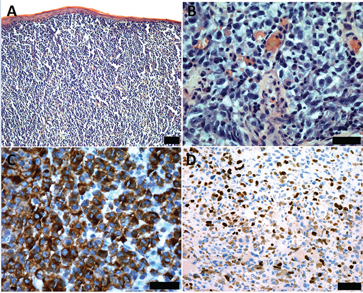
Figure 1. Urethral melanoma histology (A) The epithelioid tumour cells display a sheet-like configuration beneath the epithelium H&E, Bar = 100 µm. (B) Pleomorphic epithelioid tumour cells with pale cytoplasm are seen. H&E. Bar = 50 µm. (C) The tumour cells stained positive for MELAN-A. Bar = 50 µm. (D) A proliferative index of approximately 50% is seen when stained with Ki-67. Bar = 50 µm.
Primary treatment
The most frequent treatment of bladder melanomas was cystectomy (n = 3). For two patients, the treatment was unknown.
The treatment for urethral melanomas had great variations, but the most frequent treatment was excision of the tumour (n = 7).
Adjuvant treatment
Adjuvant treatment of the bladder melanomas consisted of chemotherapy (n = 2) or radiotherapy (n = 1). One case was not given any treatment.
Adjuvant therapy was not applied in nine cases of urethral melanomas. Three had radiotherapy, while one case was treated with immunotherapy.
Local lymph node metastasis
Local lymph node metastases were found in 4/6 cases of bladder melanomas and 9/13 cases of urethral melanomas. One case refused further tests and another case also had disseminated breast cancer and was therefore not examined further.
Lymph node status was missing in one case of bladder melanoma and in one case of urethral melanoma.
Distant metastasis
Distant metastases were found in 5/7 bladder melanomas, and they were localised to the lung (n = 1), lung and rectum (n = 1), the vagina (n = 1), the vagina and stomach (n = 1), and the liver (n = 1).
Distant metastases were found in 11/16 of the urethral melanomas, and these were localised to the vulva (n = 4), the lungs (n = 2), followed by the rectum, bladder, vertebrae, liver, and mesentery, which was found in one case each.
Survival
The patients with bladder melanoma had the poorest survival, with a mean of 22.5 months ranging from 6 to 64 months. The 5-year survival was 12.5%. For bladder melanoma, there were too few cases to predict a SMR.
The patients with urethral melanoma had a poor survival, with a mean survival of 42 months ranging from 1 to 189 months. The 5-year survival was 22.2%. For urethral melanoma, the SMR was calculated to be 4.5 (95% confidence interval [CI]: 3.4–6.1), representing a significantly increased number of deaths in the population at risk.
Genetics (Figure 2)
Next-generation sequencing was performed on eight bladder melanomas and 15 urethral melanomas giving a total number of 23 sequenced melanomas. Mutations were detected in 5/8 of the bladder melanomas and 12/15 of the urethral melanomas adding up to a total of 17 melanomas with detected mutations. The remaining six melanomas were of such bad quality that no data could be extracted.
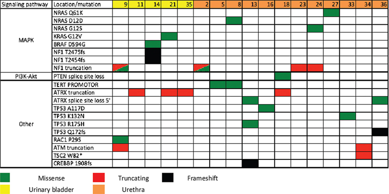
Figure 2. Mutational pattern of urinary tract melanoma in Denmark. Urethral melanoma mutations: 5 NF1, 2 TERT PROMOTOR, 4 NRAS, 3 ATRX, 3 TP53, 1 ATM, 1 TSC2, 1 CREBBP. Bladder melanoma mutations: 4 NF1, 1KRAS, 3 ATRX, 1 TP53, 1 RAC1, 1 BRAF. Most frequent mutations: NF1 (53%), ATRX (35%), TP53 (24%), NRAS (24%), and TERT PROMOTOR (12%).
The most frequent gene mutation was in NF1, which was found in 53% of mutated cases (9/17 analysed) followed by gene mutations in ATRX (6/17 analysed; 35.3%), TP53 (4/17 analysed; 23.5%), NRAS (4/17 analysed; 23.5%), TERT PROMOTOR (2/17 analysed; 11.7%), KRAS (1/17 analysed; 5.9%), RAC1 (1/17 analysed; 5.9%), BRAF (1/17 analysed; 5.9%), ATM (1/17 analysed; 5.9%), TSC2 (1/17 analysed; 5.9%), and CREBBP (1/17 analysed; 5.9%).
Discussion
This report presents a nationwide study on 26 Danish patients with primary urinary tract melanoma diagnosed between 1990 and 2019, indicating the rarity of this disease.
In this study, the incidence of primary bladder melanoma in Denmark was 0.05 cases/million/year and 0.12 cases/million/year for urethral melanomas. Other studies of primary mucosal melanomas in Denmark have found an incidence of primary melanoma of the small intestine of 0.04 cases/million/year [11], of the conjunctiva of 0.5 cases/million/year [12], and of the head and neck of 1.2 million/year [13].
A mean age at diagnosis of, respectively, 67 and 77 years was found for bladder and urethral melanomas, which is older than for other mucosal melanomas [3, 6].
All current literature agrees that the gold standard for local disease management of mucosal melanomas is surgical treatment. There is uncertainty regarding whether a conservative or radical treatment strategy provides the best prognosis. The majority of the patients in our cohort received radical surgical treatment. This surgical strategy yielded a survival after diagnosis ranging from 4 to 189 months, with a median survival of 14 months. Only urethral melanomas had a more conservative surgical approach, with seven cases treated with excision of the tumour, resulting in a survival after diagnosis ranging from 1 to 101 months.
Praveen et al. [14] suggested a conservative strategy with excision with tumour-free margins, which yielded an overall 5-year disease-specific survival of 27%. Oliva et al. [15] and DiMarco et al. [16] both advocated more radical surgeries, because they believed that the high rate of recurrences of the mucosal melanomas is due to inadequate surgical margins. This more radical surgical strategy yielded a disease-specific survival of 39%.
Previous literature has stated that the poor prognosis is due to the late stage of the disease at diagnosis, where metastatic spread to the lymph nodes and other distant sites has already occurred [9]. If the disease was detected sooner, the tumour might not be as infiltrative and the prognosis might be improved with the use of radical treatment [17].
The late stage at diagnosis results in a high presence of metastases, which has also been suggested to be a negative prognostic factor according to two studies [9, 18]. In our cohort, 13 cases had local lymph node metastases and 16 cases had distant metastases. Patients without metastases presented a favourable survival.
Von Heppt et al. [19] stated that negative prognostic factors include tumour depth > 2 mm and ulceration. In our cohort, the majority of tumours had a thickness exceeding 2 mm, since only two cases had a tumour thickness of 2 mm or less. In terms of ulceration, about half of the cases (14 cases) of the cohort were found to have microscopic ulceration, which might also play a role in the poor prognosis. Another study by Zhu et al. [20] found tumour size <2 cm to be a positive prognostic factor. In our cohort, a size of less than 2 cm was found in seven cases, and there was a tendency leading towards longer survival for tumours measuring less than 1 cm.
We performed a sequencing of the tumours and found these to be highly heterogeneous at the genetic level.
The same mutational heterogeneity was found in a recent study by Newell et al. [4], which studied mutational patterns in 67 mucosal melanomas, not including any urinary tract melanomas. This study was also able to show a certain pattern of body site-specific mutations that we were not able to find in our study.
Limitations to this study include the incomplete material, as it was not possible to retrieve all clinical journals and FFPE samples from the cases.
This study is strengthened by the unique material of a national cohort of primary melanomas of the urinary tract with long follow-up.
Future studies could include several countries to increase size of the cohort, making it more suitable for statistical analysis.
Conclusion
In conclusion, we identified 26 patients in the Danish population with primary melanoma of the urinary tract during a period of 26 years. Our study highlights the rarity of urinary tract melanomas, with an incidence of 0.05 cases/million/year for bladder melanomas and 0.12 cases/million/year for urethral melanomas. The study also shows a poor prognosis, with a median survival after diagnosis of 22.5 and 42 months for bladder and urethral melanomas, respectively. For urethral melanoma, the SMR was 4.5, indicating a poor prognosis compared with the standard population.
The most used treatment for urogenital melanomas remains surgical, while adjuvant therapy strategies are evolving. Future development of targeted therapy may increase the prognosis considerably. Next-generation sequencing of the tumours in the cohort found mutations in several genes, including NF1, RAS, and ATRX. The mutations of the tumours showed no location-specific mutational patterns.
Future studies of the genetics of the mucosal melanomas and potential novel therapy will benefit from a multicentre collaboration to include more cases due to rareness of the disease.
Acknowledgements
The authors would like to thank Asger Sand Paludan-Müller, MSc, PhD, of the Nordic Cochrane Center for his help with the survival statistics.
References
- [1] Chang AE, Karnell LH, Menck HR. The National Cancer Data Base report on cutaneous and noncutaneous melanoma: a summary of 84,836 cases from the past decade. The American College of Surgeons Commission on Cancer and the American Cancer Society. Cancer. 1998;83(8):1664–78. https://doi.org/10.1002/(SICI)1097-0142(19981015)83:8<1664::AID-CNCR23>3.0.CO;2-G
- [2] Mikkelsen LH, Larsen AC, von Buchwald C, et al. Mucosal malignant melanoma – a clinical, oncological, pathological and genetic survey. APMIS. 2016;124(6):475–86. https://doi.org/10.1111/apm.12529
- [3] Mikkelsen LH, Heegaard S. Mucosal Melanoma. In: Riker A, editor. Melanoma. Cham: Springer; 2018. https://doi.org/10.1007/978-3-319-78310-9_15
- [4] Newell F, Kong Y, Wilmott JS, et al. Whole-genome landscape of mucosal melanoma reveals diverse drivers and therapeutic targets. Nat Commun. 2019;10(1):3163. https://doi.org/10.1038/s41467-019-11107-x
- [5] Mundra PA, Dhomen N, Rodrigues M, et al. Ultraviolet radiation drives mutations in a subset of mucosal melanomas. Nat Commun. 2021;12(1):1–7. https://doi.org/10.1038/s41467-020-20432-5
- [6] Piura B. Management of primary melanoma of the female urogenital tract. Lancet Oncol. 2008;9(10):973–81. https://doi.org/10.1016/S1470-2045(08)70254-7
- [7] McLaughlin CC, Wu XC, Jemal A, et al. Incidence of noncutaneous melanomas in the U.S. Cancer. 2005;103(5):1000–7. https://doi.org/10.1002/cncr.20866
- [8] Nguyen AT, Kavolius JP, Russo P, et al. Primary genitourinary melanoma. Urology. 2001;57(4):633–8. https://doi.org/10.1016/S0090-4295(00)01107-9
- [9] El-Safadi S, Estel R, Mayser P, et al. Primary malignant melanoma of the urethra: a systematic analysis of the current literature. Arch Gynecol Obstet. 2014;289(5):935–43. https://doi.org/10.1007/s00404-013-3130-3
- [10] Barillaro F, Camilli M, Dessanti P, et al. Primary melanoma of the bladder: case report and review of the literature. Arch Ital Urol Androl. 2018;90(3):224–6. https://doi.org/10.4081/aiua.2018.3.224
- [11] Tingsgaard JK, Henriksen A, Mikkelsen LH, et al. Primary and secondary mucosal melanoma of the small intestine – a clinical, pathological, and genetic nationwide survey of Danish patients between 1980 and 2014. J Pathol Microbiol Immunol. 2018;126(9):739–45. https://doi.org/10.1111/apm.12883
- [12] Larsen AC. Conjunctival malignant melanoma in Denmark. Epidemiology, treatment and prognosis with special emphasis on tumorigenesis and genetic profile. Acta Ophthalmol. 2016;94(8):842. https://doi.org/10.1111/aos.13207
- [13] Lawaetz M, Birch-Johansen F, Friis S, et al. Primary mucosal melanoma of the head and neck in Denmark, 1982–2012: demographic and clinical aspects. A retrospective DAHANCA study. Acta Oncol (Madr). 2016;55(8):1001–8. https://doi.org/10.3109/0284186X.2016.1143117
- [14] Pandey PK, Vijay MK, Goel H, et al. Primary malignant melanoma of female urethra: a rare neoplasm. J Cancer Res Ther. 2014;10(3):758–60. https://doi.org/10.4103/0973-1482.136027
- [15] Oliva E, Quinn TR, Amin MB, et al. Primary malignant melanoma of the urethra a clinicopathologic analysis of 15 cases. Am J Surg Pathol. 2000;24(6):785–96. https://doi.org/10.1097/00000478-200006000-00003
- [16] Dimarco DS, Dimarco CS, Zincke H, et al. Outcome of surgical treatment for primary malignant melanoma of the female urethra. J Urol. 2004;171(2 I):765–7. https://doi.org/10.1097/01.ju.0000104671.20863.47
- [17] Hori T, Kato T, Komiya A, et al. Primary melanoma of the urinary bladder identified by urine cytology: a rare case report. Diagn Cytopathol. 2014;42(12):1091–5. https://doi.org/10.1002/dc.23074
- [18] Papeš D, Altarac S, Arslani N, et al. Melanoma of the glans penis and urethra. Urology. 2014;83(1):6–11. https://doi.org/10.1016/j.urology.2013.07.009
- [19] Heppt MV, Roesch A, Weide B, et al. Prognostic factors and treatment outcomes in 444 patients with mucosal melanoma. Eur J Cancer. 2017;81:36–44. https://doi.org/10.1016/j.ejca.2017.05.014
- [20] Zhu H, Dong D, Li F, et al. Clinicopathologic features and prognostic factors in patients with non-cutaneous malignant melanoma: a single-center retrospective study of 71 cases. Int J Dermatol. 2015;54(12):1390–5. https://doi.org/10.1111/ijd.12745