Erythroderma is challenging to diagnose. The aim of this single-centre retrospective study was to identify factors that can be used to improve the diagnosis of erythroderma. Among 91 patients with erythroderma, 21 were diagnosed with eczema, 17 with psoriasis, 20 with drug-induced erythroderma, 13 with erythrodermic mycosis fungoides and 20 with Sézary syndrome. Nail alterations, ear involvement, and severe scaling were significantly associated with psoriasis (p = 0.044). Fever and hypereosinophilia were associated with drug-induced erythroderma. Expression of programmed cell death protein 1 was observed in all skin biopsies. However, with Sézary syndrome, programmed cell death protein 1 expression was significantly higher than with other aetiologies. A programmed cell death protein 1 hormone receptor score (H-score) >50 was associated with Sézary syndrome (p < 0.001, sensitivity 75%, specificity 92%) as well as CXCL13 expression (p < 0.044). CD7 loss was more frequent with erythrodermic mycosis fungoides and Sézary syndrome (p = 0.022). This study reports the importance of programmed cell death protein 1 expression for the differential diagnosis of Sézary syndrome and other aetiologies, including erythrodermic mycosis fungoides.
Key words: Sézary syndrome; erythrodermic mycosis fungoides; erythrodermic psoriasis; erythrodermic eczema; erythrodermic drug-induced erythroderma; programmed cell death protein 1.
Accepted Jun 27, 2022; Epub ahead of print Jun 27, 2022
Acta Derm Venereol 2022; 102: adv00773.
DOI: 10.2340/actadv.v102.1062
Corr: Anne Pham-Ledard, Dermatology Department, CHU de Bordeaux, Bordeaux, France. E-mail: anne.pham-ledard@chu-bordeaux.fr
SIGNIFICANCE
Erythroderma is challenging to diagnose, as both specific clinical and histopathological features are often lacking. Programmed cell death protein 1 expression was observed in cutaneous T-cell lymphomas and inflammatory erythrodermas, and increased programmed cell death protein 1 expression on lymphocytes was associated with Sézary syndrome. Among other follicular T-helper lymphocyte markers, CXCL13 overexpression was associated with Sézary syndrome and mycosis fungoides, whereas inducible co-stimulator expression was non-discriminant. Based on these results, calculating an H-score for programmed cell death protein 1 expression appears to be a reproducible and effective way of diagnosing Sézary syndrome among erythroderma cases, with H-scores >50 having high specificity (92%) and sensitivity (75%).
INTRODUCTION
Erythroderma is defined as chronic, generalized erythema affecting more than 80% of the body surface area. It decreases patients’ quality of life and is potentially life-threatening (1). Erythroderma is associated with various skin disorders, which are divided into 2 categories: cutaneous T-cell lymphoma (CTCL) and inflammatory dermatoses, such as eczema, psoriasis, and drug-induced eruptions. Identifying the aetiologies of erythroderma is critical due to their different management strategies, treatments, and prognosis. However, clinical differential diagnosis of erythroderma is often challenging (2–6), even if the history of dermatosis is highly informative (7). In addition, pathological diagnosis is also complicated, as non-specific features are often seen with erythroderma (5, 6). Ram-Wolff et al. (8) showed that histological analysis provided a correct diagnosis in only 31% of 47 total skin biopsies examined. The presence of epidermotropism, intra-epidermal atypical lymphocytes, Pautrier’s micro-abscesses, and dermal cerebriform lymphocytes are criteria previously reported to differentiate erythrodermic mycosis fungoides (E-MF) from Sézary syndrome (SS) (9, 10). It was also shown that the accuracy of diagnosis could be increased by taking multiple biopsies in different areas and at different disease stages (3, 5, 11, 12). SS may also initially present as non-erythrodermic dermatitis, even though 86% of patients with SS will eventually develop erythroderma (13). Programmed cell death protein 1 (PD-1) overexpression in SS was first reported in 2010 by Samimi et al. (14), therefore associated with immunosuppressive functions of these cells as PD-1 blockade enhanced interferon γ production. Since then, PD-1 has been identified as a diagnostic marker in skin biopsies (10, 14–18). However, PD-1 expression was not assessed consistently, other inflammatory skin diseases were considered as a single category and E-MF cases were not included.
PD-1 is an inhibitory receptor in the B7/CD28 family (17, 19, 20). Both follicular T-helper lymphocytes (TFHs) and some subsets of activated T cells constitutively express PD-1. TFHs migrate to the germinal centre, where they are needed for B-cell maturation (20–23). To distinguish them from other CD4+ T cells, TFH markers include B-cell lymphoma 6 (Bcl6), CD10, PD-1, inducible T-cell co-stimulator (ICOS), and chemoattractant chemokine ligand 13 (CXCL13) (20). ICOS is structurally and functionally related to CD28 (24), whereas CXCL13 is a chemokine that selectively interacts with B lymphocytes (25). The function of PD-1 in Sézary cells remains unclear, probably associated with suppressive and/or exhausted phenotype (19). Enhanced PD-1 expression has been shown in clonal Sézary cells compared with normal CD4+ T cells from SS patients or healthy individuals (19). However, PD-1 expression patterns on leukemic cells, as detected using flow cytometry, may define heterogeneous subtypes of SS and their distinctive immune environments (26). In addition, rare PD-1 gene deletions may drive aggressive disease behaviour, preventing the development of the T-cell exhaustion phenotype observed in wild-type cases (27), and PD-1 blockade in vitro may enhance tumour T-cell proliferation (19).
The aim of this study was to search for discriminant diagnostic markers in a retrospective series of 91 patients with erythroderma of differing aetiologies by analysing clinical, biological, histopathological, and immunohistochemical data, focusing on PD-1, CXCL13, and ICOS.
MATERIALS AND METHODS
Patients and samples
Data from the electronic health records of all patients with erythroderma admitted to the Dermatology Department of the University Hospital of Bordeaux between 2010 and 2020 were reviewed. Patients with the following 5 diagnoses were selected: psoriasis, eczema, drug-induced erythroderma (DE), SS, and E-MF. All selected patients had a well-defined diagnosis, available clinical photographs, available formalin-fixed paraffin-embedded (FFPE) skin biopsy sections, and a follow-up period of at least 1 year after diagnosis. SS and E-MF were diagnosed based on the World Health Organization-European Organization for Research and Treatment of Cancer (WHO-EORTC) classification of cutaneous lymphoma (11). The data were anonymized after collection and password-protected during the study. Consent was obtained from the study participants, and the local ethics advisory board approved the study (Research Ethics comitee of the University Hospital of Bordeaux, CE-GP-2020-45).
Clinical and biological data
The patient data included their age, sex, medical history, ongoing therapies, newly introduced medications, median time from onset of erythroderma to final diagnosis, B symptoms, fever status, blood eosinophilia status, lactate dehydrogenase (LDH) level, and total IgE level. Clinical data were retrieved together with clinical photographs. Blood involvement was determined according to blood immunophenotyping data for absolute counts of CD3+CD4+CD7− and/or CD3+CD4+CD26− cells. The patients were categorized as follows: B0, <250/μl; B1, 250–1,000/μl; and B2, ≥1,000/μl plus T-cell clones in the blood (28). When available, data on CD158 expression were taken into account. Routine T-cell clonality data on skin and blood samples were also collected.
Histology and immunohistochemistry
All cases were assessed in a blinded procedure by 2 dermatopathologists (SM and BV). For each case, pathological diagnosis was based solely on the histological criteria without any clinical information. Histological parameters including the presence of psoriasiform hyperplasia, parakeratosis, neutrophil micro-abscesses, spongiosis, eosinophilic infiltration, lichenoid lesions, necrotic keratinocytes, epidermotropism, and atypical lymphocytes (lymphocytes with enlarged, irregular, and chromatic nuclei) were recorded. Automated immunostaining was performed on all FFPE skin biopsies with antibodies against PD-1 (NAT-105 clone; Bio SB, Santa Barbara, USA), ICOS (polyclonal antibody; Abcam, Amsterdam, Netherlands), CXCL13 (53610 clone; RD Systems, Minneapolis, USA), Ki-67 (MM1 clone; Leica, Nanterre, France), and CD7 (LP15 clone; Leica). The results of standard routine immunostaining (CD3, CD4, CD8) were reviewed retrospectively. The percentages of lymphocytes expressing PD-1 and ICOS were calculated from the whole lymphocytic infiltrate (epidermal and dermal) and classified into 4 categories: 0%, 1–50%, 51–75%, and ≥75%. The intensity of PD-1 and ICOS expression was scored as 0, 1+, 2++, or 3+++. The hormone receptor score (H-score) was used for semi-quantitative evaluation of PD-1 and ICOS expression, and it is calculated based on the percentages of positive cells stained at different intensities, as follows: H-score=[0 × (% cells with score 0) + 1 × (% cells with score 1+) + 2 × (% cells with score 2+) + 3 × (% cells with score 3+)] (29–31). The final score ranges from 0 to 300 (25, 26). CXCL13 was considered positive when more than 5% of the lymphocytic infiltrate was stained (32). Loss of CD7 expression was scored as the percentage of CD3+ cells (10, 33). Ki-67 expression served as an indicator of the proliferation of dermal infiltrating lymphocytes.
Statistical analysis
Statistical calculations were performed using R software (ver. 4.0.0; R Development Core Team, Vienna, Austria). Quantitative variables are provided as medians and interquartile ranges, and qualitative variables as numbers and percentages. Quantitative and qualitative variables were compared using the non-parametric Kruskal–Wallis test and Fisher’s test, respectively. The alpha level was set at 0.05, and the Bonferroni correction was used to adjust for multiple comparisons. Reproducibility between pathologists was assessed with intraclass correlation coefficients (ICCs) for H-scores and the intensity of PD-1 expression; the kappa coefficient was used for qualitative H-scores. A sensitivity analysis with elastic-net penalized multinomial regression was performed.
RESULTS
Demographic data
A total of 91 patients fulfilled the inclusion criteria: 17 with psoriasis, 21 with eczema, 20 with DE, 20 with SS, and 13 with E-MF. Median age at diagnosis was 67 years (range 20–95 years). Male-to-female ratio was 48:43. Median time from onset of erythroderma until diagnosis was 4.1 months; this was significantly longer in patients with SS (13.5 months) and E-MF (11.5 months) (p < 0.001). Thirty-seven patients (40.7%) had pre-existing dermatoses that corresponded to the final diagnosis of erythroderma. The median follow-up time was 3 years (range 1–10 years) (Table I).

Clinical findings
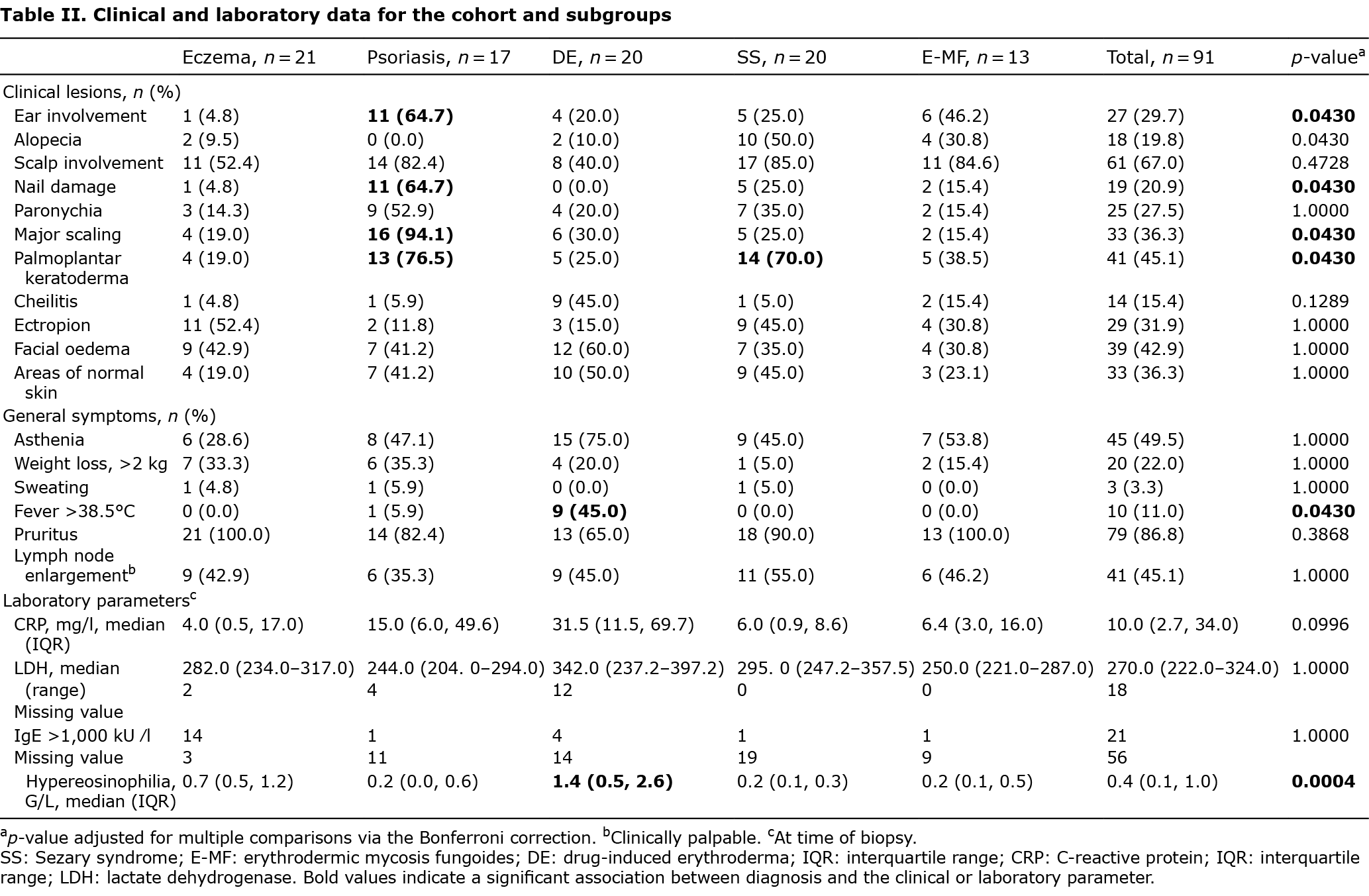
Clinical features are detailed in Table II. Nail damage was more frequent in patients with psoriasis (11/17, 64.7%, p = 0.0430). Severe scaling and ear involvement occurred more frequently in the psoriasis subgroup (p = 0.0430) (Fig. 1). Ten out of 91 patients had fever, most were in the DE subgroup (9/18, 45%).

Laboratory and T-cell clonality findings
The blood eosinophil count was higher in the DE subgroup (p = 0.0004). An elevated LDH level was observed in 52 of 73 patients, with no significant differences between subgroups. All patients with SS had B2 stage disease; 14 patients with other diseases had an abnormal immunophenotype corresponding to “B1” stage: 5 with eczema, 4 with psoriasis, and 5 with E-MF (Table II). T-cell clonality PCR analyses revealed skin T-cell clones in all 33 patients with CTCL, and 8 of 29 patients (29%) with eczema or psoriasis. Blood T-cell clones were found in 28 of 31 patients (90%) with CTCL, as well as in 8 of 26 patients (31%) with eczema or psoriasis. However, identical skin T-cell clones in different biopsies and/or identical blood and skin T-cell clones were only seen in patients with E-MF or SS (Table III).

Histopathological findings
Histopathological findings of the 91 patients are summarized in Table IV and Fig. 2. Psoriasis hyperplasia, parakeratosis, and neutrophil micro-abscesses in the stratum corneum were most frequent in the psoriasis subgroup, but were also seen in the eczema and SS subgroups. Spongiosis was observed in all subgroups. Eosinophilic infiltration was frequent in patients with eczema (57.1%), DE (50%), psoriasis (47.1%), and MF-E (38.5%), but very rare in those with SS (5%). Epidermotropism was seen in 70% of the cases with DE, 92% of E-MF and 80% of the SS cases. Atypical lymphocytes were seen in 46.2% of patients with E-MF, 55% of those with SS, and 25% of those with DE. The diagnosis suggested by histopathological analysis alone was consistent with the final diagnosis in 49 of 91 patients (53.8%).


Immunohistochemical findings
The percentage of PD-1+ lymphocytes was significantly higher in the SS subgroup (median 75%, p < 0.0001), with perivascular location. In 14 of 20 SS patients (70%), >50% of the T cells expressed PD-1, including 10 of 20 patients with >75% PD-1+ T cells. Of 20 SS patients, 1 was PD-1–. A PD-1 expression cut off 50% was associated with SS with 70% sensitivity and 93% specificity. The median H-score for the SS subgroup (115.0) was significantly higher than in any of the other subgroups (p < 0.0001) (Fig. 3, Table IV). An H-score ≥100 was associated with SS ((p < 0.001) with very high specificity, but poor sensitivity (99% and 60%, respectively). An H-score ≥ 50 was also strongly associated with SS ((p < 0.001), with higher sensitivity (75%) and high specificity (93%). H-scores were not significantly different in patients with E-MF compared with those with inflammatory dermatitis (median 30, (p = 0.4409). These data were subjected to receiver operating characteristic curve analysis (Fig. 3). The reproducibility of the interpretation of PD-1 expression was evaluated. H-scores exhibited good reproducibility with an ICC of 0.81 (95% confidence interval; 95% CI 0.62–0.91) and kappa coefficient of 0.66 (95% CI 0.33–1.00). For estimations of the intensity of PD-1 expression, ICCs of 0.72 (95% CI 0.45–0.87), 0.58 (95% CI 0.24–0.80), and 0.17 (95% CI –0.25–0.53) were obtained for the +, ++, and +++ intensities, respectively.

Both the percentage of lymphocytes expressing ICOS and the ICOS H-score were slightly, but not significantly, higher in the SS subgroup. CXCL13 positivity was significantly associated with SS (70%, p = 0.0430). CXCL13 expression was also detected in 38.5% of the E-MF patients. Among patients with inflammatory dermatitis, CXCL13 expression was rare or absent in those with eczema (19%) or psoriasis (0%), but was more common in those with DE (35%).
The median percentage CD7 loss was significantly higher in E-MF and SS patients (70% for both groups, p = 0.0220) than in those with inflammatory dermatitis. A ≥ 50% loss of CD7 expression was observed in 16 of 20 (80%) SS patients, 11 of 13 (84.6%) E-MF patients, and 27 of 58 (46.6%) inflammatory dermatitis patients.
The Ki-67 proliferative index was significantly higher for SS and DE patients (median 15.0 for both). Differences in the Ki-67 proliferative index among subgroups were relatively small (range 2–21.2). Similar results were obtained from sensitivity analysis using elastic-net penalized multinomial regression.
DISCUSSION
This study retrospectively investigated the clinical, biological, histological, and phenotypical features in a large cohort (n = 91) of erythroderma patients with well-defined diagnoses. The main discriminant marker was PD-1 expression, which may be an important tool for the differential diagnosis of erythroderma. PD-1 expression in > 50% of T cells and a PD-1 H-score ≥ 50 were strongly associated with an SS diagnosis. PD-1 was also expressed in E-MF and inflammatory erythroderma patients, but at lower levels.
Previous reports have shown that PD-1 expression is a feature of SS, but it had not been precisely quantified or compared with erythroderma cases derived from other causes, including E-MF (10, 18). To avoid heterogeneity and increase the accuracy of PD-1 expression quantification, we decided to use the H-score as a semi-quantitative method of comparing PD-1 expression among erythroderma subgroups. In this study, a high PD-1 H-score was significantly associated with a diagnosis of SS (median 115) (Fig. 3). A PD-1 H-score ≥ 50 was more sensitive, more specific, and had greater intra-rater reliability compared with standard evaluation using percentage cell expression. Nevertheless, the evaluation of PD-1 expression using the percentage of PD-1+ lymphocytes still provides valuable data. PD-1 expression was absent in only 1 case of SS in our series, which might have been due to PD-1 deletion as previously reported in aggressive cases (27). Data series on the expression of PD-1 in E-MF cases are lacking, as most reports include only patch/plaque patients (17, 20, 34, 35). In our study, PD-1 expression in E-MF patients was lower than in those with SS, with a median H-score of 30. PD-1 expression has also been observed in cases of inflammatory dermatosis, but precise data are not provided in the literature. In a previous study, 46% of the patients with mixed erythrodermic inflammatory dermatoses expressed PD-1 (10). Here, we determined that PD-1 expression was frequently seen with inflammatory dermatosis, but in patients with other pathologies, less than 50% of the lymphocytes expressed PD-1. In addition, H-scores were < 50 in most of the non-inflammatory dermatosis cases (median for eczema: 5, median for psoriasis: 2, median for DE: 10), except for 2 patients diagnosed with psoriasis.
We considered several other TFH markers in the current study. There was no significant difference in either the median expression level of ICOS or in the ICOS H-score among the patient subgroups. Conversely, CXCL13 expression was associated with SS (70%) compared with the other subgroups, including E-MF patients (38.5%), with these results similar to those reported by Picchio et al. (25) We observed that, in most of the SS patients, atypical T cells expressed both PD-1 and CXCL13, contrary to the patients with E-MF, supporting the conclusion that SS and E-MF are distinct pathologies arising from separate T-cell functional subsets (36).
There were several correlations between clinical features and erythroderma aetiology. Male patients, severe scaling, and ear involvement are more common in psoriasis cases (4, 37). Palmoplantar keratoderma was significantly associated with psoriasis and SS, as described previously (4, 5, 37, 38), but it was non-discriminant.
We also noted blood immunophenotype abnormalities in 5 eczema and 4 psoriasis cases. These patients’ blood immunophenotypes included CD3+CD4+CD7− or CD3+CD4+CD26− circulating T-cell populations, which correspond to stage B1 disease. These phenotypic aberrations have been previously described in patients with benign inflammatory dermatosis or infections (39, 40), and may lead to an incorrect diagnosis in the presence of erythroderma. Conversely, molecular data, such as those indicating the presence of identical T-cell clones in different skin biopsies and/or identical skin and blood T-cell clones, may provide important information for diagnosing CTCL (41). However, T-cell clones isolated from unique skin biopsies or blood were not taken into account.
In the current study, agreement between the pathological and final diagnoses occurred in only 53.8% of cases. However, although atypical lymphocytes and epidermotropism were frequent features of both SS (55%) and E-MF (46.2%), these were also seen in patients in other subgroups, in particular in DE patients; thus these observations must be interpreted carefully (42). Moreover, an absence or paucity of epidermotropism in erythrodermic CTCL was noted in some of our cases, as reported previously (10, 42).
In conclusion, these results support the use of a PD-1 H-score as a semi-quantitative and reproducible tool for evaluating PD-1 expression. A high H-score was associated with SS diagnosis, among other causes of erythroderma, including E-MF. Future studies should focus on PD-1-depleting antibodies for the treatment of SS (43).
ACKNOWLEDGEMENTS
We would like to thank the patients and their families. We would also like to thank the technicians from the pathology department for their help in acquiring immunohistochemistry data.
The authors have no conflicts of interest to declare.
REFERENCES
- Botella-Estrada R. Erythroderma. A clinicopathological study of 56 cases. Arch Dermatol 1994; 130: 1503–1507.
- Yuan X-Y, Guo J-Y, Dang Y-P, Qiao L, Liu W. Erythroderma: a clinical-etiological study of 82 cases. Eur J Dermatol 2010; 20: 373–377.
- Li J, Zheng H-Y. Erythroderma: a clinical and prognostic study. Dermatology 2012; 225: 154–162.
- Khaled A, Sellami A, Fazaa B, Kharfi M, Zeglaoui F, Kamoun M. Acquired erythroderma in adults: a clinical and prognostic study: acquired erythroderma in adults. J Eur Acad Dermatol Venereol 2009; 24: 781–788.
- Miyashiro D, Sanches JA. Erythroderma: a prospective study of 309 patients followed for 12 years in a tertiary center. Sci Rep 2020; 10: 9774.
- César A, Cruz M, Mota A, Azevedo F. Erythroderma. A clinical and etiological study of 103 patients. J Dermatol Case Rep 2016; 10: 1–9.
- Zip C, Murray S, Walsh NMG. The specificity of histopathology in erythroderma. J Cutan Pathol 1993; 20: 393–398.
- Ram-Wolff C, Martin-Garcia N, Bensussan A, Bagot M, Ortonne N. Histopathologic diagnosis of lymphomatous versus inflammatory erythroderma: a morphologic and phenotypic study on 47 skin biopsies. Am J Dermatopathol 2010; 32: 755–763.
- Kamarashev J, Burg G, Kempf W, Schmid MH, Dummer R. Comparative analysis of histologicai and immunohistological features in mycosis fungoides and Sezary syndrome. J Cutan Pathol 1998; 25: 407–412.
- Klemke CD, Booken N, Weiss C, Nicolay JP, Goerdt S, Felcht M, et al. Histopathological and immunophenotypical criteria for the diagnosis of Sézary syndrome in differentiation from other erythrodermic skin diseases: a European Organisation for Research and Treatment of Cancer (EORTC) Cutaneous Lymphoma Task Force Study of 97 cases. Br J Dermatol 2015; 173: 93–105.
- Willemze R, Cerroni L, Kempf W, Berti E, Facchetti F, Swerdlow SH, et al. The 2018 update of the WHO-EORTC classification for primary cutaneous lymphomas. Blood 2019; 133: 1703–1714.
- Walsh NMG, Prokopetz R, Tron VA, Sawyer DM, Kevin Walters A, Murray S, et al. Histopathology in erythroderma: review of a series of cases by multiple observers. J Cutan Pathol 1994; 21: 419–423.
- Mangold AR, Thompson AK, Davis MD, Saulite I, Cozzio A, Guenova E, et al. Early clinical manifestations of Sézary syndrome: a multicenter retrospective cohort study. J Am Acad Dermatol 2017; 77: 719–727.
- Samimi S, Benoit B, Evans K, Wherry EJ, Showe L, Wysocka M, et al. Increased programmed death-1 expression on CD4+ T cells in cutaneous T-cell lymphoma: implications for immune suppression. Arch Dermatol 2010; 146: 1382.
- Nguyen GH, Olson LC, Magro CM. Upregulation of inhibitory signaling receptor programmed death marker-1 (PD-1) in disease evolution from cutaneous lymphoid dyscrasias to mycosis fungoides and Sezary’s syndrome. Ann Diagn Pathol 2017; 28: 54–59.
- Kantekure K, Yang Y, Raghunath P, Schaffer A, Woetmann A, Zhang Q, et al. Expression patterns of the immunosuppressive proteins PD-1/CD279 and PD-L1/CD274 at different stages of cutaneous T-cell lymphoma/mycosis fungoides. Am J Dermatopathol 2012; 34: 126–128.
- Çetinözman F, Jansen PM, Vermeer MH, Willemze R. Differential expression of programmed death-1 (PD-1) in Sézary syndrome and mycosis fungoides. Arch Dermatol 2012; 148: 1379.
- Çetinözman F, Jansen PM, Willemze R. Expression of programmed death-1 in skin biopsies of benign inflammatory vs. lymphomatous erythroderma. Br J Dermatol 2014; 171: 499–504.
- Saulite I, Ignatova D, Chang Y-T, Fassnacht C, Dimitriou F, Varypataki E, et al. Blockade of programmed cell death protein 1 (PD-1) in Sézary syndrome reduces Th2 phenotype of non-tumoral T lymphocytes but may enhance tumor proliferation. Oncoimmunology 2020; 9: 1738797.
- Bosisio FM, Cerroni L. Expression of T-follicular helper markers in sequential biopsies of progressive mycosis fungoides and other primary cutaneous T-cell lymphomas. Am J Dermatopathol 2015; 37: 115–121.
- Jogdand GM, Mohanty S, Devadas S. Regulators of Tfh cell differentiation. Front Immunol 2016; 7: 520.
- Gaulard P, de Leval L. Follicular helper T cells: implications in neoplastic hematopathology. Semin Diagn Pathol 2011; 28: 202–213.
- Lunning MA, Vose JM. Angioimmunoblastic T-cell lymphoma: the many-faced lymphoma. Blood 2017; 129: 1095–1102.
- Hutloff A. Regulation of T follicular helper cells by ICOS. Oncotarget 2015; 6: 21785–21786.
- Picchio MC, Scala E, Pomponi D, Caprini E, Frontani M, Angelucci I, et al. CXCL13 is highly produced by Sezary cells and enhances their migratory ability via a synergistic mechanism involving CCL19 and CCL21 chemokines. Cancer Res 2008; 68: 7137–7146.
- Vergnolle I, Douat-Beyries C, Boulinguez S, Rieu J-B, Vial JP, Baracou R, et al. CD158k and PD-1 expressions define heterogeneous subtypes of Sezary syndrome. Blood 2022; 6: 1813–1825.
- Park J, Daniels J, Wartewig T, G. Ringbloom K. Integrated genomic analyses of cutaneous T-cell lymphomas reveal the molecular bases for disease heterogeneity. Blood 2021; 138: 1225–1236.
- Scarisbrick JJ, Prince HM, Vermeer MH, Quaglino P, Horwitz S, Porcu P, et al. Cutaneous lymphoma international consortium study of outcome in advanced stages of mycosis fungoides and Sézary syndrome: effect of specific prognostic markers on survival and development of a prognostic model. J Clin Oncol 2015; 33: 3766–3773.
- Cohen DA, Dabbs DJ, Cooper KL, Amin M, Jones TE, Jones MW, et al. Interobserver agreement among pathologists for semiquantitative hormone receptor scoring in breast carcinoma. Am J Clin Pathol 2012; 138: 796–802.
- Hirsch FR, Varella-Garcia M, Bunn PA, Di Maria MV, Veve R, Bremnes RM, et al. Epidermal growth factor receptor in non-small-cell lung carcinomas: correlation between gene copy number and protein expression and impact on prognosis. J Clin Oncol 2003; 21: 3798–3807.
- Specht E, Kaemmerer D, Sänger J, Wirtz RM, Schulz S, Lupp A. Comparison of immunoreactive score, HER2/ neu score and H score for the immunohistochemical evaluation of somatostatin receptors in bronchopulmonary neuroendocrine neoplasms. Histopathology 2015; 67: 368–377.
- Leclaire Alirkilicarslan A, Dupuy A, Pujals A, Parrens M, Vergier B, Robson A, et al. Expression of TFH markers and detection of RHOA p.G17V and IDH2 p.R172K/S mutations in cutaneous localizations of angioimmunoblastic T-cell lymphomas. Am J Surg Pathol 2017; 41: 1581–1592.
- Ryu H-J, Kim S-I, Jang H-O, Kim S-H, Oh S-H, Park S, et al. Evaluation of the International Society for Cutaneous Lymphoma Algorithm for the diagnosis of early mycosis fungoides. Cells 2021; 10: 2758.
- Wada DA, Wilcox RA, Harrington SM, Kwon ED, Ansell SM, Comfere NI. Programmed death 1 is expressed in cutaneous infiltrates of mycosis fungoides and Sézary syndrome. Am J Hematol 2011; 86: 325–327.
- Roncador G, Verdes-Montenegro J-FG, Tedoldi S, Paterson JC, Klapper W, Ballabio E, et al. Expression of two markers of germinal center T cells (SAP and PD-1) in angioimmunoblastic T-cell lymphoma. Haematologica 2007; 92: 1059–1066.
- Campbell JJ, Clark RA, Watanabe R, Kupper TS. Sézary syndrome and mycosis fungoides arise from distinct T-cell subsets: a biologic rationale for their distinct clinical behaviors. Blood 2010; 116: 767–771.
- Hawilo A, Zaraa I, Benmously R, Mebazaa A, Osman AB. Erythrodermic psoriasis: epidemiological clinical and therapeutic features about 60 cases. Tunis Med 2011; 89: 841–847.
- Martin SJ, Duvic M. Prevalence and treatment of palmoplantar keratoderma and tinea pedis in patients with Sézary syndrome. Int J Dermatol 2012; 51: 1195–1198.
- Rie MA, Catro I, Lier RAW, Bos JD. Expression of the T-cell activation antigens CD27 and CD28 in normal and psoriatic skin. Clin Exp Dermatol 1996; 21: 104–111.
- Liu L, Abken H, Pfohler C, Rappl G, Tilgen W, Reinhold U. Accumulation of CD4+CD7- T cells in inflammatory skin lesions: evidence for preferential adhesion to vascular endothelial cells. Clin Exp Immunol 2000; 121: 94–99.
- Delfau-Larue MH, Laroche L, Wechsler J, Lepage E, Lahet C, Asso-Bonnet M, et al. Diagnostic value of dominant T-cell clones in peripheral blood in 363 patients presenting consecutively with a clinical suspicion of cutaneous lymphoma. Blood 2000; 96: 2987–2992.
- Diwan AH, Prieto VG, Herling M, Duvic M, Jones D. Primary Sézary syndrome commonly shows low-grade cytologic atypia and an absence of epidermotropism. Am J Clin Pathol 2005; 123: 510–515.
- Decroos A, Giustiniani J, Pelletier L, Ingen-Housz-Oro S, Gaulard P, Ortonne N. PD1 in Sézary syndrome: a repressor of cell survival sometimes lost during progression, but a new target using depleting antibodies? Eur J Cancer 2021; 156: 14–15.