SHORT COMMUNICATION
Microneedle Electroporation for Intralesional Administration of Corticosteroid Treatment of Keloid Scar
Karina KARINA1–3*, Krista EKAPUTRI1, Hubert ANDREW2,4 and Johannes Albert BIBEN1
1Klinik Hayandra, Yayasan Hayandra Peduli, 2Hayandra Laboratory, Yayasan Hayandra Peduli, Jl. Kramat VI Number. 11, Jakarta Pusat, 3Faculty of Medicine, Universitas Pembangunan Nasional Veteran Jakarta, Jl. Rs. Fatmawati, Jakarta, 4Faculty of Medicine, Universitas Indonesia, Jl. Salemba Raya Number 6, Jakarta Pusat, Indonesia. E-mail: karina@hayandra.com
Citation: Acta Derm Venereol 2023; 103: adv13402. DOI: https://doi.org/10.2340/actadv.v103.13402.
Copyright: © Published by Medical Journals Sweden, on behalf of the Society for Publication of Acta Dermato-Venereologica. This is an Open Access article distributed under the terms of the Creative Commons Attribution-NonCommercial 4.0 International License (https://creativecommons.org/licenses/by-nc/4.0/)
Accepted: Oct 3, 2023; Published: Dec 7, 2023
INTRODUCTION
Keloids are pathological scars thought to be caused by an abnormal wound-healing process. While benign, these scars do not regress and tend to extend beyond the original site over time, an attribute that differentiates keloids from hypertrophic scars, which usually do not invade the surrounding normal skin (1). In a study of 28 patients with keloid scars, Lee et al. (2) found that 86% experienced keloid-associated pruritus and 46% experienced pain. In addition, keloid scars may also result in psychosocial issues due to undesirable cosmetic impacts and functional impairment, leading to restricted mobility and a decrease in patients’ quality of life (3).
While various modalities exist, there is no single established treatment for keloid scars (4). Currently, intradermal corticosteroid injection is the most common form of treatment. Triamcinolone is generally used for this purpose and has been the corticosteroid of choice since the 1960s (5). However, intradermal keloid injection is associated with significant pain, and the procedure itself may also be physically challenging for the operator (4).
Microneedling is a relatively new alternative in the field of transdermal and intradermal drug delivery. These needles can enhance drug penetration, through the creation of microchannels. Microneedles are also less painful compared with conventional intradermal injections. Electroporation delivers a high frequency of high-voltage pulses to disrupt the lipid bilayer of cell membranes. This leads to the rearrangement of the cell membrane and formation of aqueous pores, which facilitate the transport of molecules of various weights and properties. The combination of microneedling with electroporation can yield an even better result in skin permeation (6, 7).
We report here a case of a patient with a keloid scar that was treated successfully with intradermal triamcinolone administered via microneedle electroporation.
CASE REPORT
A 29-year-old woman presented to our clinic, Klinik Hayandra, Jakarta, Indonesia, with keloid scars on her chest acquired after contracting varicella (chickenpox) approximately 24 months previously (Fig. 1). Approximately 20 months prior to presentation the scar had started to form and continued to grow. The scar was accompanied by intermittent pain and pruritus. When symptomatic, the worst pain and itch experienced by the patient was rated as 4 and 3, respectively, on a numerical rating scale (NRS). Informed consent was obtained for this study. The scar was assessed with the Vancouver Scar Scale (VSS), which ranges from 0 (representing normal skin) to 13 (representing worst scar imaginable) (8). Initially, the scar was assessed as a 6 out of 13 (vascularity: 0, pigmentation: 0, pliability: 4, height: 2) on the VSS.
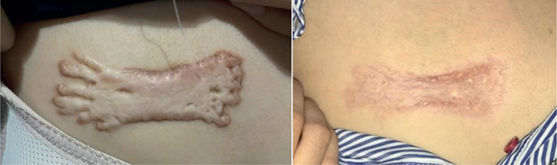
Comparison of the lesion before the initiation of microneedle electroporation corticosteroid treatment (left) and after the treatment (right). The wound measured at 4×9 cm at the beginning and 2.5×6.75 cm at the end. The result was attained after 8 months of routine treatment
At each session, approximately 30 min before the injection, topical anaesthetic cream containing lidocaine and prilocaine was applied on the keloid scar. The remaining cream was then wiped off and the injection site was cleaned with an alcohol swab. Topical application of 1 ml triamcinolone acetonide suspension (containing 40 mg of the drug) was administered to the keloid scar, drop-by-drop via a needleless syringe. Intralesional injection with the electroporation microneedling device (EPN®, Eunsung, Wonju-si, South Korea) was then performed. The multineedle system consisted of 9 33 G-sized needles, and was set at 4,500 punctures per min and the needles inserted to a depth of 0.5 mm. The electroporation system was set at level 2–3. The minimal bleeding that occurred was stopped by simple dabbing. The procedure took approximately 5 min and was repeated at 2–3-week intervals. The procedure took approximately 5 min and was repeated at 2–3-week intervals. The patient received treatment for a total of 15 times. After the procedure, antiseptic gel containing 40% polyhexanide was applied to the wound. The patient did not require painkillers such as paracetamol.
The result at the end of treatment is shown in Fig. 1, with a VSS score of 1 out of 13 (vascularity: 0, pigmentation: 0, pliability: 0, height: 1). The patient now reports the pain and itch from the scar as both very mild, i.e. NRS 1 at worst. It took approximately 26 months to attain these results (August 2020 to October 2022). The final photograph was taken in October 2022. However, it should be noted that the patient came for treatment only once in 2021 (in March) and resumed treatment only in March 2022. Adjusting for this, the amount of time the patient was routinely under treatment was approximately 8 months, with the procedure being performed a total of 15 times. Appendix S1 explains the technical aspects of this report in more detail.
DISCUSSION
The pathogenesis of keloid scars is yet to be fully elucidated, but they generally occur after injury or inflammation, such as surgery, burns, piercings, acne, or vaccination, in predisposed individuals. Previous in vitro research found that keloidal fibroblasts overproduce type I procollagen, express elevated levels of vascular endothelial growth factor, transforming growth factor (TGF)-β1 and β2, and have reduced growth factor requirements. They also have a lower rate of apoptosis due to downregulation of genes associated with apoptosis. The anatomical location of the lesion may also determine its morphology and prognosis, with facial keloids showing a higher rate of recurrence (3, 5). Genetics are also thought to play a role in keloid susceptibility, and several genetic variations, such as those in the 2q23 and 7p11 chromosomal regions, HLA-DRB1*15 genes, and multiple genes of the TGF-β pathway have been studied and proposed to be associated with keloid predisposition (1).
In general, there are 3 categories of therapy for keloid scars: modulation of inflammation processes, alteration of collagen metabolism, and physical manipulation of the lesion. Intralesional triamcinolone injections have been shown to reduce the volume and height of keloid scars, alleviate pruritus and pain, and prevent lesion recurrence. Corticosteroids tackle keloid scars in various ways. First, they are anti-inflammatory as they inhibit leukocyte migration and phagocytosis. Secondly, the anti-mitotic property of corticosteroids inhibits keratinocyte and fibroblast proliferation and function. Thirdly, corticosteroids have vasoconstrictor activity, which restricts the delivery of oxygen and nutrition to the scar (3, 4). Corticosteroids may also lower α-globulin concentrations (natural inhibitors of collagenase, which are abundant in keloid tissue) including α2-macroglobulin and alpha-1-antitrypsin (9, 10). Expression of TGF-β1 levels were also found to be significantly decreased in keloid fibroblast cell lines treated with steroids (4). TGF-β1 is able to increase tissue inhibitor of metalloproteinases and decrease matrix metalloproteinase levels (11). Together, this results in fibroblast and collagen degeneration, ultimately leading to clinical improvement of the keloid scar.
The response rates and recurrence rates for intralesional corticosteroid injections vary widely and can be unsatisfactory. The effectivity of intralesional corticosteroid injection alone for keloid scars is 50–100%, with recurrence rates of 33% and 50% after 1 and 5 years, respectively (3, 9). A previously published meta-analysis found that intralesional corticosteroid was superior to radiotherapy but equally as effective as other modalities. When combined with keloid excision, however, radio-therapy has a lower recurrence rate than corticosteroids (12). In addition to the variability of its effectiveness, the injections are painful and challenging to administer due to the dense fibrous tissue composition of keloid scars. The high pressure required to inject the drug and the resulting pain may deter patients from seeking further treatment, leading to suboptimal outcomes (13).
Microneedling in combination with topical products, such as platelet-rich plasma, stem cells, and minoxidil, as well as radiofrequency, have been explored as alternative treatment options. Previous studies have reported the use of radiofrequency microneedling with adjunct triamcinolone and onabotulinumtoxinA for hypertrophic scars and the effect of calcium electroporation in keloids (14, 15). Microneedling offers advantages, such as reduced pain, simplicity of technique, avoidance of accidental subcutaneous injection, improved patient compliance, and potentially better clinical outcomes. In the current case, pain from the procedure lasted 1 day and was described as stinging discomfort. However, the pain was very slight and there was no need for analgesics. However, further research is needed to fully evaluate the efficacy and safety of microneedling for keloids and hypertrophic scars (7).
In the current case, microneedle electroporation for the administration of intralesional corticosteroid for keloid scars was found to be safe and effective. However, this study has limitations; it includes only 1 subject and there was a period during which the patient did not receive the treatment, which may confound the results. Further studies are needed to investigate the cost-effectiveness, efficacy, and safety of microneedle electroporation, and to explore its potential impact on the pharmacokinetics of topical corticosteroid administration. Well-designed clinical trials are necessary to further validate the benefits of microneedle electroporation for intralesional corticosteroid administration in the treatment of keloids.
REFERENCES
- Tsai C-H, Ogawa R. Keloid research: current status and future directions. Scars, Burn Heal 2019; 5: 205951311986865
- Lee SS, Yosipovitch G, Chan YH, Goh CL. Pruritus, pain, and small nerve fiber function in keloids: a controlled study. J Am Acad Dermatol 2004; 51: 1002–1006.
- Perdanasari AT, Lazzeri D, Su W, Xi W, Zheng Z, Ke L, et al. Recent developments in the use of intralesional injections keloid treatment. Arch Plast Surg 2014; 41: 620–629.
- Morelli Coppola M, Salzillo R, Segreto F, Persichetti P. Triamcinolone acetonide intralesional injection for the treatment of keloid scars: patient selection and perspectives. Clin Cosmet Investig Dermatol 2018; 11: 387–396.
- Ojeh N, Bharatha A, Gaur U, Forde AL. Keloids: current and emerging therapies. Scars, Burn Heal 2020; 6: 205951312094049.
- Petchsangsai M, Rojanarata T, Opanasopit P, Ngawhirunpat T. The combination of microneedles with electroporation and sonophoresis to enhance hydrophilic macromolecule skin penetration. Biol Pharm Bull 2014; 37: 1373–1382.
- Ramaut L, Hoeksema H, Pirayesh A, Stillaert F, Monstrey S. Microneedling: where do we stand now? A systematic review of the literature. J Plast Reconstr Aesthetic Surg 2018; 71: 1–14.
- Sullivan T, Smith J, Kermode J, Mclver E, Courtemanche DJ. Rating the burn scar. J Burn Care Rehabil 1990; 11: 256–260.
- Eisen AZ, Bauer EA, Jeffrey JJ. Human skin collagenase. The role of serum alpha-globulins in the control of activity in vivo and in vitro. Proc Natl Acad Sci 1971; 68: 248–251.
- Diegelmann RF, Bryant CP, Cohen IK. Tissue alpha-globulins in keloid formation. Plast Reconstr Surg 1977; 59: 418–423.
- Lin P-S, Chang H-H, Yeh C-Y, Chang M-C, Chan C-P, Kuo H-Y, et al. Transforming growth factor beta 1 increases collagen content, and stimulates procollagen I and tissue inhibitor of metalloproteinase-1 production of dental pulp cells: role of MEK/ERK and activin receptor-like kinase-5/Smad signaling. J Formos Med Assoc 2017; 116: 351–358.
- Wang R, Danielsen PL, Ågren MS, Duke J, Wood F, Zeng XX, et al. Corticosteroid injection alone or combined with surgical excision of keloids versus other therapies including ionising radiotherapy: a systematic review and meta-analysis of randomised controlled trials. Eur Burn J 2021; 2: 41–54.
- Vishwanath G, Sridhar J. Microneedle-assisted steroid delivery therapy in the management of hypertrophic scars: a split-lesion study. J Mar Med Soc 2022; 24: 84–88.
- Juhasz M, Fackler N, Pham C, Mesinkovska NA. Combination therapy using radiofrequency microneedling and corticosteroids for hypertrophic scars: a case report. J Clin Aesthet Dermatol 2020; 13: 27–28.
- Falk H, Vissing M, Wooler G, Gehl J. Calcium electroporation for keloids: a first-in-man phase I study. Dermatology 2021; 237: 961–969.