QUIZ SECTION
A Solitary Pinkish Nodule on the Abdomen: A Quiz
Roberto RUSSO1,2, Emanuele COZZANI1,2*, Federica D’AGOSTINO1,2, Antonio GUADAGNO3 and Aurora PARODI1,2
1Department of Health Sciences, University of Genoa, Genoa, 2Dermatology Unit, and 3Pathology Unit, San Martino Polyclinic Hospital, Genoa, Italy. *E-mail: emanuele.cozzani@unige.it
Citation: Acta Derm Venereol 2024; 104: adv18458. DOI https://doi.org/10.2340/actadv.v104.18458.
Copyright: © Published by Medical Journals Sweden, on behalf of the Society for Publication of Acta Dermato-Venereologica. This is an Open Access article distributed under the terms of the Creative Commons Attribution-NonCommercial 4.0 International License (https://creativecommons.org/licenses/by-nc/4.0/)
Published: Apr 3, 2024
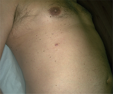
A 60-year-old Caucasian man presented with a solitary pinkish nodular asymptomatic lesion on his abdomen (Fig. 1). He reported that the lesion had developed during the previous month. He denied any personal or familiar history of skin cancer, and his medical history was unremarkable. He was not on immunosuppressive treatments.

Fig. 1. Pinkish nodule on the abdomen.
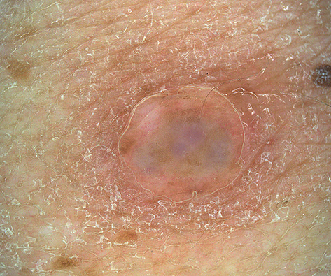
Clinical examination revealed a solitary pinkish, firm, tender nodule, 0.5 cm in size, on his right abdomen. No openings were visible on the skin. Dermoscopy revealed pink and white areas with small linear vessels (Fig. 2). An excisional biopsy was performed.

Fig. 2. Dermoscopic appearance of the lesion: 5 mm diameter nodule with pink and white areas, together with small linear vessels.
What is your diagnosis?
Differential diagnosis 1: Apocrine cystadenoma
Differential diagnosis 2: Spitzoid melanoma
Differential diagnosis 3: Skin metastasis from internal malignancy
Differential diagnosis 4: Dermatofibrosarcoma protuberans
See next page for answer.
ANSWERS TO QUIZ
A Solitary Pinkish Nodule on the Abdomen: A Commentary
Diagnosis: Apocrine Cystadenoma
Microscopic examination revealed a multiloculated intradermal cystic lesion lined with multiple layer of cuboidal epithelial cells without atypia, showing a characteristic apocrine-type secretion.
Apocrine cystadenoma (AC) is a relatively uncommon benign naevoid tumour of the skin, first described by Mehregan in 1964 (1). It represents an adenomatous cystic proliferation derived from apocrine glands and not a mere retention cyst. The tumour generally occurs as a well-defined, dome-shaped, translucent, asymptomatic and solitary papule or nodule. AC most commonly appears on the face, especially on the periorbital region and cheek, where apocrine glands occur with considerable regularity. The tumour usually ranges from 0.5 to 1 cm in size, although larger AC have been reported. AC may be skin-coloured, but also brown-greyish or dark bluish-black; thus it must be considered in the differential diagnosis of pigmented skin lesions (1–3). AC seems to be more common in female patients (4).
Dermoscopic examination reveals the presence of translucent to opaque skin-coloured lesion with pinkish-blue-grey central areas and linear-irregular vessels.
The presence of dark colour in AC seems to be due to a Tyndall effect or lipofuscin-rich fluid content of the cyst, considering the lack of melanocytes or melanophages within the stroma on histological examination (3).
Pink nodules are often a diagnostic challenge for dermatologists, since they may represent aggressive lesions, such as dermatofibrosarcoma protuberans or spitzoid melanoma. In addition, an internal malignancy might metastasize to the skin, appearing as a pink nodule. Dermoscopy is useful in differential diagnosis, but histological examination is warranted.
Histopathology shows the presence of a multiloculated intradermal cyst lined by 1 or more layer of cuboidal to columnar cells with eosinophilic cytoplasm, Periodic Acid Schiff-positive, diastase-resistant granules and basally located nuclei. The adluminal surface of the cells shows blebbing, which is indicative of decapitation secretion that is considered the hallmark of apocrine secretion. Fusiform myoepithelial Smooth Muscle Actin-positive cells are scattered peripherally beneath the secretory epithelium (1, 2, 4, 5) (Figs S1 and S2).
Although, in the past, AC and apocrine hidrocystoma (AH) have been used interchangeably to designate cystic lesions of apocrine glands, nowadays a histopathological distinction between “proliferative” AC and “non-proliferative” AH has been established (4).
This classification is based on the presence in AC of true papillary proliferative projections into the cystic cavity, which have a central fibrovascular core, a variable grade of cytological atypia, and increased Ki67 staining. These papillary projections are lacking in AH, which represent a simple retention cyst (4). Differential diagnosis of AC includes non-pigmented and pigmented lesions, such as sebaceous or epidermoid cyst, basal cell carcinoma, angiomas, melanocytic naevi, particularly blue naevi, or even malignant melanoma (2).
Usually, ACs are surgically excised. A wide, complete excision is particularly recommended for apocrine cystic lesion with florid papillary proliferation, especially when showing cytological evidence of atypia (4).
The current case was difficult to diagnose, as the occurrence of this lesion on the truncal region seems to be rare, since apocrine glands are normally found on the eyelids, axilla, groin and anogenital region. Also, its appearance was as a subcutaneous, firm, nodular lesion without specific features. The lesion was surgically excised with no recurrence.
REFERENCES
- Mehregan AH. Apocrine cystadenoma; a clinicopathological study with special reference to the pigmented variety. Arch Dermatol 1964; 90: 274–279.
- Hunter GA, Donald GF. Apocrine cystadenoma. Australas J Dermatol 1970; 11: 82–86.
- Mitsuishi T, Nogita T, Kawashima M. Apocrine cystadenoma arising on the ear. J Dermatol 1996; 23: 583–584.
- Sugiyama A, Sugiura M, Piris A, Tomita Y, Mihm MC. Apocrine cystadenoma and apocrine hidrocystoma: examination of 21 cases with emphasis on nomenclature according to proliferative features. J Cutan Pathol 2007; 34: 912–917.
- Charles NC, Patel P, Belinsky I, Oami S. Apocrine cystadenoma of the eyelid: a rare palpebral neoplasm. Report of 2 cases. Ophthalmic Plast Reconstr Surg 2018; 34: e67–e69.