Diagnosis and Management of Inherited Palmoplantar Keratodermas

Bjorn R. Thomas1,2 and Edel A. O’Toole1,2

1Centre for Cell Biology and Cutaneous Research, The Blizard Institute, Barts and the London School of Medicine and Dentistry, Queen Mary University of London, and 2Department of Dermatology, Royal London Hospital, Barts Health NHS Trust, ERN-Skin, London, UK

ABSTRACT

Inherited monogenic palmoplantar keratodermas are a heterogeneous group of conditions characterised by persistent epidermal thickening of the palmoplantar skin. Palmoplantar keratodermas are grouped depending on the morphology of the keratoderma into diffuse, focal/striate or papular/punctate. Some palmoplantar keratodermas just affect the skin of the palms and soles and others have associated syndromic features which include changes in hair, teeth, nails, hearing loss or cardiomyopathy. Next generation sequencing has helped discover genes involved in many of these conditions and has led to reclassification of some palmoplantar keratodermas. In this review, we discuss the diagnostic features of palmoplantar keratodermas and management options.

Key words: keratoderma; palmoplantar; keratin; genetic; inherited.

Accepted Feb 12, 2020; Epub ahead of print Mar 9, 2020

Acta Derm Venereol 2020; 100: adv00094.

Corr: Prof. Edel A. O’Toole, Centre for Cell Biology and Cutaneous Research, The Blizard Institute, Barts and the London School of Medicine and Dentistry, 4 Newark Str, London E1 2AT, UK. E-mail: e.a.otoole@qmul.ac.uk

SIGNIFICANCE

The palmoplantar keratodermas are a complex group of diseases where the main feature is thickening of the skin of the palms and soles. Genetic testing has given insight into the biology of these conditions and has allowed experts to reclassify them. In this review, we present a summary of the key features of the major types of palmoplantar keratodermas and discuss their management.

INTRODUCTION

The palmoplantar keratodermas (PPK) are a complex group of conditions that are characterised by persistent epidermal thickening (hyperkeratosis) of palmoplantar skin. The PPK are traditionally classified as hereditary (HPPK) or acquired. The main feature distinguishing hereditary from acquired PPK is the presence of a positive family history, early onset of disease, associated syndromic features and relative treatment resistance (1). Sporadic (spontaneous) mutations need to be considered in those without a family history or late onset disease (2).

Next generation sequencing has given us a better understanding of HPPK pathophysiology and has shown that one genotype can have several phenotypes. This has led to reclassification of some PPK thought previously to be distinct entities. Laboratory investigation shows that palmoplantar skin is a site at which multiple molecular pathways converge: gap junctions via connexins, intracellular adhesion through desmosomes and mechanical stability by means of the keratin cytoskeleton amongst others (3).

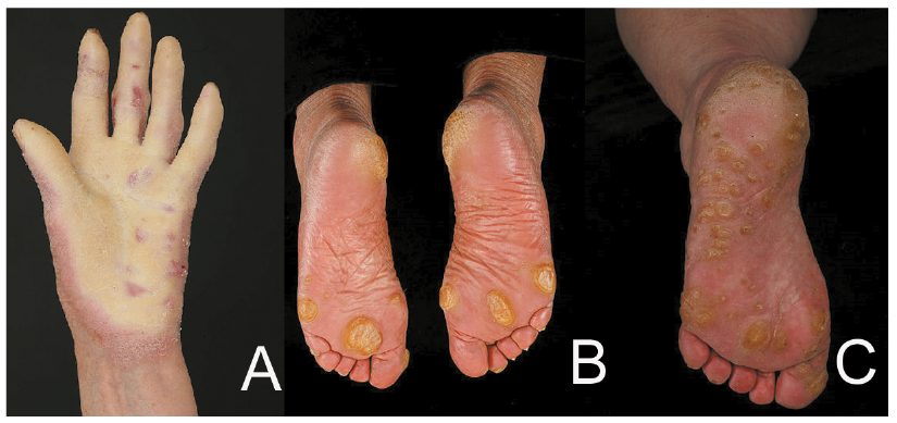
An initial approach to PPK is to take a history asking about age of onset, palmoplantar pain and/or blistering, sweating and infection and other associated features including hearing loss, abnormal hair, nail or teeth/ mucosal problems, cysts and family history including family history of cancer. Clinical examination can usually differentiate PPK into 3 groups: diffuse, focal or punctate (Fig. 1). The clinical features and management will be discussed in this review and are summarized in Table I.


Fig. 1. Patterns of palmoplantar keratodermas. A) diffuse, B) focal and C) punctate.
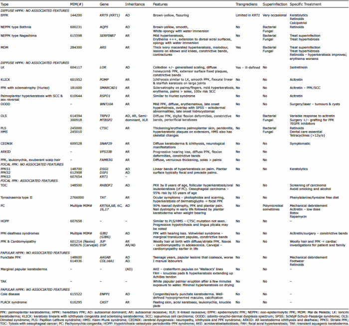

Table I. Summary of hereditary palmoplantar keratodermas (HPPK)

DIFFUSE HEREDITARY PALMOPLANTAR KERA­TODERMAS: NO ASSOCIATED FEATURES

Diffuse epidermolytic PPK (EPPK; MIM# 144200, KRT9, KRT1) is the most common diffuse PPK with epidermolytic changes in suprabasal keratinocytes seen on histology (4). It is inherited in an autosomal dominant (AD) fashion due to mutations in KRT9 and sometimes KRT1 (5, 6). The KRT9 gene encodes for the type I keratin, keratin 9, which is mainly expressed in suprabasal palmoplantar skin. Type I keratins form heterodimers with type II keratins, in this instance, possibly keratin 1, found in the epidermis including the palms and soles, to form intermediate filaments which provide strength to the skin (7, 8).

This PPK develops in infancy and in adults the hyperkeratosis is brown-yellow and confluent with fissuring, confined to the palmoplantar surfaces with an erythematous edge. Limited transgradient lesions or flexural hyperkeratosis may indicate KRT1 mutations (9). There may be a history of blistering and knuckle pads have been reported.

Treatment is mainly by mechanical debridement and use of keratolytics like urea, salicylic acid and lactic acid in emollient, sometimes under occlusion. Oral retinoids can help but pain from increased fragility limits their use (10, 11). Topical calcipotriol has been reported to be of benefit (12). Small inhibitory RNA therapy may be a possibility for the future (13).

DIFFUSE NON-EPIDERMOLYTIC PALMO­PLANTAR KERATODERMAS

Non-epidermolytic PPK type Bothnia (MIM# 600231, AQP5) was first described in Northern Sweden and is due to heterozygous missense mutations in AQP5 (14). This gene encodes for the water-channel protein aquaporin-5, which is expressed in exocrine glands but also the plasma membrane of palmar stratum granulosum. Mutations in the gene allow these cells to transport water by forming open water channels at this site.

This PPK usually starts in the first few months of life and is classically a brown-yellow, smooth keratoderma with an erythematous edge. Due to the defect in aquaporins, water immersion leads to a white spongy appearance which lasts for about 30 min. Pitted keratolysis and dermatophyte superinfection is common and can be treated with topical erythromycin or oral anti-fungals (15). Acitretin at low doses can be helpful.

NEPPK type Nagashima (MIM# 615598, SERPINB7) is an autosomal recessive (AR) PPK due to mutations in SERPINB7 described in Japanese and Chinese patients. Mutations in this gene may cause uncontrolled activity of proteases in the stratum corneum leading to increased water permeation (16, 17).

The condition presents in early life and is characterised by mild hyperkeratosis and striking redness extending to the dorsum fingers/feet and anterior wrist (18). A white spongy appearance after water immersion is seen (19) and associated hyperhidrosis and bacterial/fungal superinfection can be present.

Mal de Meleda (MDM; MIM#248300, ARS) is an eponymous AR PPK named after the Island of Mljet (née Meleda) (20). Mutations in ARS which encodes SLURP-1 cause MDM (21). SLURP-1 stimulates nicotinic acetylcholine receptors which regulate keratinocyte growth. When SLURP-1 is not functioning, it is thought that there is a reduction in keratinocyte apoptosis regulation (22).

MDM is characterised by a diffuse, ivory-yellow macerated hyperkeratosis with a characteristic malodour and striking erythematous trangradiens that extends to dorsal surfaces. A key feature includes lesions on the elbows and knees (21). Perioral hyperkeratosis and erythema can be present (23). The disease starts in infancy and progresses through life. Flexion contractures can occur and constrictive bands can lead to spontaneous amputation (24). Nail thickening, subungual hyperkeratosis and koilonychia can be present. The diffuse keratoderma of Gamborg-Nielsen also due to ARS mutations is likely a mild variant of MDM (25). Interestingly female heterozygotes can also present with a mild phenotype (26).

Treatment of bacterial/fungal superinfection and the hyperhidrosis is helpful, although the mainstay of treatment is oral retinoids which improve the hyperkeratosis although the erythema may worsen (27, 28).

DIFFUSE HEREDITARY PALMOPLANTAR KERA­TODERMAS: WITH ASSOCIATED FEATURES

Loricrin keratoderma (LK; MIM# 604117, LOR) is AD and starts in early childhood. It is due to a mutation in LOR which interferes with the regulation of epidermal cornification (29). Some children are born with a collodion membrane and generalised scaling from birth may be noted (30). During childhood the PPK develops with a characteristic diffuse, honeycomb pattern which can extend to wrist/ankles and is associated with non-migratory red plaques on the extensor surfaces of joints (31). Trangradiens is present but the edges of the hyperkeratosis are ill-defined. Constrictive bands can develop. Knuckle pads may be present (32) and hearing is intact.

Isotretinoin has been reported as helpful (33). In the future, there may be a role for treating LK with vascular endothelial growth factor 2 receptor inhibitors (34).

Keratosis linearis with ichthyosis congenita and sclerosing keratoderma (KLICK, MIM#601952, POMP) presents in early childhood and similar to LK, starts with generalised erythema and fine scaling with the subsequent diffuse, smooth PPK (35). Inheritance of this PPK is AR caused by mutations in the POMP gene, which lead to endoplasmic reticular stress and subsequent dysfunctional profilaggrin processing (36, 37). Flexural linear and starfish keratoses overlying large joints are distinctive (35). Acitretin can be helpful for both the ichthyosis and keratoderma (38).

PPK with scleroatrophy (Huriez syndrome, MIM #181600) is a cancer-related PPK caused by haploinsufficiency in SMARCAD1 (39). Scleroatrophy is seen across the entire palm and fingers (40) with mild hyperkeratosis of the palms. The affected skin is often red and the palms are usually more severely affected than the soles. Hypoplastic nail changes may be present and 50% experience hypohidrosis. The most important characteristic is the 100-fold increased risk of developing squamous cell carcinoma (SCC) in the affected skin. Acitretin may be helpful for the PPK and prevention of SCC (41).

Palmoplantar hyperkeratosis with squamous cell carcinoma of skin and sex reversal (MIM#610644, RSPO1) is similar to Huriez syndrome as it is a mild PPK with sclerodactyly and nail hypoplasia (42). This condition is AR caused by mutations in RSPO1 (43). This gene is responsible for stabilising β-catenin in the Wnt signaling pathway which antagonises SRY/SOX9 actions for sex determination (44). A characteristic feature is the female to male sex reversal seen in females. The karyotype is 46, XX. Predisposition for cutaneous SCC and also laryngeal SCC is noted (45). Periodontitis with loss of teeth may be present.

Odonto-onycho-dermal dysplasia spectrum (OODD) is an AR condition caused by mutations in WNT10A which starts in early life (46). In absence of WNT10A, β-catenin pathway activity and epithelial progenitor proliferation are reduced. In these patients, Wnt-active stem cells are seen in sweat ducts, hair follicles, nails and taste buds and there are differentiation abnormalities in palmoplantar skin (47).

Typically the PPK is mild, diffuse and erythematous with late onset palmoplantar hyperhidrosis (48). There is overlap with Schöpf-Schulz-Passarge syndrome (SSPS) and patients can have hypodontia with abnormal teeth, nail hypoplasia, smooth tongue and hypotrichosis (49). Eyelid hidrocystomas and other benign adnexal tumours can present at a later age (50). Biopsy of palmoplantar skin shows eccrine syringoadenomatosis (46). Tumours/cysts may need treatment with surgery or laser.

Olmsted syndrome (OLS; MIM# 614594 - TRPV3, 300918 - MTBSP2) typically presents as a severe mutilating transgradient keratoderma. AD, AR, semi-dominant and X-linked recessive (XLR) forms have been described caused by mutations in TRPV3 (AD, AR) and MBTPS2 (XLR) (51, 52) The Ca2+-permeable cation channel TRPV3 is expressed abundantly in keratinocytes, associated with TGF-α/EGFR signalling and may play a role in keratinocyte differentiation by elevating Ca2+ within these cells (53). MBTPS2 mutations in skin may cause a decrease in responsiveness to sterols subsequent to depletion of proteases (54).

The keratoderma is diffuse and can be associated with digital flexion deformities and constrictive bands. Periorificial/ear/nose/umbilical keratoses can also be present. Dystrophy of teeth, nails and cornea, alopecia, erythromelalgia and joint laxity have also been reported. A milder phenotype can simulate pachyonychia congenita (PC) (55). Melanoma and SCC have been reported in OLS (56).

Treatment in general is difficult with variable response to systemic retinoids. Topical anti-inflammatories can be helpful for hyperkeratosis and itching. Surgery with excision and grafting of the keratoderma can lead to more favourable long-term outcomes (2). Finally, there has been one report of a patient treated with the EGFR inhibitor, erlotinib, which gave a transient improvement (57).

PPK with periodontitis (MIM#245000, allelic disease: Haim-Munk #245010, CTSC) encapsulates both Papillon-Lefèvre syndrome (PLS) and Haim-Munk syndrome (HMS). Both conditions are caused by homozygous mutations in CTSC. CTSC is expressed in the palms, soles, alveolar bone and keratinized gingiva; it plays a role in immune cell protease activation and possibly has a role in epidermal differentiation leading to this particular phenotype (58, 59).

Patients have thickening and erythema of the palmoplantar skin, associated with bacterial skin infections and periodontitis (60). The PPK typically starts/worsens with the breakthrough of the deciduous teeth and actually improves after tooth loss/reduction of gingival inflammation (61). Hyperkeratotic plaques on the extensor surfaces are seen. PLS is associated with pyogenic liver abscesses (62). HMS has the same features with arachnodactyly, onychogryphosis and acro-osteolysis, mainly described in Cochin Jews (63).

Retinoids have shown to improve the PPK and oral disease (62). Specialist dental care is essential. For those above the age of 12, low dose tetracycline may be helpful for gingivitis, even at subtherapeutic doses (64).

Cerebral dysgenesis, neuropathy, ichthyosis and PPK syndrome (CEDNIK; MIM#609528, SNAP29) is a PPK with neurological manifestations which starts in infancy. This AR condition is caused by mutations in SNAP29 which lead to abnormal lamellar granule formation with subsequent aberrant epidermal differentiation (65). Around one year of age a diffuse keratoderma and ichthyosis become apparent (65, 66). Histology of CEDNIK demonstrates clear vesicles in the top 3 layers of the epidermis. Treatment is symptomatic.

Autosomal recessive keratoderma ichthyosis and deafness (ARKID) is caused by mutations in VPS33B. Mutations in this gene can lead to abnormal lamellar body morphology and function and impaired barrier formation. Patients present with progressive hearing loss (normal at birth) and delayed development. The PPK that develops is diffuse and associated with flexion deformities and autoamputation (67).

PPK, leukonychia and exuberant scalp hair is caused by AR mutations in FAM83G. FAM83G may have a role as a suppressor of the Wnt signalling pathway. Diffuse, verrucous thickening of soles and mild palmar involvement is noted. Leukonychia/dystrophy of the toenails and rapid hair growth are also seen (68).

FOCAL HEREDITARY PALMOPLANTAR KERATO­DERMAS: NO ASSOCIATED FEATURES

Striate PPK (PPKS) can be separated into PPKS1 (MIM# 148700, DSG1) (69), PPKS2 (MIM# 612908, DSP1) (70), and PPKS3 (MIM# 607654, KRT1) (71). The DSG1 (desmoglein 1) and DSP1 (desmoplakin) genes encode for desmosomal proteins required for intercellular adhesion of keratinocytes (72). Mutations in the V2 domain of KRT1 cause PPKS3 and disrupt the intermediate filament network.

Classically, striate PPK presents with linear bands of hyperkeratosis on the palmar surface (73). Diffuse or focal changes may also be present. It is usual for plantar changes to be focal and to present early in life (i.e. first or second year) and the palmar changes follow (71). If patients exhibit woolly/curly hair or abnormal dentition associated cardiomyopathy should be considered. Histology in PPKS can be helpful as it will demonstrate acantholysis of keratinocytes pointing to a desmosomal mutation (74).

FOCAL HEREDITARY PALMOPLANTAR KERATO­DERMAS: WITH ASSOCIATED FEATURES

Tylosis with oesophageal cancer (TOC; MIM#148500, RHBDF2) is rare condition that is AD and caused by gain of function mutations in RHBDF2 which create a hyperproliferative phenotype through continuous EGFR signalling (75). Patients present with focal keratoderma at sites of pressure usually by 8 years of age. Patients also have follicular hyperkeratosis and oral leukokeratosis similar to PC (76). Most patients with tylosis have a family history of oesophageal carcinoma and carry a risk of oesophageal cancer of 95% by age 65 years (77). Regular screening for oesophageal dysplasia is required and smoking and alcohol should be avoided.

Tyrosinaemia type II (MIM#276600, TAT) (78) is a very rare AR condition that initially presents in the first few months of life with ocular symptoms including photophobia and pain and subsequent ocular scarring (79). Hyperkeratosis of the palms that follows the fingerprints develops prior to a focal plantar keratoderma (80). About 50% of patients will have some form of intellectual disability and neurological signs. Increased tyrosine levels found in the bloods/urine, due to abnormal tyrosine aminotransferase function, can aid diagnosis (79) and symptoms may be prevented by a phenylalanine/tyrosine free diet.

Pachyonychia congenita (PC; Multiple MIM#) is a heterogeneous groups of conditions characterised by nail dystrophy and a painful focal keratoderma. The Pachyonychia Project (www.pachyonychia.org) has collected extensive data on PC collated in the International PC Research Registry (IPCRR). The current classification is based on keratin gene mutation: PC-6a, PC-6b, PC-6c, PC-16 and PC-17 (81). These 5 subtypes have replaced the original PC type 1 and 2 classification. Mutations in these genes lead to increased palmoplantar skin fragility due to disruption of keratin filament formation, nail changes and changes in the pilosebaceous unit.

The IPCRR data has shown that 90% of patients > 3 years old will have 3 clinical features: toenail dystrophy, plantar keratoderma and plantar pain (82). The hypertrophic nail dystrophy starts in the first few months of life up to 9 years. KRT6A mutations are associated with early onset disease. All nails need not be affected. The focal plantar keratoderma starts when children begin to weight bear with blistering under the calluses (83). Plantar pain has a neuropathic component and can be severe enough to require ambulatory aids. The palmar lesions are usually less prominent than the plantar lesions, except in the case of PC-KRT16 with striate lesions (82).

Follicular hyperkeratoses are seen in areas of friction. Oral leukokeratosis can resemble oral candidiasis and laryngeal involvement can lead to hoarseness and infantile respiratory obstruction (83). Cysts occur in all subtypes of PC although KRT17 mutations are usually associated with more steatocystomas/pilosebaceous cysts and natal teeth (less commonly KRT6A) (83). KRT6A mutations can also be associated with ear pain with feeding difficulties in infants/toddlers. PC-6C has a limited keratoderma and mild nail dystrophy (84).

Current treatment is largely mechanical paring of the calluses, assisted by a podiatrist, if necessary. Low dose acitretin can help in some patients but is associated with increased pain. Botox injections can also help reduce pain (85). The IPCC has had some promising results with siRNA and rapamycin (86, 87). Clinical trials of topical rapamycin are ongoing. As with most PPKs, comfortable foot wear and customised insoles are helpful.

Hypotrichosis-osteolysis-periodontitis-palmoplantar keratoderma syndrome (HOPP; MIM 607658) is a rare syndrome with a phenotype similar to PLS/HMS although CTSC mutations were not found. There is a striking keratoderma with a reticular pitted/punctate pattern (88). Progressive hypotrichosis from 6 years of age is seen sometimes with pili annulati. Lingua plicata can be noted at an early age (89).

PPK-deafness syndromes are mainly caused by GJB2 and rarely GJB6 mutations. Numerous gap junctions are present in the skin and inner ear. Mutations in gap junction genes lead to abnormal keratinocyte differentiation/growth and dyfunctional inner ear potassium ion recycling required for hearing (90).

Phenotypically these PPK-deafness syndrome are distinct and still carry their eponymous names. Despite having mutations in the same gene, keratitis-ichthyosis-deafness-like (MIM# 148210), hystrix-like ichthyosis-deafness (MIM#602540), palmoplantar keratoderma-deafness (MIM#148350), Bart-Pumphrey (MIM# 149200) and Vohwinkel (MIM#124500) syndromes have phenotypic differences likely explained by mutations in particular domains of connexin 26 (GJB2) (91). Cardinal features are PPK and hearing loss of varying severity. For example, Vohwinkel syndrome has marginal translucent papules which become confluent over time. It also has the ‘classic’ starfish keratoses on the knuckles and extensor surfaces of joints and pseudoainhum (92).

Oral retinoids are helpful for the constricting bands seen in Vohwinkel syndrome (93) but surgery may be required.

PPK and cardiomyopathy are similar to PPKS1&2 as they are also associated with keratinocyte disadhesion. Naxos syndrome (AR) caused by mutations in JUP (MIM# 601214) encoding plakoglobin presents with woolly hair at birth followed by a diffuse/striate keratoderma in the first year of life. Cardiomyopathy presents in adolescence and has 100% penetrance (94). Carvajal-Heurta syndrome (CHS), caused by mutations in DSP (MIM# 605676), is like Naxos although the cardiomyopathy presents earlier in the teens and is usually biventricular. Some patients with CHS have short woolly hair and keratoses on the elbows/knees (95, 96). The DSP and JUP genes encode desmosomal proteins required for formation of cell junctions in hair, skin and cardiac tissue (97). Mutations in KANK2 can cause woolly hair, hypotrichosis and a PPK without cardiac involvement (98). The KANK2 gene regulates steroid receptor coactivators. Patients with a striate keratoderma/PPK and woolly hair should have cardiac investigations. Family members should also be screened as these can have AR or AD inheritance.

PAPULAR HEREDITARY PALMOPLANTAR KERATODERMAS: NO ASSOCIATED FEATURES

Punctate PPK occurs in 1 in 100,000 people and has AD inheritance. Mutations in the AAGAB gene occur in about 1/3 (99). The AAGAB gene is involved in recycling of EGFR proteins and impairment in this function leads to keratinocyte proliferation (100). Also, mutations in COL14A1, encoding collagen XIV required for fibrillogenesis, have been found in Chinese families (101). Lesions seem to develop after the teenage years. Lesions are typically papular sometimes coalescing into plaques (102). The lesions are worse in manual labourers. Rarely, there is an association with malignancy (99). Treatment with mechanical debridement is helpful. Comfortable shoes are key. Acitretin and alitretinoin can be helpful for some (103).

Marginal papular keratoderma describes acrokeratoelastoidosis (AKE) and focal acral hyperkeratosis (FAH), thought to be inherited in an AD manner. AKE is characterized by small crateriform papules along ‘Wallace’s’ line on the medial aspect of the foot and the border of the palmar thenar/hypothenar eminences (104). FAH, differentiated by the lack of fragmented dermal elastic fibres on histology, is associated with knuckle pads and hyperkeratosis extending onto the Achilles tendon (105) presenting in the teenage years in individuals of African or Afro-Caribbean ethnicity.

Transient Aquagenic keratoderma (TAK) is an unusual keratoderma that mainly effects the palms and is triggered by contact with water or sweat. Patients are typically young women and after a few minutes of exposure to water a fine white papular eruption is present on the palms (106). The eruption resolves after drying, leaving minimal hyperkeratosis. TAK can be differentiated from hereditary papulotranslucent acrokeratoderma (MIM 101840) as the papules in TAK do not persist. Aquagenic wrinkling of the palms, seen in 50% of patients with cystic fibrosis and 10% of CFTR mutation heterozygotes, can also look similar (107).

PAPULAR HEREDITARY PALMOPLANTAR KERA­TODERMAS: WITH ASSOCIATED FEATURES

Cole disease (MIM# 615522, ENPP1) is a very rare genodermatosis characterised by congenital or early-onset punctate keratoderma (108). The condition can be AD and AR and is due to ENPP1 mutations which impair homodimerization of the ENPP1 protein leading to impaired melanocyte regulation and function (109).

Over time children develop well-defined hypopigmented macules which are most prominent on the extremities. Cases of associated calcinosis cutis or tendon calcification have been reported.

PLACK syndrome (MIM# 616295, CAST) is an AR disorder characterised by peeling skin, acral keratoses, leukonychia, cheilitis and knuckle pads causes by mutations in CAST which causes dysregulation of keratinocyte adhesion and apoptosis (110).

CONCLUSION

The PPK are a heterogeneous group of conditions with a biologically fascinating diversity of genetic mutations. Modern sequencing techniques have aided our ability to re-classify these conditions. Targeted gene sequencing and keratoderma specific gene panels will aid in confirming diagnoses.

ACKNOWLEDGEMENTS

EAO is on the MSAB of Pachyonychia Project.

REFERENCES
    1. Schiller S, Seebode C, Hennies HC, Giehl K, Emmert S. Palmoplantar keratoderma (PPK): acquired and genetic causes of a not so rare disease. J Dtsch Dermatol Ges 2014; 12: 781–788.
      View article    Google Scholar
    2. Duchatelet S, Hovnanian A. Olmsted syndrome: clinical, molecular and therapeutic aspects. Orphanet J Rare Dis 2015; 10: 33.
      View article    Google Scholar
    3. Guerra L, Castori M, Didona B, Castiglia D, Zambruno G. Hereditary palmoplantar keratodermas. Part I. Non-syndromic palmoplantar keratodermas: classification, clinical and genetic features. J Eur Acad Dermatol Venereol 2018; 32: 704–719.
      View article    Google Scholar
    4. Navsaria HA, Swensson O, Ratnavel RC, Shamsher M, McLean WH, Lane EB, et al. Ultrastructural changes resulting from keratin-9 gene mutations in two families with epidermolytic palmoplantar keratoderma. J Invest Dermatol 1995; 104: 425–429.
      View article    Google Scholar
    5. Reis A, Hennies HC, Langbein L, Digweed M, Mischke D, Drechsler M, et al. Keratin 9 gene mutations in epidermolytic palmoplantar keratoderma (EPPK). Nat Genet 1994; 6: 174–179.
      View article    Google Scholar
    6. Smith F. The molecular genetics of keratin disorders. Am J Clin Dermatol 2003; 4: 347–364.
      View article    Google Scholar
    7. Kuster W, Reis A, Hennies HC. Epidermolytic palmoplantar keratoderma of Vorner: re-evaluation of Vorner’s original family and identification of a novel keratin 9 mutation. Arch Dermatol Res 2002; 294: 268–272.
      View article    Google Scholar
    8. Morley S, Lane E. The keratinocyte cytoskeleton. The keratinocyte handbook. Cambridge University Press is the publisher, Cambridge UK. 1994: p. 293–321.
      View article    Google Scholar
    9. Oji V MD, Traupe H Inherited Disorders of Cornification. Rook’s Textbook of Dermatology. 9th ed: Wiley-Blackwell, Oxford, UK, 2016.
      View article    Google Scholar
    10. Braun-Falco M. Hereditary palmoplantar keratodermas. J Dtsch Dermatol Ges 2009; 7: 971–984; quiz 984–975.
      View article    Google Scholar
    11. Happle R, van de Kerkhof PC, Traupe H. Retinoids in disorders of keratinization: their use in adults. Dermatologica 1987; 175 Suppl 1: 107–124.
      View article    Google Scholar
    12. Lucker GP, van de Kerkhof PC, Steijlen PM. Topical calcipotriol in the treatment of epidermolytic palmoplantar keratoderma of Vorner. Br J Dermatol 1994; 130: 543–545.
      View article    Google Scholar
    13. Leslie Pedrioli DM, Fu DJ, Gonzalez-Gonzalez E, Contag CH, Kaspar RL, Smith FJ, et al. Generic and personalized RNAi-based therapeutics for a dominant-negative epidermal fragility disorder. J Invest Dermatol 2012; 132: 1627–1635.
      View article    Google Scholar
    14. Blaydon DC, Lind LK, Plagnol V, Linton KJ, Smith FJ, Wilson NJ, et al. Mutations in AQP5, encoding a water-channel protein, cause autosomal-dominant diffuse nonepidermolytic palmoplantar keratoderma. Am J Hum Gen 2013; 93: 330–335.
      View article    Google Scholar
    15. Lind L, Lundstrom A, Hofer PA, Holmgren G. The gene for diffuse palmoplantar keratoderma of the type found in northern Sweden is localized to chromosome 12q11-q13. Hum Mol Genet 1994; 3: 1789–1793.
      View article    Google Scholar
    16. Kabashima K, Sakabe J, Yamada Y, Tokura Y. “Nagashima-ype” keratosis as a novel entity in the palmoplantar keratoderma category. Arch Dermatol 2008; 144: 375–379.
      View article    Google Scholar
    17. Yin J, Xu G, Wang H, Zhao J, Duo L, Cao X, et al. New and recurrent SERPINB7 mutations in seven Chinese patients with Nagashima-type palmoplantar keratosis. J Invest Dermatol 2014; 134: 2269–2272.
      View article    Google Scholar
    18. Kubo A, Shiohama A, Sasaki T, Nakabayashi K, Kawasaki H, Atsugi T, et al. Mutations in SERPINB7, encoding a member of the serine protease inhibitor superfamily, cause Nagashima-type palmoplantar keratosis. Am J Hum Genet 2013; 93: 945–956.
      View article    Google Scholar
    19. Mizuno O, Nomura T, Suzuki S, Takeda M, Ohguchi Y, Fujita Y, et al. Highly prevalent SERPINB7 founder mutation causes pseudodominant inheritance pattern in Nagashima-type palmoplantar keratosis. Br J Dermatol 2014; 171: 847–853.
      View article    Google Scholar
    20. Bakija-Konsuo A, Basta-Juzbasic A, Rudan I, Situm M, Nardelli-Kovacic M, Levanat S, et al. Mal de Meleda: genetic haplotype analysis and clinicopathological findings in cases originating from the island of Mljet (Meleda), Croatia. Dermatology 2002; 205: 32–39.
      View article    Google Scholar
    21. Fischer J, Bouadjar B, Heilig R, Huber M, Lefevre C, Jobard F, et al. Mutations in the gene encoding SLURP-1 in Mal de Meleda. Hum Mol Genet 2001; 10: 875–880.
      View article    Google Scholar
    22. Akbar A, Prince C, Payne C, Fasham J, Ahmad W, Baple EL, et al. Novel nonsense variants in SLURP1 and DSG1 cause palmoplantar keratoderma in Pakistani families. BMC Med Gen 2019; 20: 145.
      View article    Google Scholar
    23. Lestringant GG, Hadi SM, Qayed KI, Blayney BJ. Mal de Meleda: recessive transgressive palmoplantar keratoderma with three unusual facultative features. Dermatology 1992; 184: 78–82.
      View article    Google Scholar
    24. Degos R, Delort J, Charlas J. Ainhum aveckératodermie palmoplantaire. Bull Soc Fr Dermatol Syphiligr 1963; 70: 136–138.
      View article    Google Scholar
    25. Zhao L, Vahlquist A, Virtanen M, Wennerstrand L, Lind LK, Lundstrom A, et al. Palmoplantar keratoderma of the Gamborg-Nielsen type is caused by mutations in the SLURP1 gene and represents a variant of Mal de Meleda. Acta Derm Venereol 2014; 94: 707–710.
      View article    Google Scholar
    26. Mokni M, Charfeddine C, Ben Mously R, Baccouche D, Kaabi B, Ben Osman A, et al. Heterozygous manifestations in female carriers of Mal de Meleda. Clin Genet 2004; 65: 244–246.
      View article    Google Scholar
    27. Park HK, Kim EJ, Ko JY. Alitretinoin: treatment for refractory palmoplantar keratoderma. Br J Dermatol 2016; 174: 1143–1144.
      View article    Google Scholar
    28. van de Kerkhof PC, van Dooren-Greebe RJ, Steijlen PM. Acitretin in the treatment of mal de Meleda. Br J Dermatol 1992; 127: 191–192.
      View article    Google Scholar
    29. Maestrini E, Monaco AP, McGrath JA, Ishida-Yamamoto A, Camisa C, Hovnanian A, et al. A molecular defect in loricrin, the major component of the cornified cell envelope, underlies Vohwinkel’s syndrome. Nat Genet 1996; 13: 70–77.
      View article    Google Scholar
    30. Matsumoto K, Muto M, Seki S, Saida T, Horiuchi N, Takahashi H, et al. Loricrin keratoderma: a cause of congenital ichthyosiform erythroderma and collodion baby. Br J Dermatol 2001; 145: 657–660.
      View article    Google Scholar
    31. Gedicke MM, Traupe H, Fischer B, Tinschert S, Hennies HC. Towards characterization of palmoplantar keratoderma caused by gain-of-function mutation in loricrin: analysis of a family and review of the literature. Br J Dermatol 2006; 154: 167–171.
      View article    Google Scholar
    32. Pohler E, Cunningham F, Sandilands A, Cole C, Digby S, McMillan JR, et al. Novel autosomal dominant mutation in loricrin presenting as prominent ichthyosis. Br J Dermatol 2015; 173: 1291–1294.
      View article    Google Scholar
    33. Camisa C, Rossana C. Variant of keratoderma hereditaria mutilans (Vohwinkel’s syndrome). Treatment with orally administered isotretinoin. Arch Dermatol 1984; 120: 1323–1328.
      View article    Google Scholar
    34. Yoneda K, Demitsu T, Nakai K, Moriue T, Ogawa W, Igarashi J, et al. Activation of vascular endothelial growth factor receptor 2 in a cellular model of loricrin keratoderma. J Biol Chem 2010; 285: 16184–16194.
      View article    Google Scholar
    35. Vahlquist A, Ponten F, Pettersson A. Keratosis linearis with ichthyosis congenita and sclerosing keratoderma (KLICK-syndrome): a rare, autosomal recessive disorder of keratohyaline formation? Acta Derm Venereol 1997; 77: 225–227.
      View article    Google Scholar
    36. Dahlqvist J, Klar J, Tiwari N, Schuster J, Torma H, Badhai J, et al. A single-nucleotide deletion in the POMP 5’ UTR causes a transcriptional switch and altered epidermal proteasome distribution in KLICK genodermatosis. Am J Hum Genet 2010; 86: 596–603.
      View article    Google Scholar
    37. Dahlqvist J, Torma H, Badhai J, Dahl N. siRNA silencing of proteasome maturation protein (POMP) activates the unfolded protein response and constitutes a model for KLICK genodermatosis. PLoS One 2012; 7: e29471.
      View article    Google Scholar
    38. van Steensel MA, van Geel M, Steijlen PM. A new type of erythrokeratoderma. Br J Dermatol 2005; 152: 155–158.
      View article    Google Scholar
    39. Gunther C, Lee-Kirsch MA, Eckhard J, Matanovic A, Kerscher T, Ruschendorf F, et al. SMARCAD1 Haploinsufficiency Underlies Huriez Syndrome and Associated Skin Cancer Susceptibility. J Invest Dermatol 2018; 138: 1428–1431.
      View article    Google Scholar
    40. Huriez C, Agache P, Bombart M, Souilliart F. Epitheliomas spinocellulaires sur atrophie cutanée congénitale dans deux familles à morbidité cancéreuse élevée. Bull Soc Fr Dermatol Syphiligr 1963; 70: 24–28.
      View article    Google Scholar
    41. Delaporte E, N’Guyen-Mailfer C, Janin A, Savary JB, Vasseur F, Feingold N, et al. Keratoderma with scleroatrophy of the extremities or sclerotylosis (Huriez syndrome): a reappraisal. Br J Dermatol 1995; 133: 409–416.
      View article    Google Scholar
    42. Vernole P, Terrinoni A, Didona B, De Laurenzi V, Rossi P, Melino G, et al. An SRY-negative XX male with Huriez syndrome. Glin Genet 2000; 57: 61–66.
      View article    Google Scholar
    43. Parma P, Radi O, Vidal V, Chaboissier MC, Dellambra E, Valentini S, et al. R-spondin1 is essential in sex determination, skin differentiation and malignancy. Nat Genet 2006; 38: 1304–1309.
      View article    Google Scholar
    44. Tomaselli S, Megiorni F, Lin L, Mazzilli MC, Gerrelli D, Majore S, et al. Human RSPO1/R-spondin1 is expressed during early ovary development and augments beta-catenin signaling. PLoS One 2011; 6: e16366.
      View article    Google Scholar
    45. Micali G, Nasca MR, Innocenzi D, Frasin LA, Radi O, Parma P, et al. Association of palmoplantar keratoderma, cutaneous squamous cell carcinoma, dental anomalies, and hypogenitalism in four siblings with 46,XX karyotype: a new syndrome. J Am Acad Dermatol 2005; 53: S234–239.
      View article    Google Scholar
    46. Bohring A, Stamm T, Spaich C, Haase C, Spree K, Hehr U, et al. WNT10A mutations are a frequent cause of a broad spectrum of ectodermal dysplasias with sex-biased manifestation pattern in heterozygotes. Am J Hum Genet 2009; 85: 97–105.
      View article    Google Scholar
    47. Xu M, Horrell J, Snitow M, Cui J, Gochnauer H, Syrett CM, et al. WNT10A mutation causes ectodermal dysplasia by impairing progenitor cell proliferation and KLF4-mediated differentiation. Nat Commun 2017; 8: 15397.
      View article    Google Scholar
    48. Fadhil M, Ghabra TA, Deeb M, Der Kaloustian VM. Odontoonychodermal dysplasia: a previously apparently undescribed ectodermal dysplasia. Am J Med Genet 1983; 14: 335–346.
      View article    Google Scholar
    49. Schopf E, Schulz HJ, Passarge E. Syndrome of cystic eyelids, palmo-plantar keratosis, hypodontia and hypotrichosis as a possible autosomal recessive trait. Birth Defects Orig Artic Ser 1971; 7: 219–221.
      View article    Google Scholar
    50. Burket JM, Burket BJ, Burket DA. Eyelid cysts, hypodontia, and hypotrichosis. J Am Acad Dermatol 1984; 10: 922–925.
      View article    Google Scholar
    51. Cao X, Wang H, Li Y, Lee M, Jiang L, Zhou Y, et al. Semidominant Inheritance in Olmsted Syndrome. J Invest Dermatol 2016; 136: 1722–1725.
      View article    Google Scholar
    52. Eytan O, Fuchs-Telem D, Mevorach B, Indelman M, Bergman R, Sarig O, et al. Olmsted syndrome caused by a homozygous recessive mutation in TRPV3. J Invest Dermatol 2014; 134: 1752–1754.
      View article    Google Scholar
    53. Lin Z, Chen Q, Lee M, Cao X, Zhang J, Ma D, et al. Exome sequencing reveals mutations in TRPV3 as a cause of Olmsted syndrome. Am J Hum Genet 2012; 90: 558–564.
      View article    Google Scholar
    54. Oeffner F, Fischer G, Happle R, Konig A, Betz RC, Bornholdt D, et al. IFAP syndrome is caused by deficiency in MBTPS2, an intramembrane zinc metalloprotease essential for cholesterol homeostasis and ER stress response. Am J Hum Genet 2009; 84: 459–467.
      View article    Google Scholar
    55. Wilson NJ, Cole C, Milstone LM, Kiszewski AE, Hansen CD, O’Toole EA, et al. Expanding the phenotypic spectrum of Olmsted syndrome. J Invest Dermatol 2015; 135: 2879–2883.
      View article    Google Scholar
    56. Tao J, Huang CZ, Yu NW, Wu Y, Liu YQ, Li Y, et al. Olmsted syndrome: a case report and review of literature. Int J Dermatol 2008; 47: 432–437.
      View article    Google Scholar
    57. Kenner-Bell BM, Paller AS, Lacouture ME. Epidermal growth factor receptor inhibition with erlotinib for palmoplantar keratoderma. J Am Acad Dermatol 2010; 63: e58–59.
      View article    Google Scholar
    58. Hart TC, Hart PS, Michalec MD, Zhang Y, Marazita ML, Cooper M, et al. Localisation of a gene for prepubertal periodontitis to chromosome 11q14 and identification of a cathepsin C gene mutation. J Med Genet 2000; 37: 95–101.
      View article    Google Scholar
    59. Toomes C, James J, Wood AJ, Wu CL, McCormick D, Lench N, et al. Loss-of-function mutations in the cathepsin C gene result in periodontal disease and palmoplantar keratosis. Nat Genet 1999; 23: 421–424.
      View article    Google Scholar
    60. Ullbro C, Crossner CG, Nederfors T, Alfadley A, Thestrup-Pedersen K. Dermatologic and oral findings in a cohort of 47 patients with Papillon-Lefevre syndrome. J Am Acad Dermatol 2003; 48: 345–351.
      View article    Google Scholar
    61. Posteraro AF. Papillon-Lefevre syndrome. Journal - Alabama Dental Association 1992; 76: 16–19.
      View article    Google Scholar
    62. Bergman R, Friedman-Birnbaum R. Papillon-Lefevre syndrome: a study of the long-term clinical course of recurrent pyogenic infections and the effects of etretinate treatment. Br J Dermatol 1988; 119: 731–736.
      View article    Google Scholar
    63. Hart TC, Hart PS, Michalec MD, Zhang Y, Firatli E, Van Dyke TE, et al. Haim-Munk syndrome and Papillon-Lefevre syndrome are allelic mutations in cathepsin C. J Med Genet 2000; 37: 88–94.
      View article    Google Scholar
    64. French D, Scott H, Overall CM. Papillon-Lefevre syndrome associated early onset periodontitis: a review and case study. J Can Dent Assoc 1995; 61: 432–438.
      View article    Google Scholar
    65. Sprecher E, Ishida-Yamamoto A, Mizrahi-Koren M, Rapaport D, Goldsher D, Indelman M, et al. A mutation in SNAP29, coding for a SNARE protein involved in intracellular trafficking, causes a novel neurocutaneous syndrome characterized by cerebral dysgenesis, neuropathy, ichthyosis, and palmoplantar keratoderma. Am J Hum Genet 2005; 77: 242–251.
      View article    Google Scholar
    66. Fuchs-Telem D, Stewart H, Rapaport D, Nousbeck J, Gat A, Gini M, et al. CEDNIK syndrome results from loss-of-function mutations in SNAP29. Br J Dermatol 2011; 164: 610–616.
      View article    Google Scholar
    67. Gruber R, Rogerson C, Windpassinger C, Banushi B, Straatman-Iwanowska A, Hanley J, et al. Autosomal recessive keratoderma-ichthyosis-deafness (ARKID) syndrome is caused by VPS33B mutaitons affecting rab protein interaction and collagen modification. J Invest Dermatol 2017; 137: 845–854.
      View article    Google Scholar
    68. Maruthappu T, McGinty LA, Blaydon DC, Fell B, Maatta A, Duit R, et al. Recessive Mutation in FAM83G Associated with Palmoplantar Keratoderma and Exuberant Scalp Hair. J Invest Dermatol 2018; 138: 984–987.
      View article    Google Scholar
    69. Rickman L, Simrak D, Stevens HP, Hunt DM, King IA, Bryant SP, et al. N-terminal deletion in a desmosomal cadherin causes the autosomal dominant skin disease striate palmoplantar keratoderma. Hum Mol Genet 1999; 8: 971–976.
      View article    Google Scholar
    70. Armstrong DK, McKenna KE, Purkis PE, Green KJ, Eady RA, Leigh IM, et al. Haploinsufficiency of desmoplakin causes a striate subtype of palmoplantar keratoderma. Hum Mol Genet 1999; 8: 143–148.
      View article    Google Scholar
    71. Whittock NV, Smith FJ, Wan H, Mallipeddi R, Griffiths WA, Dopping-Hepenstal P, et al. Frameshift mutation in the V2 domain of human keratin 1 results in striate palmoplantar keratoderma. J Invest Dermatol 2002; 118: 838–844.
      View article    Google Scholar
    72. Hardman MJ, Liu K, Avilion AA, Merritt A, Brennan K, Garrod DR, et al. Desmosomal cadherin misexpression alters beta-catenin stability and epidermal differentiation. Mol Cell Biol 2005; 25: 969–978.
      View article    Google Scholar
    73. Lucker GP, Steijlen PM. Keratosis palmoplantaris varians et punctata Klinische Variabilität eines einzigen genetischen Defektes? Hautarzt 1996; 47: 858–859.
      View article    Google Scholar
    74. Bergman R, Hershkovitz D, Fuchs D, Indelman M, Gadot Y, Sprecher E. Disadhesion of epidermal keratinocytes: a histologic clue to palmoplantar keratodermas caused by DSG1 mutations. J Am Acad Dermatol 2010; 62: 107–113.
      View article    Google Scholar
    75. Blaydon DC, Etheridge SL, Risk JM, Hennies HC, Gay LJ, Carroll R, et al. RHBDF2 mutations are associated with tylosis, a familial esophageal cancer syndrome. Am J Hum Genet 2012; 90: 340–346.
      View article    Google Scholar
    76. Ellis A, Risk JM, Maruthappu T, Kelsell DP. Tylosis with oesophageal cancer: Diagnosis, management and molecular mechanisms. Orphanet J Rare Dis 2015; 10: 126.
      View article    Google Scholar
    77. Ellis A, Field JK, Field EA, Friedmann PS, Fryer A, Howard P, et al. Tylosis associated with carcinoma of the oesophagus and oral leukoplakia in a large Liverpool family – a review of six generations. Eur J Cancer B Oral Oncol 1994; 30b: 102–112.
      View article    Google Scholar
    78. Natt E, Westphal EM, Toth-Fejel SE, Magenis RE, Buist NR, Rettenmeier R, et al. Inherited and de novo deletion of the tyrosine aminotransferase gene locus at 16q22.1----q22.3 in a patient with tyrosinemia type II. Hum Genet 1987; 77: 352–358.
      View article    Google Scholar
    79. Pena-Quintana L, Scherer G, Curbelo-Estevez ML, Jimenez-Acosta F, Hartmann B, La Roche F, et al. Tyrosinemia type II: Mutation update, 11 novel mutations and description of 5 independent subjects with a novel founder mutation. Clin Genet 2017; 92: 306–317.
      View article    Google Scholar
    80. Rossi LC, Santagada F, Besagni F, Cambiaghi S, Colombo E, Brena M, et al. Palmoplantar hyperkeratosis with a linear disposition along dermatoglyphics: a clue for an early diagnosis of tyrosinemia type II. G Ital Dermatol Venereol 2017; 152: 182–183.
      View article    Google Scholar
    81. McLean WH, Hansen CD, Eliason MJ, Smith FJ. The phenotypic and molecular genetic features of pachyonychia congenita. J Invest Dermatol 2011; 131: 1015–1017.
      View article    Google Scholar
    82. Eliason MJ, Leachman SA, Feng BJ, Schwartz ME, Hansen CD. A review of the clinical phenotype of 254 patients with genetically confirmed pachyonychia congenita. J Am Acad Dermatol 2012; 67: 680–686.
      View article    Google Scholar
    83. Shah S, Boen M, Kenner-Bell B, Schwartz M, Rademaker A, Paller AS. Pachyonychia congenita in pediatric patients: natural history, features, and impact. JAMA Dermatol 2014; 150: 146–153.
      View article    Google Scholar
    84. Wilson NJ, Messenger AG, Leachman SA, O’Toole EA, Lane EB, McLean WH, et al. Keratin K6c mutations cause focal palmoplantar keratoderma. J Invest Dermatol 2010; 130: 425–429.
      View article    Google Scholar
    85. Swartling C, Karlqvist M, Hymnelius K, Weis J, Vahlquist A. Botulinum toxin in the treatment of sweat-worsened foot problems in patients with epidermolysis bullosa simplex and pachyonychia congenita. Br J Dermatol 2010; 163: 1072–1076.
      View article    Google Scholar
    86. Hickerson RP, Leake D, Pho LN, Leachman SA, Kaspar RL. Rapamycin selectively inhibits expression of an inducible keratin (K6a) in human keratinocytes and improves symptoms in pachyonychia congenita patients. J Dermatol Sci 2009; 56: 82–88.
      View article    Google Scholar
    87. Leachman SA, Hickerson RP, Schwartz ME, Bullough EE, Hutcherson SL, Boucher KM, et al. First-in-human mutation-targeted siRNA phase Ib trial of an inherited skin disorder. Mol Ther 2010; 18: 442–446.
      View article    Google Scholar
    88. Van Steensel MA, Van Geel M, Steijlen PM. New syndrome of hypotrichosis, striate palmoplantar keratoderma, acro-osteolysis and periodontitis not due to mutations in cathepsin C. Br J Dermatol 2002; 147: 575–581.
      View article    Google Scholar
    89. Brun AM, Van Steensel MAM. A third case of HOPP syndrome – onfirmation of the phenotype. Br J Dermatol 2004; 150: 1032–1033.
      View article    Google Scholar
    90. Kelsell DP, Di WL, Houseman MJ. Connexin mutations in skin disease and hearing loss. Am J Hum Genet 2001; 68: 559–568.
      View article    Google Scholar
    91. van Steensel MA. Gap junction diseases of the skin. Am J Med Genet C Semin Med Genet 2004; 131c: 12–19.
      View article    Google Scholar
    92. Sensi A, Bettoli V, Zampino MR, Gandini E, Calzolari E. Vohwinkel syndrome (mutilating keratoderma) associated with craniofacial anomalies. Am J Med Genet 1994; 50: 201–203.
      View article    Google Scholar
    93. Bondeson ML, Nystrom AM, Gunnarsson U, Vahlquist A. Connexin 26 (GJB2) mutations in two Swedish patients with atypical Vohwinkel (mutilating keratoderma plus deafness) and KID syndrome both extensively treated with acitretin. Acta Derm Venereol 2006; 86: 503–508.
      View article    Google Scholar
    94. Protonotarios N, Tsatsopoulou A. Naxos disease: cardiocutaneous syndrome due to cell adhesion defect. Orphanet J Rare Dis 2006; 1: 4.
      View article    Google Scholar
    95. Norgett EE, Hatsell SJ, Carvajal-Huerta L, Cabezas JC, Common J, Purkis PE, et al. Recessive mutation in desmoplakin disrupts desmoplakin-intermediate filament interactions and causes dilated cardiomyopathy, woolly hair and keratoderma. Hum Mol Genet 2000; 9: 2761–2766.
      View article    Google Scholar
    96. Norgett EE, Lucke TW, Bowers B, Munro CS, Leigh IM, Kelsell DP. Early death from cardiomyopathy in a family with autosomal dominant striate palmoplantar keratoderma and woolly hair associated with a novel insertion mutation in desmoplakin. J Invest Dermatol 2006; 126: 1651–1654.
      View article    Google Scholar
    97. Uzumcu A, Norgett EE, Dindar A, Uyguner O, Nisli K, Kayserili H, et al. Loss of desmoplakin isoform I causes early onset cardiomyopathy and heart failure in a Naxos-like syndrome. J Med Genet 2006; 43: e5.
      View article    Google Scholar
    98. Ramot Y, Molho-Pessach V, Meir T, Alper-Pinus R, Siam I, Tams S, et al. Mutation in KANK2, encoding a sequestering protein for steroid receptor coactivators, causes keratoderma and woolly hair. J Med Genet 2014; 51: 388–394.
      View article    Google Scholar
    99. Pohler E, Mamai O, Hirst J, Zamiri M, Horn H, Nomura T, et al. Haploinsufficiency for AAGAB causes clinically heterogeneous forms of punctate palmoplantar keratoderma. Nature Genet 2012; 44: 1272–1276.
      View article    Google Scholar
    100. Furniss M, Higgins CA, Martinez-Mir A, Horev L, Petukhova L, Stanimirovic A, et al. Identification of distinct mutations in AAGAB in families with type 1 punctate palmoplantar keratoderma. J Invest Dermatol 2014; 134: 1749–1752.
      View article    Google Scholar
    101. Guo BR, Zhang X, Chen G, Zhang JG, Sun LD, Du WD, et al. Exome sequencing identifies a COL14A1 mutation in a large Chinese pedigree with punctate palmoplantar keratoderma. J Med Genet 2012; 49: 563–568.
      View article    Google Scholar
    102. Emmert S, Kuster W, Hennies HC, Zutt M, Haenssle H, Kretschmer L, et al. 47 patients in 14 families with the rare genodermatosis keratosis punctata palmoplantaris Buschke-Fischer-Brauer. Eur J Dermatol 2003; 13: 16–20.
      View article    Google Scholar
    103. Hesse S, Berbis P, Privat Y. Keratodermia palmo-plantaris papulosa (Buschke-Fischer’s disease): efficacy of acitretin. Br J Dermatol 1993; 128: 104–105.
      View article    Google Scholar
    104. Moulin C, Petiot-Roland A, Chouvet B, Faure M, Claudy A. Acrokératoélastoïdose unilatérale. Ann Dermatol Venereol 1996; 123: 654–656.
      View article    Google Scholar
    105. Dowd PM, Harman RR, Black MM. Focal acral hyperkeratosis. Br J Dermatol 1983; 109: 97–103.
      View article    Google Scholar
    106. Cabrol C, Bienvenu T, Ruaud L, Girodon E, Noacco G, Delobeau M, et al. Aquagenic palmoplantar keratoderma as a CFTR-related disorder. Acta Derm Venereol 2016; 96: 848-849.
      View article    Google Scholar
    107. Weil B, Chaillou E, Troussier F, Pelatan C, Chiffoleau M, Darviot E, et al. Kératodermie palmoplantaire aquagénique chez l’enfant mucoviscidosique. Arch Pediatr 2013; 20: 1306–1309.
      View article    Google Scholar
    108. Cole LA. Hypopigmentation with punctate keratosis of the palms and soles. Arch Dermatol 1976; 112: 998–100.
      View article    Google Scholar
    109. Chourabi M, Liew MS, Lim S, H’Mida-Ben Brahim D, Boussofara L, Dai L, et al. ENPP1 Mutation Causes Recessive Cole Disease by Altering Melanogenesis. J Invest Dermatol 2018; 138: 291–300.
      View article    Google Scholar
    110. Lin Z, Zhao J, Nitoiu D, Scott CA, Plagnol V, Smith FJ, et al. Loss-of-function mutations in CAST cause peeling skin, leukonychia, acral punctate keratoses, cheilitis, and knuckle pads. Am J Hum Genet 2015; 96: 440–447.
      View article    Google Scholar