Atopic dermatitis (AD) is a chronic, relapsing, pruritic skin disease, with a prevalence of 15~30% in children and 2~10% in adults in industrialized countries (1). Type 2 skin inflammation involving interleukin (IL)-4, IL-5, IL-13 and other cytokines is the main pathogenesis of AD. Dupilumab, an antagonist of IL-4 receptor α (IL-4Rα), is a first-line treatment for AD that acts by binding to IL-4Rα to inhibit IL-4 and IL-13 signalling and blocking the type 2 inflammatory pathways (2, 3). To date, dupilumab has been shown to be effective in patients with moderate-to-severe AD with a low profile of side-effects (4), including conjunctivitis, reactivation of oral herpes simplex, injection site reaction, etc. (2, 5, 6).
CASE REPORT
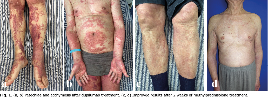
We report here a case of an 85-year-old male patient with hypertension and AD who developed purpuric drug eruption during treatment with dupilumab. The patient had a 40-year history of AD with insufficient response to conventional therapy. As he developed systemic erythema, papules and severe pruritus in May 2021, with eosinophils elevating to 3.21×10⁹/l and total IgE reaching 162.2 IU/ml, dupilumab 600 mg was first administered subcutaneously on 11 June 2021. He presented with non-painful petechiae and ecchymosis on his bilateral lower extremities the next day, and the rash gradually extended to the trunk and upper extremities, without blisters, blood blisters, nodules, necrosis, exudation, or mucosal injury (Fig. 1 a, b). There was no abnormality in systemic physical examination. The patient had no symptoms of itch, fever, rigor, joint swelling and pain, limb weakness, abdominal pain, diarrhoea, vomiting, haematochezia, haematuria, oral haemorrhage, or gingival haemorrhage during the onset of purpura. Laboratory tests showed a platelet count of 210 before and 272 after the onset of purpura (reference range 125–350 ×109 g/l), urinalysis was 0–1/high-power field (hpf) (reference range 0–3/hpf). Coagulation function, renal function, liver function, electrolytes, immune globulins and erythrocyte sedimentation rate (ESR) were all within the normal range. Antinuclear antibody, extractable nuclear antigen, antineutrophil cytoplasmic antibodies, treponema pallidum particle agglutination, tolulized red unheated serum and human immunodeficiency virus tests were all negative.
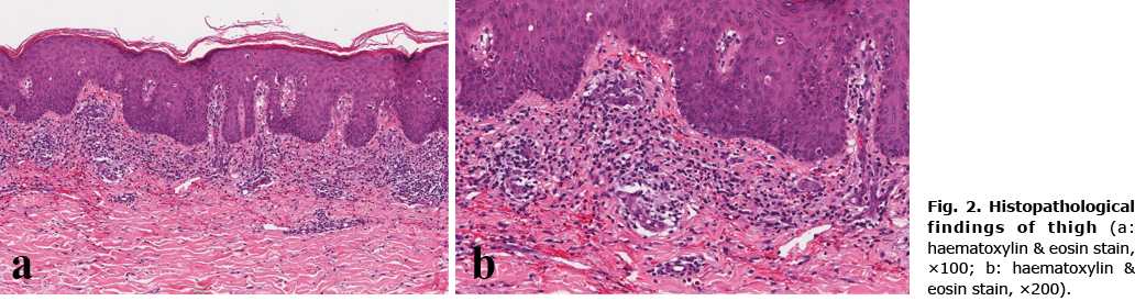
The pathological results showed focal parakeratosis and dyskeratosis cell in the epidermis, with irregular hyperplasia of the epidermis. Liquefied degeneration of the basal layer cells was also observed. Lymphocytes, histomorphic cells, and a small number of eosinophils were observed in the superficial dermis and surrounding blood vessels, showing a lichenoid pattern. Some blood vessels were swollen with extravasated erythrocytes. The results were consistent with the pathological features of purpura, and suggested that it was related to drug reaction (Fig. 2).
The patient’s longstanding medications included losartan potassium for hypertension and finasteride for prostatic hyperplasia. He took the intermittent use of thalidomide for AD. However, the patient had used these drugs for more than 2 years without adverse reactions. A detailed medical history did not reveal any other potential triggers for purpuric drug eruption.
During the subsequent 2-week hospitalization, the patient received methylprednisolone pulse therapy (40 mg/day for 8 days, 32 mg/day for 4 days, and 24 mg/day for 2 days, sequentially). His AD and purpura were both well controlled, and the rash improved visibly before discharge (Fig. 1c, d). Eosinophilic granulocyte levels finally fell below 0.06×10⁹/l. After discharge, the patient continued oral methylprednisolone in a gradually decreasing dose for prevention of recurrence.
DISCUSSION
We report here a patient with AD who developed a purpuric drug eruption during treatment with dupilumab. Purpuric drug eruption is relatively rare clinically, accounting for approximately 1.17% of drug eruption (7). The pathogenesis is complex, which can be divided into thrombocytopaenia and vasculitis. The former may be non-inflammatory purpura caused by thrombocytopaenia or dysfunction, which is caused by direct toxicity of drugs or allergic reaction, and is involved in type II allergic reaction. The first case of immune thrombocytopaenic purpura caused by dupilumab was reported in 2019 (8). The latter may be caused by the direct effect of drug toxicity on capillaries, causing damage and purpura, which is involved in type III allergy, but has not reached vascular necrosis, and may be the early manifestation of vasculitis drug eruption (7). This effect of drug toxicity on capillaries may be the pathogenesis of this case.
Multi-morbidity and frailty are likely to increase susceptibility to adverse drug reaction (9). The current patient is 85 years old and has had hypertension for 2 years. The target of dupilumab action is the skin. Therefore, frailty, hypertension and the target of dupilumab action may be the reasons for the rapid emergence of drug-induced purpura only 1 day after administration.
ACKNOWLEDGEMENTS
We thank the patient for his approval and support in publishing this case report. We also thank for the financial support of Three-year Action Plan (ZY(2018–2020)-ZYBZ-38) and Shanghai Sailing Program (21YF1441500).
The authors have no conflicts of interest to declare.
REFERENCES
- Ariëns LFM, van der Schaft J, Stades AME, van de Woestijne AA, De Bruin-Weller MS. Successful treatment with dupilumab in a patient with severe, difficult to treat atopic dermatitis: beware of symptomatic adrenal insufficiency due to abrupt discontinuation of potent topical corticosteroids. Acta Derm Venereol 2018; 98: 601.
- Steve C. Dupilumab for the treatment of atopic dermatitis. Prescriber 2018; 29: 35–37.
- Simpson EL, Bieber T, Guttman-Yassky E, Beck LA, Blauvelt A, Cork MJ, et al. Two phase 3 trials of dupilumab versus placebo in atopic dermatitis. N Engl J Med 2016; 375: 2335–2348.
- de Wijs LEM, Bosma AL, Erler NS, Hollestein LM, Gerbens LAA, Middelkamp-Hup MA, et al. Effectiveness of dupilumab treatment in 95 patients with atopic dermatitis: daily practice data. Br J Dermatol 2020; 182: 418–426.
- Thomson J, Wernham AGH, Williams HC. Long-term management of moderate-to-severe atopic dermatitis with dupilumab and concomitant topical corticosteroids (LIBERTY AD CHRONOS): a critical appraisal. Br J Dermatol 2018; 178: 897–902.
- Lommatzsch M, Stoll P, Winkler J, Zeise-Wehry D, Tronnier M, Weber MA, Virchow JC. Eosinophilic pleural effusion and stroke with cutaneous vasculitis: two cases of dupilumab-induced hypereosinophilia. Allergy 2021; 76: 2920–2923.
- Changsha Jia, Yongyan Chen, Wen Yan, et al. A case of purpura drug eruption. The Chin J Dermatovenereol 2016; 30: 397–399.
- Frey S, Kendziora B, Holch JW, Lindner L, French LE, Wollenberg A. Immune thrombocytopenic purpura in a patient with atopic dermatitis treated with dupilumab. Acta Derm Venereol 2021; 101: adv00409.
- Young JWS, Shear NH. Cutaneous drug reactions in the elderly. Drugs Aging 2017; 34: 655–672.