SHORT COMMUNICATION
Penile Pyoderma Gangrenosum, a Rare Encounter in STI Healthcare: A Case Report and a Review of the Literature
Kirsten SALADO-RASMUSSEN1-3*, Kasper Køhler ALSING4, Nina Løth MÅRTENSSON3,5, Ewa Anna BURIAN1,4 and Helle Kiellberg LARSEN1,3
1Department of Dermato-Venereology, Bispebjerg University Hospital, 2Department of Bacteria, Parasites and Fungi, Statens Serum Institut, 3Department of Clinical Medicine, University of Copenhagen, 4Copenhagen Wound Healing Center, Department of Dermato-Venereology, Bispebjerg University Hospital, 5Department of Pathology, Rigshospitalet University Hospital, Copenhagen, Denmark.
Citation: Acta Derm Venereol 2024; 104: adv32160. DOI: https://doi.org/10.2340/actadv.v104.32160.
Copyright: © Published by Medical Journals Sweden, on behalf of the Society for Publication of Acta Dermato-Venereologica. This is an Open Access article distributed under the terms of the Creative Commons Attribution-NonCommercial 4.0 International License (https://creativecommons.org/licenses/by-nc/4.0/)
Submitted: Nov 27, 2023; Accepted: Feb 27, 2024; Published: Mar 29, 2024
*E-mail: Kirsten.salado-rasmussen.02@regionh.dk
Competing interests and funding: The authors have no conflicts of interest to declare.
INTRODUCTION
Penile pyoderma gangrenosum is a rare manifestation and a diagnosis of exclusion. The patient first presented at an STI clinic because of the localization of the ulcer, and in this setting this diagnosis can easily be missed, increasing the risk of mutilating lesions. This patient was treated successfully with immunosuppressive drugs but still suffered from disfiguration and urine leakage from the lesion because of the delayed diagnosis. The case and a review of the literature are reported.
CASE REPORT
A 26-year-old man from India presented at a sexually transmitted infection (STI) clinic in Copenhagen with a painful ulcer on the glans penis. The patient was undergoing treatment with rifampicin and isoniazid due to a positive quantiferon test performed in his home country, but he exhibited no other signs or symptoms of tuberculosis and was otherwise healthy. The lesion had developed slowly over 8 months, starting as a papule on the glans. The lesion progressed and eventually involved the urethra, leading to urine leakage from the ulcer. Three months of treatment with anti-tuberculosis drugs had no effect. Clinical examination showed an irregular purulent ulceration with undermined borders (Fig. 1A). The patient tested negative for the following STIs: syphilis (serologically and by Treponema pallidum specific PCR from the ulcer), Haemophilus ducreyi, Neisseria gonorrhoeae, Chlamydia trachomatis, HIV, and hepatitis B and C, and was referred to a Department of Urology for urinary catheterization. After 4 weeks the patient returned to the STI clinic with a fever and elevated C-reactive protein (CRP) of 92, showing clinical signs of infection localized to the penis and unilateral lymphadenopathy (Fig. 1B). The infection was treated effectively with moxifloxacin for 7 days.
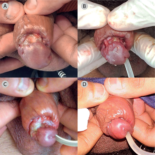
Fig. 1. Clinical case photos of penile pyoderma gangrenosum. (A) First visit. (B) The patient returned after 4 weeks due to fever and signs of infection and was treated with moxifloxacin. (C) After 4 weeks of systemic immunosuppression. (D) Improvement after 12 weeks of systemic immunosuppression.
The patient then underwent further investigations: 2 punch biopsies from the border of the ulcer were negative for leishmaniasis and mycobacteria (PCR and microscopy). The histopathological examinations showed unspecific, ulcer-related histological changes with chronic lymphoplasmacytic inflammation and underlying acute dense neutrophilic infiltrate (Fig. 2). There were no spirochetes, acid-fast bacilli, or fungus, and no signs of malignancy. Due to continuous suspicion of malignancy, the patient had three resections performed at the Department of Urology. However, again the histopathological examinations showed unspecific, ulcer-related findings including areas of ulceration, necrosis, acute neutrophilic infiltration, granulation tissue, and variable lymphoplasmacytic inflammation and fibrosis. There were no signs of malignancy. A computed tomography (CT) scan of the thorax and abdomen was normal. Additional laboratory findings including bacterial cultures from the ulcer, immunoglobulins, and blood glucose were negative/normal. The urine tested positive for carbapenemase-producing Klebsiella pneumoniae, and the patient was isolated during hospitalization. A microbiome analysis (16S/18S) was performed on the tissue from the glans biopsy but was negative. Further, there were no clinical or histopathological signs of lichen sclerosus and the patient was uncircumcised and had never had any urogenital surgery.
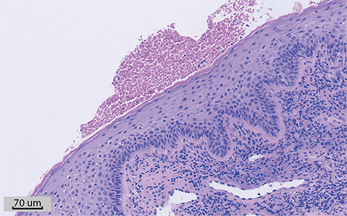
Fig. 2. Biopsy from the glans penis. Shows lymphoplasmacytic inflammation and underlying acute dense neutrophilic infiltrate. Hematoxylin and eosin staining.
Based on the clinical presentation and the exclusion of other diagnoses, the patient was diagnosed with penile pyoderma gangrenosum (PG) and started a treatment regimen with prednisolone. The initial 37.5 mg daily was reduced to 25 mg daily after 2 days due to side effects. After 2 weeks of treatment with prednisolone, cyclosporine was added (75 mg twice daily). After 4 weeks of treatment, the patient reported reduced pain, but clinically only minimal improvement was observed (Fig. 1C). The patient tolerated cyclosporine well, and after 8 weeks, the dose was increased to 100 mg twice daily. After 3 months, the lesion had markedly improved, but the patient still suffered from disfiguration and urine leakage requiring continuous catheterization (Fig. 1D). At that time the patient returned to India. He was still receiving prednisolone 10 mg daily and cyclosporine 100 mg twice daily. He was strongly recommended to seek medical assistance in India because of the ongoing systemic immunosuppressive treatment and the need for future reconstructive surgery.
A review of the literature was performed on PubMed on 2 February 2024, using the following search terms: “pyoderma” and “penile” or “genital”, with no retrospective time limit. Only male patients were included. Articles were included if at least the abstract was in English. A total of 45 cases from 43 articles were identified (Table SI). The age of the patients ranged from 16–89. The majority of the cases received systemic treatment with corticosteroids and only very few were treated with biologics. Most patients had a favorable outcome with complete healing of the lesions although patients with urethral fistulas required surgery.
DISCUSSION
Penile PG is a rare manifestation and a diagnosis of exclusion. In this case, the patient first presented at an STI clinic because of the localization of the ulcer. In this setting, an ulcerative lesion would most likely be infectious, such as a syphilitic chancre, herpes simplex virus, lymphogranuloma venereum (Chlamydia trachomatis, serovars L1, L2, and L3), ulcus molle (caused by Haemophilus ducreyi), or regular skin bacteria. PG is a rare and painful autoinflammatory neutrophilic skin condition, most often affecting the legs (1). Penile involvement is rare, and the diagnosis may therefore be easily missed, increasing the risk of mutilating lesions. In our case, the patient’s young age and ethnicity were also misleading factors as PG most often affects those 50–65 years of age (2). A painful rapidly progressing ulcer with violaceus borders, with negative microbiology, a neutrophilic infiltrate on biopsy, and non-responsiveness to normal wound care may be suggestive of PG. Associated diseases can be identified in 50–75% of patients with PG, e.g. inflammatory bowel disease, arthritis, hematologic malignancies, or solitary cancers (3). The rest remain idiopathic, as in our case. Despite a positive quantiferon test prior to the development of the penile ulcer, there were no other laboratory or clinical findings indicating tuberculosis. Diagnostic criteria such as the PARACELSUS score (4) or the criteria by Maverakis et al. (5) can aid the diagnosis of PG, and in our case both criteria supported the diagnosis.
The genital location of PG is troublesome due to pain, difficulties with applying appropriate wound dressings, and the risk of bacterial contamination. A urinary catheter may alleviate some of these symptoms, but the mechanical trauma can in theory also trigger pathergy, a phenomenon of aggressive deterioration of the wound, or new wounds, by mechanical trauma. This is usually seen 7 days after a mechanical procedure (6). Despite the risk of pathergy, biopsies are advocated in cases with suspected PG to exclude important differential diagnoses such as malignancy, Fournier’s gangrene, calciphylaxis, and vasculitis.
Management of penile PG follows the general treatment of PG and is largely based on 2 randomized clinical trials (7, 8), retrospective studies, and expert opinions (1). Smaller PG lesions may be treated with topical immunosuppression, such as group III or IV steroids or topical tacrolimus. In rapidly evolving PG, systemic immunosuppression is usually required. The best evidence of efficacy and safety involves prednisolone, cyclosporin, and TNF inhibitors (1, 7–9). As the treatment duration may involve several months, steroid-sparing options are often used, which is the reason cyclosporine was added in our case.
Remarkably, our literature review demonstrated that only a minority of patients with penile PG were treated with biologics. The patients seemed to have favorable outcomes on systemic corticosteroids; however, patients with deeper lesions in some cases needed reconstructive surgery, which is not always feasible. In conclusion, early diagnosis is crucial due to the potentially mutilating consequences.
REFERENCES
- Alavi A, French LE, Davis MD, Brassard A, Kirsner RS. Pyoderma gangrenosum: an update on pathophysiology, diagnosis and treatment. Am J Clin Dermatol 2017; 18: 355–372.
- Burian EA, Karlsmark T, Fogh K, Bech R. Pyoderma gangraenosum. Ugeskr Laeger 2021; 183: 1–10.
- Al Ghazal P, Herberger K, Schaller J, Strölin A, Hoff N-P, Goerge T, et al. Associated factors and comorbidities in patients with pyoderma gangrenosum in Germany: a retrospective multicentric analysis in 259 patients. Orphanet J Rare Dis 2013; 8: 136.
- Jockenhöfer F, Wollina U, Salva KA, Benson S, Dissemond J. The PARACELSUS score: a novel diagnostic tool for pyoderma gangrenosum. Br J Dermatol 2019; 180: 615–620.
- Maverakis E, Ma C, Shinkai K, Fiorentino D, Callen JP, Wollina U, et al. Diagnostic criteria of ulcerative pyoderma gangrenosum: a Delphi Consensus of International Experts. JAMA Dermatol 2018; 154: 461–466.
- Tolkachjov SN, Fahy AS, Cerci FB, Wetter DA, Cha SS, Camilleri MJ. Postoperative pyoderma gangrenosum: a clinical review of published cases. Mayo Clin Proc 2016; 91: 1267–1279.
- Brooklyn TN, Dunnill MGS, Shetty A, Bowden JJ, Williams JDL, Griffiths CEM, et al. Infliximab for the treatment of pyoderma gangrenosum: a randomised, double blind, placebo controlled trial. Gut 2006; 55: 505–509.
- Ormerod AD, Thomas KS, Craig FE, Mitchell E, Greenlaw N, Norrie J, et al. Comparison of the two most commonly used treatments for pyoderma gangrenosum: results of the STOP GAP randomised controlled trial. BMJ 2015; 350: h2958.
- Partridge ACR, Bai JW, Rosen CF, Walsh SR, Gulliver WP, Fleming P. Effectiveness of systemic treatments for pyoderma gangrenosum: a systematic review of observational studies and clinical trials. Br J Dermatol 2018; 179: 290–295.