SHORT COMMUNICATION
Trident-shaped Dermatitis in a Child
Céline RASO1, Haude COGO2, Camille BRÉHIN1,2 and Isabelle CLAUDET1*
1Pediatric emergency unit, and 2General medicosurgical pediatric and infectiology ward, Hôpital des Enfants, 330, avenue de Grande-Bretagne, TSA 70034, FR-31059 Toulouse cedex 9, France. *E-mail: claudet.i@chu-toulouse.fr
Citation: Acta Derm Venereol 2024; 104: adv33016. DOI: https://doi.org/10.2340/actadv.v104.33016.
Copyright: © Published by Medical Journals Sweden, on behalf of the Society for Publication of Acta Dermato-Venereologica. This is an Open Access article distributed under the terms of the Creative Commons Attribution-NonCommercial 4.0 International License (https://creativecommons.org/licenses/by-nc/4.0/)
Submitted: Nov 30, 2023; Accepted: Jan 30, 2024; Published: Mar 4, 2024
Competing interests and funding: The authors have no conflicts of interest to declare.
INTRODUCTION
Mites can cause significant human skin lesions, although establishing a link with a specific pathogen can be problematic (1). Sometimes a specific dermatological aspect can suggest a particular pathogen. We report here a case of paediatric dermatitis with an unusual aspect caused by an ectoparasite.
CASE REPORT
A 3-year-old child with a previous medical history of acute community-acquired pneumonia, atopic dermatitis, and asthma was admitted to our paediatric emergency unit in June for inflammatory erythematous dermatitis of her right foot (Fig. 1). The condition was discovered in the afternoon, was extremely itchy, and local inflammation led to transient functional weakness. The lesion was characterized by a central red spot on the heel with 3 centrifugal serpiginous tracks. There was no recent history of stings or bites. She had no other symptoms, fever, satellite adenopathy or other similar lesions. There was no family history of similar dermatitis. She had no previous contact with animals and had not travelled recently or stayed anywhere she was not in the habit of staying. She lived with her family in a house with old wooden floors (Fig. 2). A dermatology consultation confirmed the diagnosis of Pyemotes ventricosus dermatitis due to so-called “comet sign”. She was discharged with a combined treatment of acetaminophen and an antihistamine (desloratadine). The dermatitis resolved within the subsequent 12 h.
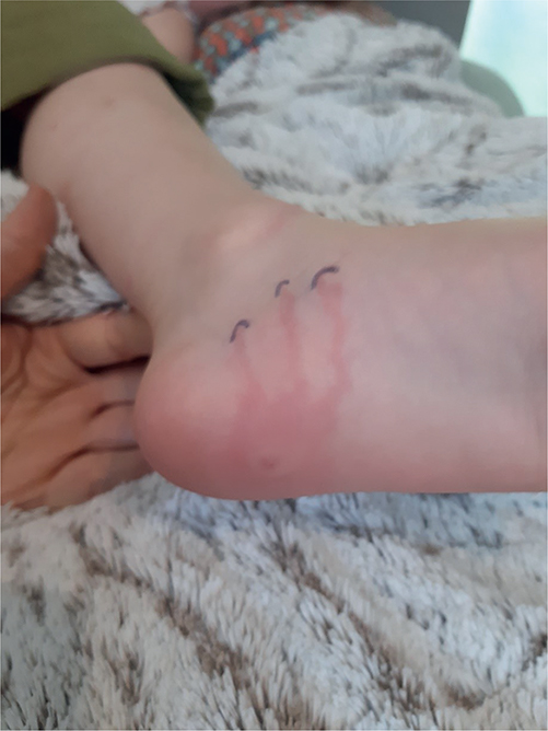
Fig. 1. Erythema associated with 3 inflammatory tracks in a 3-year-old girl.
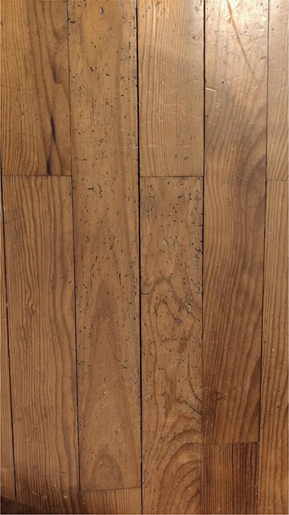
Fig. 2. Photograph showing indirect proof (holes) of the presence of the wood-boring beetle (Anobium punctatum) in the old wooden floor of the child’s bedroom.
DISCUSSION
Pyemotes ventricosus belongs to the phylum Arthropoda, order Acari, sub-order Prostigmata, genus Pyemotes. Over 30 different species of Pyemotes have been identified (1). The P. ventricosus group is commonly known as “straw itch mites” or “grain mites” (2). P. ventricosus is an ectoparasite that lives on different hosts, such as the larvae of Anobium punctatum (furniture beetle), a type of wood-boring beetle. This mite is only just visible to the naked eye (length 0.16 mm (male) to 0.22 mm (female)). Human infestation occurs when infested materials (wood, wooden furniture, grain, feed, hay bales) are held or handled (2, 3). Such infestation occurs in seasonal outbreaks and is more frequent during the summer, with a peak between May and September (4). Outbreaks usually end within 1 week (2). Although well-known since the mid-19th century, particularly in workers exposed to agricultural products, foresters, antique dealers, school or house occupants, very few published cases have involved children (5–10). After a usually painless bite, the patients present with highly itchy lesions consisting of a pinhead-sized central vesicle/urticarial lesion (the site of the mite’s bite) surrounded by a rapidly expanding erythematous macule (8). Cutaneous manifestations can be limited to a single lesion or manifest through multiple aspects (7). When a linear or serpiginous erythematous track called a “comet sign” is present (30% of cases) (2), the diagnosis is oriented toward Pyemotes ventricosus dermatitis and a clinical differentiation with other arthropod bites or stings (2, 3, 5, 9). Usually there is 1 inflammatory track per lesion. The current case is unusual because there were 3 separate tracks, comparable to a “trident” rather than a comet feature. The origin of this linear feature has been associated with a specific sign of self-limiting lymphangitis (5, 11), while the hypothesis of a parasite migration was excluded by the histological normality of the epidermis along the erythematous track (7). The upper limbs (71%), back (67%), chest and abdomen (56%) are the sites most involved (1). A history of atopy was observed in 22% of the patients (1). The lesions evolve within 24 h and typically resolve in 1–3 weeks. Other symptoms have been reported, such as fever, nausea, vomiting, arthralgia, and headache (1, 12). Most patients are treated with a combination of topical corticosteroids and oral antihistamines (7).
The suspected furniture or materials infested by the Pyemotes host insects can be removed from the environment and/or the expertise of a medical entomology professional can be solicited to confirm the infestation and provide the most adapted control measures (1, 13, 14).
In conclusion, pyemotes-related dermatitis has been rarely reported in children. The current case has an unusual dermatological aspect (“trident-shaped tracks”) and location (plantar). The comet sign is not pathognomonic and not very frequent. Its presence helps to make a distinction in the diagnostic challenge of similar skin reactions after arthropod bites or stings. If absent, the diagnosis can be based on a set of arguments: the seasonality (summer months), the customary appearance (central lesion with lymphangitis), the complaints (pruritus) and furniture or wooden ceiling beams as source of parasitized A. punctatum (Fig. 2).
REFERENCES
- Stingeni L, Bianchi L, Hansel K, Neve D, Foti C, Corazza M, et al. Dermatitis caused by arthropods in domestic environment: an Italian multicentre study. J Eur Acad Dermatol Venereol 2017; 31: 1526–1533.
- Vellere I, Di Felice A, Botta A, Angheben A, Bartoloni A, Zammarchi L. Pyemotes ventricosus dermatitis: a case report and an extensive review of outbreaks and clinical cases. Infect Dis Rep 2022; 14: 668–674.
- Laghi A, Ongaro C, Moliterni E, Malvindi S, Roberti V, Iacovino C, et al. Mite bites, comet signs and possible mammary prosthesis rejection after returning to a vacation home: a diagnostic challenge. Travel Med Infect Dis 2021; 42: 102077.
- Principato M. Observation on the spread of pyemotes ventricosus in houses in Umbria, Central Italy. In: Bernini F, Nannelli R, Nuzzaci G, de Lillo E. (editors). Acarid phylogeny and evolution: adaptation in mites and ticks. Springer, Dordrecht. [accessed 2023 October 30]. Available from: https://doi.org/10.1007/978-94-017-0611-7_44.
- Del Giudice P, Blanc-Amrane V, Bahadoran P, Caumes E, Marty P, Lazar M, et al. Pyemotes ventricosus Dermatitis, South-eastern France. Emerg Infect Dis 2008; 14: 1759–1761.
- Tognetti L, Cinotti E, Pianigiani E, Rubegni P. Pyemotes ventricosus detection in a baby skin folds. J Eur Acad Dermatol Venereol 2019; 33: e93–94.
- Del Giudice P, Caumes E, Boissy C, Leduff F, Delaunay P, Blanc-Amrane V, et al. An outbreak of creeping eruption in southern France. Br J Dermatol 2007; 157: 824–825.
- Corazza M, Tassinari M, Pezzi M, Ricci M, Borghi A, Minghetti S, Leis M. Multidisciplinary approach to Pyemotes ventricosus papular urticaria dermatitis. Acta Derm Venereol 2014; 94: 248–249.
- Berenger JM, Parola P. Pyemotes ventricosus dermatitis. N Engl J Med 2023; 29: e80.
- Neumayr A, Kuenzli E. Pyemotes ventricosus dermatitis: a serpiginous skin lesion due to a mite that parasitizes a wood-boring beetle. Am J Trop Med Hyg 2019; 100: 1041–1042.
- Zelin E, Di Meo N, Maronese CA, Zalaudek I. Pyemotes ventricosus dermatitis: ‘comet sign’. Clin Exp Dermatol 2021; 46: 980–983.
- Tomczyk-Socha M, Jedrzejewska-Jurga K, Limburska J, Tomczyk J. Outbreak of occupational dermatitis associated with pyemotes ventricosus. JAMA Dermatol 2017; 153: 686.
- Bianchi L, Hansel K, Tramontana M, Principato M, Moretta I, Principato S, et al. Occupational papular urticaria due to Pyemotes ventricosus in antique dealers. Contact Dermatitis 2020; 82: 181–183.
- Sevestre J, Marty P, Hubiche T, Pomares C, Delaunay P. Groundbreaking outpatient activity in medical entomology in France: an eight-year experience in a french university hospital. Infect Dis Now 2023; 53: 104728.