Verrucous Plaques on the Left Lower Extremity: A Quiz
Yuxian Lai, Xiqing Li, Liangchun Wang and Zhenrui Shi*
Department of Dermatology, Sun Yat-sen Memorial Hospital, Sun Yat-sen University, 107 Yanjiang Rd W, Guangzhou 510120, China. *E-mail: zrshi1989@outlook.com
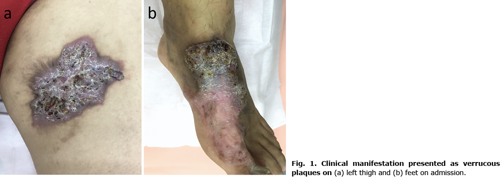
A man in his 40s was referred to our hospital for a 30-year history of asymptomatic cutaneous lesions. The patient was born and had continuously resided in Guangdong, the southernmost mainland province of China. He injured his left foot with a fishbone as a child. He reported that a lesion was initially observed on his left foot after the injury and had extended slowly thereafter. A similar lesion occurred on his left inner thigh 6 years ago. Skin biopsy suggested infectious granulomas with suspected presence of “sclerotic” cells and he was diagnosed with chromoblastomycosis 5 years ago. The patient had been treated with systemic antifungal agents, including itraconazole, terbinafine, and diflucan, for several rounds, but with no improvement. He also underwent a surgical excision, but the lesions recurred quickly. Physical examination revealed 2 well-circumscribed irregular verrucous plaques with brownish crusts on his left inner thigh and the dorsum of his left foot (Fig. 1). Atrophic scarring was observed across and around the plaques.
What is your diagnosis? See next page for answer.
Verrucous Plaques on the Left Lower Extremity: A Commentary
Acta Derm Venereol 2022; 102: adv00770.
DOI: 10.2340/actadv.v102.3480
Diagnosis: Tuberculosis verrucosa cutis
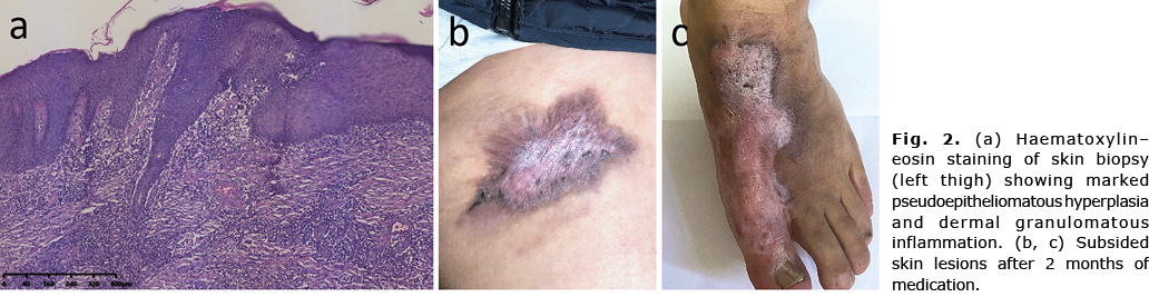
Mantoux test was strongly positive. Hepatitis B antibody testing showed positivity for HbsAg, anti-HBe and anti-HBc. There were no signs of systemic infection, as evidenced by absence of fever, lymphadenopathy, leukocytosis and a normal chest radiograph. Skin biopsy showed marked pseudoepitheliomatous hyperplasia and dermal granulomatous inflammation composed of histiocytes and mixed inflammatory cells without caseous necrosis (Fig. 2a). Tissue culture was positive for acid-fast bacillus, and the colony was further identified as Mycobacterium tuberculosis. He was diagnosed with tuberculosis verrucosa cutis (TVC) and treated with isoniazid, rifampin and ethambutol instead of the standard regimen, in order to avoid hepatotoxicity. Skin lesions progressively resolved after 2 months of therapy (Fig. 2b, c). The patient is currently being followed up with same medication.
Cutaneous tuberculosis (TB) is relatively uncommon, comprising 1–2% of extra-pulmonary TB manifestations (1). However, given its global prevalence, it is imperative for dermatologists to distinguish the masquerading infections in order to preclude misdiagnosis and consequent disease progression. The clinical manifestations were varied with the morphological patterns of the lesions, route of acquisition, and host immune status. Based on a study conducted in Brazil in 2019, the most common variant of cutaneous tuberculosis was scrofuloderma (50.7%), followed by erythema induratum of Bazin (18.7%), tuberculous gumma (13.3%), lupus vulgaris (8%), TVC (5%), orificial TB (2.7%) and associated forms (2.7%) (2). TVC occurred in previously sensitized patients as a result of direct inoculation of M. tuberculosis bacillus. It was often observed in children and skin lesions usually develop in the extremities due to trauma. Among adults, it is associated with occupational exposure, and the hands, ankles, or buttocks are the common affected sites. Clinical manifestations can resemble warts, hypertrophic lichen planus, chromoblastomycosis (3), resulting in a diagnostic dilemma, as in the current case.
The management of cutaneous TB follows the guidelines of the extra-pulmonary form of TB, 2RHZE/4RH (4). This scheme consists of 2 months of rifampin(R), isoniazid (H), pyrazinamide (Z), and ethambutol (E), followed by another 4 months of treatment with rifampicin and isoniazid. The long duration of treatment, 6 months on average, means it is likely that patients will be non-compliant with therapy. Treatment interruption is one of the main risk factors for drug resistance, and treatment adherence is therefore a must.
The authors have no conflicts of interest to declare.
REFERENCES
- Dias MF, Bernardes Filho F, Quaresma MV, Nascimento LV, Nery JA, Azulay DR. Update on cutaneous tuberculosis. An Bras Dermatol 2014; 89: 925–938.
- Mann D, Sant’Anna FM, Schmaltz CAS, Rolla V, Freitas DFS, Lyra MR, et al. Cutaneous tuberculosis in Rio de Janeiro, Brazil: description of a series of 75 cases. Int J Dermatol 2019; 58: 1451–1459.
- Khadka P, Koirala S, Thapaliya J. Cutaneous tuberculosis: clinicopathologic arrays and diagnostic challenges. Dermatol Res Pract 2018; 2018: 7201973.
- Handog EB, Gabriel TG, Pineda RT. Management of cutaneous tuberculosis. Dermatol Ther 2008; 21: 154–161.