QUIZ SECTION
A 20-year-old Woman with a Red Nodule on Her Right Knee: A Quiz
Jingwei ZHOU#, Chen HUA#, Hongrui CHEN, Yuxi CHEN and Xiaoxi LIN
Department of Plastic and Reconstructive Surgery, Shanghai Ninth People’s Hospital, Shanghai Jiao Tong University School of Medicine, Shanghai, China. E-mail: linxiaoxi@126.com
#These authors contributed equally to this work and should considered co-first authors.
Citation: Acta Derm Venereol 2024; 104: adv35391. DOI: https://doi.org/10.2340/actadv.v104.35391.
Copyright: © 2024 The Author(s). Published by MJS Publishing, on behalf of the Society for Publication of Acta Dermato-Venereologica. This is an Open Access article distributed under the terms of the Creative Commons Attribution-NonCommercial 4.0 International License (https://creativecommons.org/licenses/by-nc/4.0/).
Published: Jun 26, 2024
Competing interests and funding: The authors have no conflicts of interest to declare.
A 20-year-old woman presented with a red nodule on the inner side of her right knee joint (Fig. 1a). The lesion was first noticed more than 1 year ago. There was no history of trauma or relevant family history. She previously received local cryotherapy and sclerotherapy in another centre. However, no obvious improvement was made after the treatment and the lesion kept growing during the following 9 months. A preoperative magnetic resonance imaging scan with contrast was performed. T1-weighted image showed that while the border was relatively clear, the lesion had a highly abundant blood supply signal (Fig. 1b). After informed consent was obtained, surgical excision was carried out, and histopathological examination was performed.
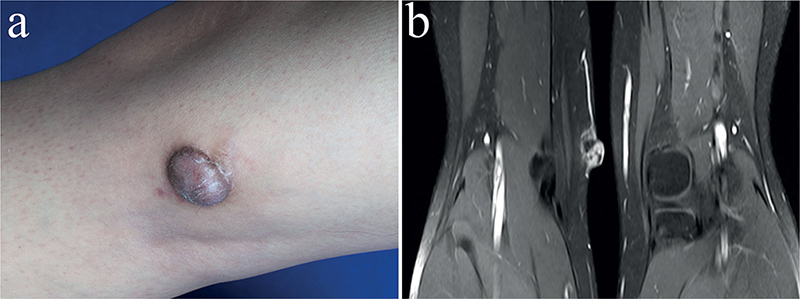
Fig. 1. (a) Red nodule at the inner side of the knee joint. (b) T1-weighted magnetic resonance imaging scan image with contrast.
What is your diagnosis?
Differential diagnosis 1: Haemangioma
Differential diagnosis 2: Angiosarcoma
Differential diagnosis 3: Spindle cell haemangioendothelioma
Differential diagnosis 4: Aneurysmal fibrous histiocytoma
See next page for answer.
ANSWERS TO QUIZ
A 20-year-old Woman with a Red Nodule on Her Right Knee: A Commentary
Diagnosis: Aneurysmal fibrous histiocytoma
Aneurysmal fibrous histiocytoma (AFH), first reported in 1981 by Santa–Cruz and Kyriakos, is a rare type of cutaneous fibrous histiocytoma that represents less than 2% of all cases (1, 2). Lesions of AFH are usually discovered in the lower extremities and characterized by blue or dark-red nodules (3). Reports on the disease are limited. Due to its clinical manifestation and blood-filled mass tissue, preoperative diagnosis is challenging, and it could be confused with vascular-origin tumours such as haemangioma.
The differential diagnosis of AFH includes many other vascular diseases, such as spindle cell haemangioendothelioma, angiosarcoma, and especially haemangioma in children, which is much more common (4). Clinically the tumour mass of AFH is similar to that of a mixed infantile haemangioma, presenting as a dark-red nodule, while angiographic or anatomic results might also reveal a distinct blood supply connecting to the lesion (i.e. a feeding artery), the same as some haemangiomas (5, 6). According to the International Society for the Study of Vascular Anomalies (ISSVA) classification, there are two kinds of haemangiomas: The first is congenital haemangioma, which presents at birth and does or does not involute with growth. The second is infantile haemangioma, which appears shortly after birth, and usually grows rapidly during the first year, then followed by a gradual involution during the next 4–5 years (6). In this case, although morphologically similar, the diagnosis cannot be haemangioma by definition, considering it was a newly discovered lesion in a young adult with no related medical history.
As an intermediate-grade tumour, studies have shown higher possibilities of recurrence than ordinary fibrous histiocytoma or even metastasis, which never happens in haemangiomas or vascular malformations, if the lesion has not been removed completely (4, 7). Thus, accurate diagnosis of AFH is crucial, especially when it affects young children, in which case the differential diagnosis becomes harder. Different from haemangiomas, complete excision with long-term follow-up is also necessary for AFH patients to avoid potential recurrence or metastasis. Otherwise, improper treatment could lead to serious consequences. Histologic diagnosis will thus take a more important place.
Despite the abundant blood supply of both AFH and haemangioma lesion, different from haemangioma, blood-filled spaces surrounded by spindled histiocytoid cells and sometimes giant cells may be found in AFH (3). AFH also shares similar histologic appearance with spindle cell haemangioendothelioma. Both tumours can show blood-filled spaces. Spindle cell haemangioendothelioma is primarily a vascular tumour with endothelial cell proliferation consisting of real cavernous vascular spaces, whereas aneurysmal fibrous histiocytoma is a fibrohistiocytic tumour with secondary haemorrhagic features (8, 9). An immuno-histochemical test might also provide more information, showing as positive for CD68 and Vimentin in the tumour cells and negative for CD31 or CD34, which helps exclude other vascular tumours such as Kaposi’s sarcoma (1, 10).
In the current case, the patient was previously diagnosed as haemangioma in another centre and received treatment with no obvious improvement. After admission to our department, surgical excision was performed and during the operation a distinct feeding artery connecting to the tumour was discovered, as in some haemangiomas (Fig. 2a). Histologically, under microscopy the lesion consisted of characteristic spindled histiocytes among which scattering irregular blood-filled tissue spaces lacking endothelial cell coverage, which is one of the histological diagnostic criteria (Fig. 2b) (7). Immunohistochemical results were positive for Vimentin, while negative for CD31 and S-100 in tumour cells, further confirming the diagnosis. No complication or recurrence was observed at 12 months’ follow-up.
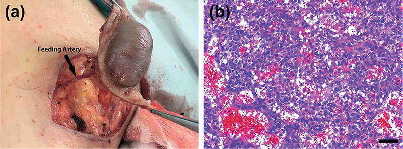
Fig. 2. (a) Intraoperative image showed a distinct feeding artery connecting to the tumour. (b) Histomorphology demonstrated characteristic spindled histiocytes with blood-filled spaces within the lesion (H&E, Scale bar: 40 μm).
In conclusion, given the very similar appearance to haemangioma in children, suspected AFH lesions require extra caution and complete excision with histologic analysis always being necessary, considering the higher possibilities of recurrence and even metastasis, especially for young patients.
Acknowledgement
This study was supported by Clinical Research Program of Shanghai Municipal Health Commission.
REFERENCES
- Munekata Y, Kitamura S, Yanagi T, Shimano M, Ujiie H. Dermoscopic features of aneurysmal dermatofibroma: a case report and review of the literature. J Dermatol 2022; 49: e169–e170.
- Ichikawa N, Kobayashi M, Kimoto M, Tanikawa A, Tanaka M. A case of multiple aneurysmal fibrous histiocytomas. Br J Dermatol 2005; 153: 664–665.
- Santa Cruz DJ, Kyriakos M. Aneurysmal (“angiomatoid”) fibrous histiocytoma of the skin. Cancer 1981; 47: 2053–2061.
- Kandal S, Ozmen S, Demir HY, Tuncer S, Ekinci O, Atabay K. Aneurysmal fibrous histiocytoma of the skin: a rare variant of dermatofibroma. Plast Reconstr Surg 2005; 116: 2050.
- Colangeli M, Rimondi E, Spinnato P, Donati DM, Manfrini M. Difficult diagnosis of angiomatoid fibrous histiocytoma of the leg mimicking a benign condition. J Radiol Case Rep 2019; 13: 38–45.
- Wang MX, Kamel S, Elsayes KM, Guillerman RP, Habiba A, Heng L, et al. Vascular anomaly syndromes in the ISSVA classification system: imaging findings and role of interventional radiology in management. Radiographics 2022; 42: 1598–1620.
- Maharjan S, Satyal B, Baidya R, Joshi A, Baral P. Angiomatoid fibrous histiocytoma mimicking a lymph nodal lesion: a case report. JNMA J Nepal Med Assoc 2022; 60: 200–203.
- McKenna DB, Kavanagh GM, McLaren KM, Tidman MJ. Aneurysmal fibrous histiocytoma: an unusual variant of cutaneous fibrous histiocytoma. J Eur Acad Dermatol Venereol 1999; 12: 238–240.
- Sheehan KM, Leader MB, Sexton S, Cunningham F, Leen E. Recurrent aneurysmal fibrous histiocytoma. J Clin Pathol 2004; 57: 312–313.
- Thway K, Fisher C. Angiomatoid fibrous histiocytoma: the current status of pathology and genetics. Arch Pathol Lab Med 2015; 139: 674–682.