Mycosis fungoides (MF) is the most common variant of cutaneous T-cell lymphoma. Besides the classic type, 3 subtypes of MF have been individualized in the 2018 World Health Organization – European Organisation for Research and Treatment of Cancer (WHO-EORTC) classification (1). The superficial pagetoid Woringer-Kolopp form; granulomatous slack-skin disease; and the folliculotropic form of MF. An additional, very rare, variant reported in the 2005 WHO-EORTC classification (2) among the folliculotropic variant is syringotropic MF (STMF), defined by a particular tropism of the T-cell lymphocytic infiltrate for the eccrine epithelium.
We report here a case of STMF presenting with infiltrated livedoid and ulcerated plaques of the legs and feet, clinically first suggestive of a vasculopathy. Pathological findings were specific due to the presence of major vascular alterations associated with the syringotropic infiltrate, and by a positivity of T-follicular-helper (T-FH) cell markers.
CASE REPORT
A 66-year-old Caucasian man was referred to our dermatology clinic for a 3-year history of plantar keratosis, recently associated with necrotic ulcers. He had no past medical history, was an active smoker (50 pack-years), occasionally consuming cannabis, but no cocaine or other illicit drugs.
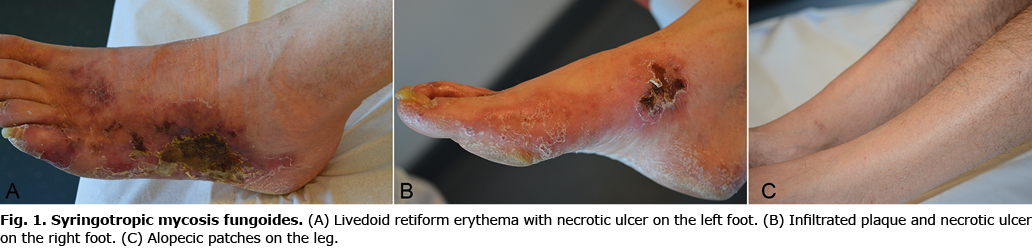
On physical examination, the patient had erythematous, infiltrated plaques and retiform livedoid lesions of the dorso-lateral and medial faces of the feet, with central necrotic ulcers (Fig. 1A, B). He also had alopecic patches on the lower third of both legs (Fig. 1C) and erythematous patches on the hands. No lymphadenopathy was present.
Biopsy specimens taken from the lesions of the feet revealed a dense dermal lymphoid infiltrate of small cells with moderate atypical features, surrounding the eccrine glands with focal infiltration of the eccrine coils and ducts and the blood vessels (Fig. 2A and inset). The perivascular infiltrate was granulomatous, associated with some thromboses and extravasated erythrocytes. A biopsy specimen taken from a patch of the hand showed a similar syringotropic aspect with mild folliculotropic and epidermotropic infiltrate.

Immunohistochemical stainings demonstrated a predominance of T-cells with high expression of CD3+ (Fig. 2B), CD5+, CD7+, CD4+, and the T-FH markers PD1+ (Fig. 2C) and CXCL13. In addition, Bcl-6 (< 10%), PD-L1 (10%) and Ki67 (15%) stainings were slightly positive, while ICOS, CD10, CD20, CD23, CD25, CD30, CD56, perforin and granzyme were negative. A T-cell receptor gene rearrangement was demonstrated in skin lesions.
Other causes of vasculopathic ulcers were ruled out by comprehensive negative investigations for small- and medium-vessels vasculitis and coagulopathies, including cryoglobulins, anti-nuclear antibodies, anti-neutrophil cytoplasmic antibodies, antiphospholipid antibodies, lupus anticoagulant, antithrombin III, proteins C and S, homocysteine, and factor V Leiden. In addition, an embologenic heart disease and a myeloproliferative disorder were ruled out by appropriate investigations, and serological assays for syphilis, HIV, HBV and HCV were negative.
Appropriate dressings associated with topical mechlorethamine on non-ulcerated lesions and subcutaneous methotrexate 20 mg per week resulted in total ulcer healing and a partial response of approximately 80% after 3 months.
DISCUSSION
We report here a patient with STMF associated vasculopathy and, to our knowledge, the first detailed pathological and immunohistochemical description of vasculopathy associated with STMF.
STMF mainly affects the limbs, but occasionally the trunk, head and neck (3), which correlates with the location of eccrine glands on the entire body skin with the highest density on the palms and soles. It usually presents clinically as erythematous lesions with patches, papules and/or small plaques (3, 4). Biopsies show a prominent involvement of eccrine glands by a dense lymphoid infiltrate without prominent atypical features, and variable degrees of syringometaplasia, often with conspicuous epitheliotropism (3). Epidermis and hair follicles can be involved. In the current patient, the prominent location of skin lesions, the presence of infiltrated plaques and the dense CD4+ T-cell infiltrate around hyperplastic eccrine glands were typical aspects of STMF, leading to this diagnosis being retained on clinical and histopathological correlation.
However, the atypical presentation with livedoid retiform erythema and necrotic ulcers was misleading and initially suggestive of a necrotizing vasculitis. Vasculopathic SMTF has been described in 5 patients out of 19, in the largest series of STMF published to date (3). Four of these patients had ulcerated lesions and 1 had a livedoid presentation. A biopsy performed on the periphery of the ulcerated skin lesions in 2 cases confirmed the diagnosis of STMF, with no evidence for large cell transformation. No vasculopathy was mentioned and the aspect of blood vessels and their relationship with the lymphoid infiltrate was not described. More recently, Yonan et al. (5) reported a case of STMF with lower limb ulcers and histological findings of small-vessel vasculopathy with dense tumour infiltration of vessels, thickened walls with an onion skin appearance, narrowed lumina and fibrin deposits. These findings, including granulomatous vasculitis and thrombosis within the lymphoid infiltrate, also argue for a direct role of the lymphoma in vascular alterations and resulting livedoid lesions and ulcers.
Surprisingly, the T-cell infiltrate in the current patient showed a positive expression of T-FH markers. Although the expression of some T-FH markers has been previously reported in MF (6), this is, to our knowledge, the first report of a STMF with a T-FH phenotype. Whether this may be explained by the fact that T-FH markers have rarely been evaluated in this very rare condition, or by a true particularity of the current case will have to be determined by further reports. The expression of T-FH markers in cutaneous lymphoproliferative disorders has been mainly described in angioimmunoblastic T-cell lymphoma with cutaneous involvement (7), primary cutaneous CD4+ small/medium lymphoproliferative disorder, or peripheral T-cell lymphoma, not otherwise specified (8). Clinical and histological features in the current patient were not suggestive of these diagnoses. The clinical improvement under local and systemic therapy was in accordance with an indolent condition, suggesting that vasculopathic STMF, although impressive at presentation, could share the usual good prognosis of typical early-stage MF.
In conclusion, dermatologists should be aware that STMF may be considered when vasculopathic lesions are present in a patient with adnexotropic MF, and that T-FH markers may be present in this very rare variant of MF.
ACKNOWLEDGEMENT
The patient in this manuscript has given written informed consent to the publication of his case details.
The authors have no conflicts of interest to declare.
REFERENCES
- Willemze R, Cerroni L, Kempf W, Berti E, Facchetti F, Swerdlow SH, et al. The 2018 update of the WHO-EORTC classification for primary cutaneous lymphomas. Blood 2019; 133: 1703–1714.
- Willemze R. WHO-EORTC classification for cutaneous lymphomas. Blood 2005; 105: 3768–3785.
- de Masson A, Battistella M, Vignon-Pennamen M-D, Cavelier-Balloy B, Mouly F, Rybojad M, et al. Syringotropic mycosis fungoides: clinical and histologic features, response to treatment, and outcome in 19 patients. J Am Acad Dermatol 2014; 71: 926–934.
- Pileri A, Boi S. Syringotropic mycosis fungoides: a rare variant of the disease with peculiar clinicopathologic features. Am J Surg Pathol 2011; 35: 10.
- Yonan YA, Cumsky HJL, Costello CM, Maly CJ, Rosenthal AC, Reeder CB, et al. Syringotropic and folliculotropic mycosis fungoides with mycosis fungoides–associated vasculopathic ulcers. JAAD Case Rep 2019; 5: 231–233.
- Park J-H, Han JH, Kang HY, Lee E-S, Kim YC. Expression of follicular helper T-cell markers in primary cutaneous T-cell lymphoma. Am J Dermatopathol 2014; 36: 6.
- Ortonne N, Dupuis J, Plonquet A, Martin N, Copie-Bergman C, Bagot M, et al. Characterization of CXCL13+ neoplastic T cells in cutaneous lesions of angioimmunoblastic T-cell lymphoma (AITL). Am J Surg Pathol 2007; 31: 1068–1076.
- Battistella M, Beylot-Barry M, Bachelez H, Rivet J, Vergier B, Bagot M. Primary cutaneous follicular helper T-cell lymphoma: a new subtype of cutaneous T-cell lymphoma reported in a series of 5 cases. Arch Dermatol 2012; 148: 725.