QUIZ SECTION
Multiple Papules and Nodules Over the Body with Diffuse Thickening of the Facial Skin: A Quiz
Aravind REDDY, Yash BUCCHA, Rohan MANOJ, Namratha PUTTUR and Kshitiz LAKHEY
Dr. D. Y. Patil Medical College, Hospital & Research Centre, Pimpri, Pune, A4 1701, Mahindra Antheia, Nehru Nagar, Pimpri, Pune, Maharashtra, India. E-mail: drkshitizlakhey@gmail.com
Citation: Acta Derm Venereol 2024; 104: adv40206. DOI: https://doi.org/10.2340/actadv.v104.40206.
Copyright: © 2024 The Author(s). Published by MJS Publishing, on behalf of the Society for Publication of Acta Dermato-Venereologica. This is an Open Access article distributed under the terms of the Creative Commons Attribution-NonCommercial 4.0 International License (https://creativecommons.org/licenses/by-nc/4.0/).
Published: Jul 15, 2024
A 48-year-old man had been admitted to the psychiatry ward in a tertiary care hospital to be treated for alcohol dependence. His psychiatrist had noted multiple nodular lesions distributed all over his body. The patient and his family members stated that they had first noticed a few raised, light-coloured lesions over the abdomen 2 years earlier, which gradually disseminated over his entire body. He also gave a history of occasional stuffiness of the nose and dryness of the lower limbs. On examination, diffuse thickening of his facial skin with madarosis and nodular infiltrations over his eyebrows were seen (Fig. 1A, B). Multiple papules and nodules were also seen scattered across his upper and lower limbs, buttocks, and trunk (Fig. 1C). The patient had not sought treatment earlier because he had attributed his skin changes to the adverse effects of excessive alcohol consumption. On palpation, the infraorbital, radial, common peroneal, and anterior and posterior tibial nerves were thickened. A sensory examination revealed impaired temperature sensation over his feet up to the ankles. After the psychiatrists had prescribed him medication for his alcohol dependence, they agreed to transfer him to the dermatology ward for further evaluation using a slit skin smear and biopsy.
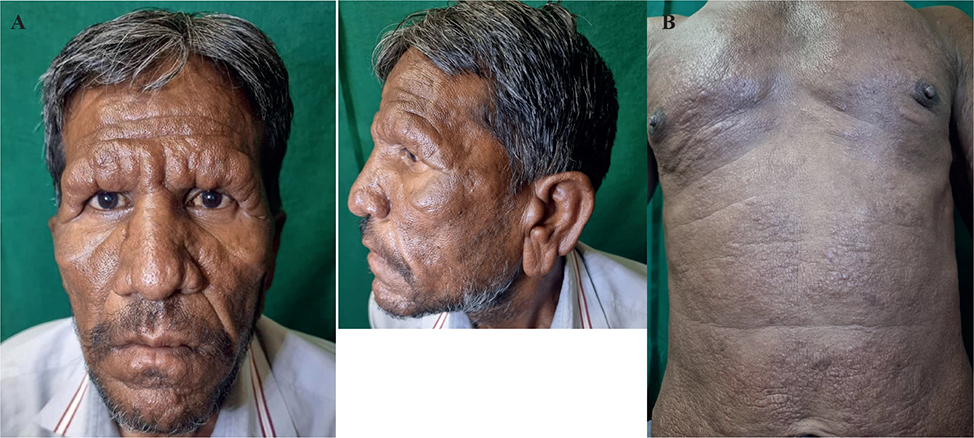
Fig. 1. A: Leonine facies with madarosis and sagging of the earlobe. B: Multiple well-defined skin-coloured papules and a few nodules over the chest and abdomen.
What is your diagnosis?
Differential diagnosis 1: lepromatous leprosy
Differential diagnosis 2: actinic reticuloid
Differential diagnosis 3: mycosis fungoides
Differential diagnosis 4: cutaneous mucinosis
Differential diagnosis 5: diffuse cutaneous leishmaniasis
See next page for answer.
ANSWERS TO QUIZ
Multiple Papules and Nodules Over the Body with Diffuse Thickening of the Facial Skin: A Commentary
Diagnosis: Lepromatous leprosy
Leprosy is a mycobacterial disease primarily of skin with cutaneous and peripheral nerve involvement (1). It is caused by Mycobacterium leprae, which is an acid-fast bacillus that has an affinity to affect cooler parts of the body such as the earlobes, fingers, toes, nose, and testes (1). Leprosy is also known to affect other parts of the body such as the mucosa, eyes, bones, muscles, kidneys, liver, adrenal glands, and lymph nodes. Mycobacterium lepromatosis, which was first identified in 2008, is now considered as a second causative organism of the disease (2). Risk factors for developing this disease include genetics and poverty, as overcrowding, poor ventilation, and malnutrition are associated with an increased incidence of leprosy (3–5). This disease has a long incubation period usually ranging from 5 years to 20 years (1).
There are two widely recognized classifications of leprosy: the World Health Organization (WHO) (1) and the Ridley–Jopling (RJ) classification (6). The RJ classification stratified leprosy into 5 subtypes based on clinical, immunological, and histopathological features, namely tuberculoid (TT), borderline tuberculoid (BT), mid-borderline (BB), borderline lepromatous (BL), and lepromatous leprosy (LL). Rare types of leprosy, which are not grouped into the aforementioned classification, include histoid leprosy, indeterminate leprosy, lepra bonita, and pure neuritic leprosy. Among these, indeterminate leprosy is not yet classifiable. Similarly, pure neuritic leprosy, which usually occurs in multibacillary leprosy, can be seen in any form but is not mentioned in the Ridley–Jopling classification. On the other hand, histoid leprosy and lepra bonita are included in the Ridley–Jopling classification under atypical presentations of lepromatous leprosy.
The WHO classification divides the disease into paucibacillary (5 or fewer lesions and a negative slit skin smear report) or multibacillary leprosy (5 or more lesions and/or associated nerve thickening or impairments and/or a positive slit skin smear report) (1). This classification is a valuable tool in the field, as it provides essential guidance towards treatment decisions (1).
The tuberculoid pole of this disease often displays few erythematous or hypopigmented patches. These patches may be flat or have a raised border due to bacillary infiltration. The lesions have diminished sensation to light touch, which is examined with a cottonwool wisp. The lesions may be dry due to reduced sweating secondary to autonomic nerve involvement. The lepromatous pole of this disease may have widespread nodular or diffuse skin lesions with multiple nerve involvement. This is due to the dissemination of the bacilli over multiple regions in the body. The bacilli have a predilection for skin and nervous tissue, hence dermatological and neurological complications are at the forefront of this disease. However, extracutaneous manifestations can also be seen due to direct bacillary infiltration into various bones and viscera. Nasal septal resorption and perforation can present as a “saddle nose”. Ophthalmological complications such as corneal xerosis, ulcers, and opacification can lead to blindness (1, 6). Nasal involvement is common, particularly in the lepromatous pole, with patients complaining of nasal congestion or recurrent episodes of epistaxis (7). Digital resorption of the toes and fingers can drastically impact the patient’s chances of employment and their quality of life.
Hypersensitivity reactions to the antigenic determinants of the lepra bacilli can further complicate the disease by causing constitutional symptoms, pedal oedema, and neuritis, which can eventually lead to permanent nerve impairments (1, 8). Type 1 lepra reaction is a delayed type IV hypersensitivity reaction only seen in the borderline spectrum. It is not seen in the polar forms of tuberculoid and lepromatous leprosy, which are stable. It is characterized by the erythematous and oedematous transformation of the lesions. Neuritis is the most prominent feature of this reaction, which requires the use of systemic corticosteroids to prevent permanent nerve damage (1, 8). Type 2 lepra reaction is a type III hypersensitivity reaction that is caused by antigen–antibody complexes and is characterised by novel red, raised, evanescent tender crops of nodules of erythema nodosum leprosum occurring all over the body with associated systemic features like arthritis, fever, malaise, uveitis, and orchitis (1, 8).
We undertook an extensive physical examination to rule out any neuritis and associated deformities of the eyes, nose, or digits. He did, however, display the characteristic clinical features of leonine facies such as madarosis and generalized thinning of the facial skin along with drooping of the earlobes. A slit skin smear revealed a high bacillary index (6+), and a skin biopsy aided to confirm the diagnosis.
Leprosy is treated using multi-drug therapy (MDT), introduced by the WHO in 1981 (9). The multibacillary MDT pack (MB-MDT) contains a monthly dose of 600 mg of rifampicin and 300 mg of clofazimine, followed by a daily dose of 50 mg of clofazimine and 100 mg of dapsone, which is started on the next day (10). The modified WHO 2018 regimen states that paucibacillary leprosy should be treated with 6 monthly doses and multibacillary leprosy with 12 monthly doses of all the 3 aforementioned drugs with the maximum permissible duration for completion of therapy being 9 and 18 months respectively (10). Our patient was counselled concerning the associated adverse effects of the medication and strongly advised to adhere to the treatment to ensure complete disease remission.
Leprosy remains a prominent health concern in certain parts of the world. Every medical practitioner should be aware of the 3 cardinal signs of leprosy, which are erythematous or hypopigmented patches with loss of sensation, enlarged peripheral nerves, and a positive skin smear. Finding any 2 of the 3 cardinal signs can establish the diagnosis even in settings where resources are limited.
REFERENCES
- White C, Franco-Paredes C. Leprosy in the 21st century. Clin Microbiol Rev 2015; 28: 80-94. https://doi.org/10.1128/CMR.00079-13
- Sharma R, Singh P, McCoy RC, Lenz SM, Donovan K, Ochoa MT, et al. Isolation of mycobacterium lepromatosis and development of molecular diagnostic assays to distinguish mycobacterium leprae and M. lepromatosis. Clin Infect Dis 2020; 71: e262-269. https://doi.org/10.1093/cid/ciz1121
- Moet FJ, Pahan D, Schuring RP, Oskam L, Richardus JH. Physical distance, genetic relationship, age, and leprosy classification are independent risk factors for leprosy in contacts of patients with leprosy. J Infect Dis 2006; 193: 346-353. https://doi.org/10.1086/499278
- Fava VM, Dallmann-Sauer M, Schurr E. Genetics of leprosy: today and beyond. Hum Genet 2020; 139: 835-846. https://doi.org/10.1007/s00439-019-02087-5
- Nery JS, Ramond A, Pescarini JM, Alves A, Strina A, Ichihara MY, et al. Socioeconomic determinants of leprosy new case detection in the 100 Million Brazilian Cohort: a population-based linkage study. Lancet Glob Health 2019; 7: e1226-1236. https://doi.org/10.1016/S2214-109X(19)30260-8
- Maymone MBC, Laughter M, Venkatesh S, Dacso MM, Rao PN, Stryjewska BM, et al. Leprosy: clinical aspects and diagnostic techniques. J Am Acad Dermatol 2020; 83: 1-14. https://doi.org/10.1016/j.jaad.2019.12.080
- Scollard DM, Skinsnes OK. Oropharyngeal leprosy in art, history, and medicine. Oral Surg Oral Med Oral Pathol Oral Radiol Endodontology 1999; 87: 463-470. https://doi.org/10.1016/S1079-2104(99)70246-5
- Froes LAR, Sotto MN, Trindade MAB. Leprosy: clinical and immunopathological characteristics. An Bras Dermatol 2022; 97: 338-347. https://doi.org/10.1016/j.abd.2021.08.006
- Rodrigues LC, Lockwood DN. Leprosy now: epidemiology, progress, challenges, and research gaps. Lancet Infect Dis 2011; 11: 464-470. https://doi.org/10.1016/S1473-3099(11)70006-8
- Maymone MBC, Venkatesh S, Laughter M, Abdat R, Hugh J, Dacso MM, et al. Leprosy: treatment and management of complications. J Am Acad Dermatol 2020; 83: 17-30. https://doi.org/10.1016/j.jaad.2019.10.138