SHORT COMMUNICATION
Facial Discoid Dermatosis, a Still Unknown Entity
Clotilde FERA1, Mélanie CHAMAILLARD2, Marie BEYLOT-BARRY2–4 and Matthieu LEVAVASSEUR1*
1Dermatology Department, Annecy Genevois Hospital Center, Annecy, 2Dermatology Department, Bordeaux University Hospital Center, Bordeaux, 3INSERM U 1312, University of Bordeaux, Bordeaux, 4Facial Dermatoses Study Group of French Society of Dermatology (DEFI), Paris, France. *E-mail: mlevavasseur@ch-annecygenevois.fr
Citation: Acta Derm Venereol 2025; 105: adv40911. DOI: https://doi.org/10.2340/actadv.v105.40911.
Copyright: © 2025 The Author(s). Published by MJS Publishing, on behalf of the Society for Publication of Acta Dermato-Venereologica. This is an Open Access article distributed under the terms of the Creative Commons Attribution-NonCommercial 4.0 International License (https://creativecommons.org/licenses/by-nc/4.0/).
Submitted: May 31, 2024; Accepted after revision: Dec 4, 2024. Published: Jan 3, 2025
Competing interests and funding: The authors have no conflicts of interest to declare.
INTRODUCTION
Facial discoid dermatosis (FDD) is a rarely described dermatological condition presenting both diagnostic and therapeutic challenges. This report highlights 2 cases, illustrating its chronic nature, distinct features, and promising treatment options. We aim to emphasize the importance of early diagnosis and effective management strategies for this enigmatic condition.
CASE REPORT
A 40-year-old man presented with chronic facial lesions for over 7 years, which were stable and asymptomatic. Clinical examination revealed erythematous, round, discreetly papular lesions limited to forehead and cheeks (Fig. 1). No triggering factors and no photosensitivity were identified. Skin biopsy showed an acanthotic and slightly spongiotic epidermis with parakeratosis and an irregular granular layer. Follicular ostia were keratotic with some demodex. The underlying dermis showed minimal lympho-histiocytic infiltrate (Fig. 1).
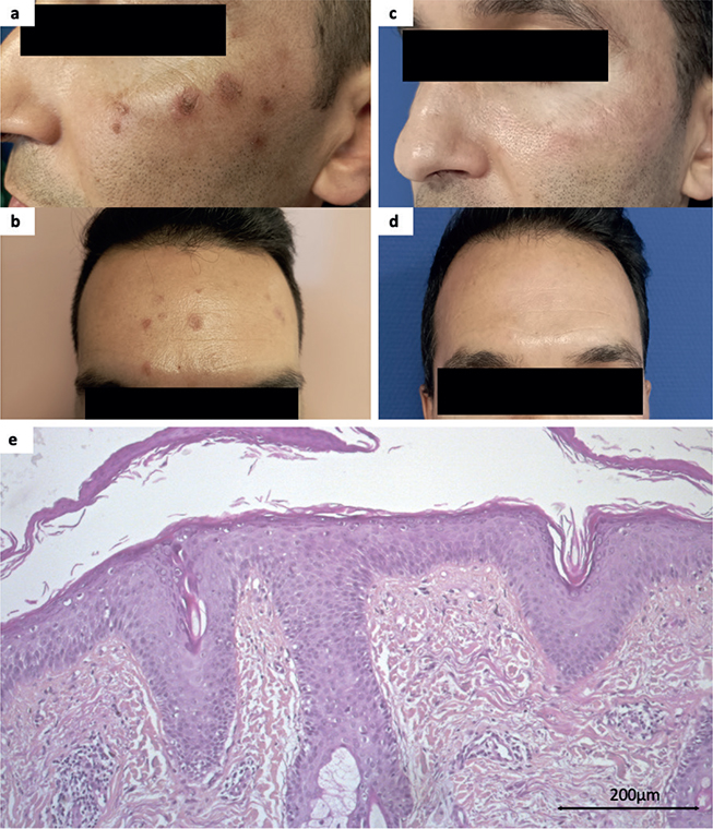
Fig. 1. First case (A, B) before and (C, D) after 6 months with acitretin); cheek lesion cutaneous biopsy (HESx20).
PAS staining and direct immunofluorescence were negative. In the hypothesis of cutaneous lupus, seborrheic dermatitis, or atypic psoriasis, different treatments were introduced, all ineffective (topical steroids, calcineurin inhibitors and imidazole derivatives, hydroxychloroquine for 6 months). Following therapeutic failures and absence of a formal diagnosis, we discussed the case within the national committee for challenging cases of the national Facial Dermatoses Study Group, leading to the diagnosis of facial discoid dermatosis (FDD). Treatment with acitretin 25 mg/day resulted in significant improvement, with lesions nearly disappeared after 6 months of treatment. The dose was reduced to 10 mg/day without recurrence after 12 months (Fig. 1).
A 54-year-old woman presented in 2020 with very similar lesions progressing over 3 years, remaining stable, asymptomatic, but noticeable, affecting the forehead, cheeks and chin (Fig. 2). Several skin biopsies performed provided limited information, with discrete acanthosis, focal parakeratosis, and mild inflammatory infiltrate in the superficial dermis. Various topical (topical steroids, calcineurin inhibitors, imidazole derivatives, ivermectin) and systemic treatments (tetracycline, hydroxychloroquine, acitretin, ivermectin, metronidazole, isotretinoin, UVB phototherapy) were tried without success. We also presented the case within our national challenging cases meeting leading to the diagnosis of FDD. Considering previous therapeutic failures and the literature, we introduced treatment with ustekinumab 45 mg, leading to initial rapid improvement after 3 injections in 6 months (Fig. 2). However, a few rare lesions remain under ongoing 12-month treatment.
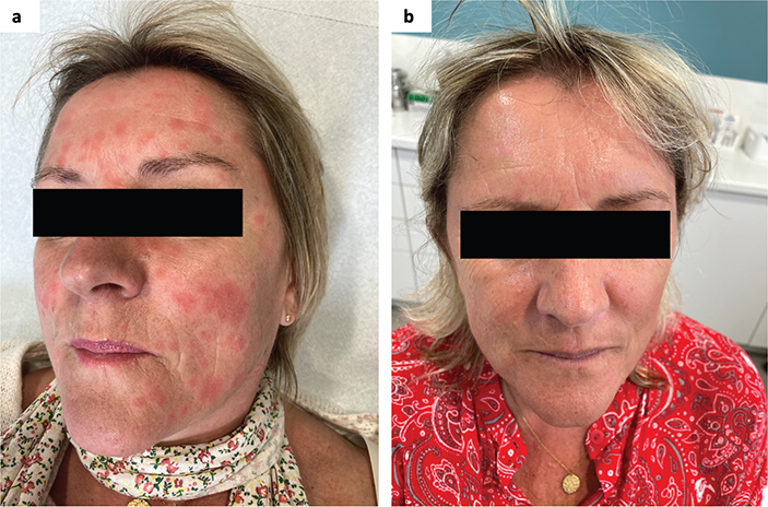
Fig. 2. Second case (A) before and (B) after 6 months with ustekinumab. Written permission is given by the patient to publish these photos.
DISCUSSION
FDD is an entity described only since 2010 (1), of unknown aetiology. It presents as well-defined, annular, pink to orange maculopapules, finely scaly, exclusively affecting the face (cheeks, chin, forehead) and neck. It tends to affect mostly women, over 30 years of age. Differential diagnoses to consider include lupus, seborrheic dermatitis, and psoriasis (1–3). Histologically, although nonspecific, it suggests similarities to pityriasis rubra pilaris (PRP): hyperkeratosis with dry parakeratosis, follicular plugging, and moderate interstitial and perifollicular inflammatory infiltrate of the superficial dermis (1). Demodex may be found in hair follicles (4). Additional histological characteristics of sub-corneal acantholysis have been observed (5). The course is usually chronic and stable for several years. One reported case progressed to authentic PRP after a long follow-up period, reinforcing the hypothesis of a link between PRP and FDD (5). This dermatosis is often refractory to many topical and systemic treatments, as in our cases (1, 3). Acitretin and the combination of topical steroids and vitamin D analogues are reported as most effective, but only in isolated clinical cases (6). Additionally, treatment with ustekinumab has been proposed, due to similarity to PRP, in a unique case, leading, as in our case, to improvement in cutaneous symptoms and quality of life (2).
Table SI provides a comprehensive overview of 24 reported cases of FDD, detailing the demographic, clinical, and pathological characteristics of the patients, along with the treatment approaches and their outcomes.
These observations highlight the diagnostic and therapeutic challenges of FDD. We draw attention to this poorly understood facial dermatosis, likely underdiagnosed but worthy of recognition. However, in our experience and on analysis of published cases, FDD has a fairly characteristic and recognizable presentation. Earlier diagnosis and management could thus reduce the impact of this displaying dermatosis. Acitretin and ustekinumab may represent effective options, as in our patients. We plan to confirm this in a large series within our national facial dermatosis group.
REFERENCES
- Ko CJ, Heald P, Antaya RJ, Bolognia JL. Facial discoid dermatosis. Int J Dermatol 2010; 49: 189–192. https://doi.org/10.1111/j.1365-4632.2009.04206.x
- Rypka KJ, Fulk TS, Afsaneh A, Miller DD, Goldfarb NI. Improvement of facial discoid dermatosis with ustekinumab treatment. JAMA Dermatol 2022; 158: 1079–1780. https://doi.org/10.1001/jamadermatol.2022.2478
- Rahmatulla S, Batta K, Tatnall F, Sandhu D, Brown V. Facial discoid dermatosis: a cosmetically disfiguring and challenging condition to treat. Skin Health Dis 2021; 1: e56. https://doi.org/10.1002/ski2.56
- Condal L, Quer A, Ferrándiz C, Bielsa I. Dermatosis discoide facial, una entidad enigmática. Actas Dermosifiliogr 2021; 112: 934–935. https://doi.org/10.1016/j.ad.2020.01.009
- Gan EY, Ng SK, Goh CL, Lee SSJ. Recalcitrant psoriasiform dermatosis of the face: is it related to pityriasis rubra pilaris? J Cutan Pathol 2018; 45: 491–497. https://doi.org/10.1111/cup.13148
- Allegue F, Fachal C, Iglesias B, Zulaica A. Facial discoid dermatosis: a new variant of pityriasis rubra pilaris? Actas Dermosifiliogr 2022; 113: T728–T731. https://doi.org/10.1016/j.ad.2022.05.015
- Huang Y-W, Tsai T-F. Topical rapamycin (sirolimus) for the treatment of facial discoid dermatosis. Eur J Dermatol 2024; 34: 201–203. https://doi.org/10.1684/ejd.2024.4651
- Amarnani R, Hughes S, Morris-Jones R, Kanwar AJ, Bunker CB. Persistent facial discoid dermatosis successfully treated with topical calcipotriol. Clin Exp Dermatol 2022; 47: 229–231. https://doi.org/10.1111/ced.14945
- Welborn M, Fletcher D, Motaparthi K. Atrophy of sebaceous lobules in facial discoid dermatosis: a link to psoriasis and seborrheic dermatitis? J Cutan Pathol 2021; 49: 320–323. https://doi.org/10.1111/cup.14190
- Bohdanowicz M, DeKoven JG. Improvement in facial discoid dermatosis with calcipotriol/betamethasone ointment and low-dose acitretin. Clin Exp Dermatol 2018; 43: 820–821. https://doi.org/10.1111/ced.13611
- Salman A, Tekin B, Berenjian A, Cinel L, Demirkesen C. Facial discoid dermatosis: a further case of a novel entity. J Dermatol 2015; 42: 1132–1133. https://doi.org/10.1111/1346-8138.13045