QUIZ SECTION
A 4-year-old Boy with a Red Nodule on His Hand: A Quiz
Riku ISHII1, Noriyuki OTSUKA2, Takashi ANAN3, Yuhei YAMAMOTO1 and Taku MAEDA1*
1Department of Plastic and Reconstructive Surgery, Faculty of Medicine and Graduate School of Medicine, Hokkaido University, Hokkaido, 2Department of Surgical Pathology, Hokkaido University Hospital, Sapporo, and 3Sapporo Dermatopathology Institute, Sapporo, Japan. *E-mail: takumaeda1105@yellow.plala.or.jp
Citation: Acta Derm Venereol 2025; 105: adv41071. DOI: https://doi.org/10.2340/actadv.v105.41071.
Copyright: © 2025 The Author(s). Published by MJS Publishing, on behalf of the Society for Publication of Acta Dermato-Venereologica. This is an Open Access article distributed under the terms of the Creative Commons Attribution-NonCommercial 4.0 International License (https://creativecommons.org/licenses/by-nc/4.0/).
Published: Jan 28, 2025
A 4-year-old boy was noticed by his mother to have a red nodule on the dorsum of his left hand. The nodule gradually increased in size. He was referred to a local clinic 3 months later, when the red nodule was 2 mm in diameter. The decision at that time was to keep him under observation. At a visit to our department 1 month later, the nodule had enlarged further. Physical examination revealed a 9×8 mm dome-shaped red nodule on the dorsum of the left hand (Fig. 1). The nodule had clear borders, was not adherent to the base, and was not tender. Echographic examination showed a well-defined, well-developed tumour extending from the epidermis to the dermis, with a homogeneous interior and abundant blood flow. There was no bleeding from the mass during treatment. The patient was otherwise in good health, with no history of any other medical problems other than allergic rhinitis, and he had no growth or developmental abnormalities.
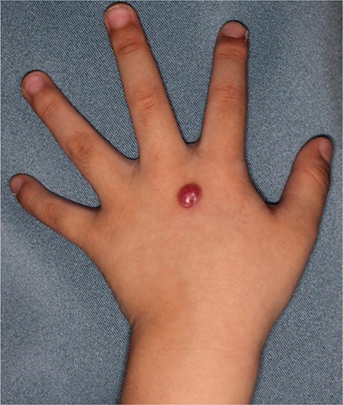
Fig. 1. A 9×8-mm dome-shaped red nodule on the dorsum of the left hand.
What is your diagnosis?
Differential diagnosis 1: Atypical Spitz tumour with ALK fusions
Differential diagnosis 2: Haemangioma
Differential diagnosis 3: Pilomatrixoma
Differential diagnosis 4: Cutaneous lymphoid hyperplasia
See next page for answer.
ANSWERS TO QUIZ
A 4-year-old Boy with a Red Nodule on His Hand: A Commentary
Diagnosis: Atypical Spitz tumour with ALK fusions
A magnetic resonance scan revealed a dome-like structure on the dorsum of the left hand with a hyperintense signal on T2-weighted imaging (Fig. 2). The tumour was completely excised, and petrolatum was applied for wound healing. Sentinel lymph node biopsy (SLNB) was not performed.
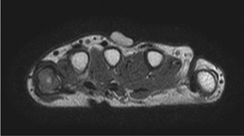
Fig. 2. T2-weighted magnetic resonance imaging shows a dome-like structure on the dorsum of left hand with a hyperintense signal.
Microscopic examination revealed spindle-shaped melanocyte-like cells distributed in sheets and bundles with increased deposition of stromal collagen fibres. Some of the tumour cells had a large nucleus with a prominent nucleolus. Mitotic figures were scattered (1/mm2), and no definite atypical mitotic figures were detected. The presence of spherical spaces or clefts between the cells was noted. These histological features suggested a Spitz tumour with ALK fusions. Immunohistochemical studies showed the tumour cells were positive for ALK (diffuse) and Melan-A (diffuse), while lacking immunoreactivity for HMB45, indicating the maturation of the melanocytic lesion. The Ki-67 labelling index was approximately 30–40% in the superficial areas, whereas overall index of the lesion was 10%. There was no loss of p16 expression in the tumour cells. Fluorescence in situ hybridization revealed 2p23 translocation where the ALK gene is located. Based on these findings, the patient was diagnosed with an atypical Spitz tumour with ALK fusions (Fig. 3).
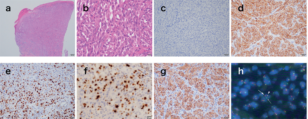
Fig. 3. Pathological examination shows findings typical of atypical Spitz tumour with ALK fusion. (a, b) Spindle-shaped, confluent melanocyte-like cells are distributed in sheets and bundles within a stroma with increased collagen fibres. Some of these cells show mitotic figures and some have large nucleus with prominent nucleolus. Small spherical spaces or clefts between the cells. In (b), mitotic figures are observed. The number of mitotic cells is 1/mm2. (HE stain, Scale bar: a = 200 μm, b = 20 μm) (c) The HMB45 indicates a maturation pattern with dermal descent. (d) Tumour cells show immunoreactivity for ALK. (e, f) The Ki-67 labelling rate is 30–40% in the superficial areas and 10% in the deeper parts of the lesion. (c~e: Scale bar = 50 μm, f: Scale bar = 20 μm) (g) Melan-A is positive. (g: Scale bar = 50 μm) (h) Fluorescence in situ hybridization shows ALK fusion in 92.5% of cells. The yellow point (triangle) is a pseudo-colour signal, the red point (thick arrow) is the 3′ ALK probe, and the green point (thin arrow) is the 5′ ALK probe.
The patient is currently undergoing follow-up with periodic imaging examinations. At 1 year postoperatively, there is no evidence of recurrence or lymph node metastasis.
Spitz tumours are melanocytic skin lesions that occur mainly in children and often resemble malignant melanoma on pathological examination. When benign and pathologically distinct from malignant melanoma, these tumours are called Spitz nevi; those with some features of malignant melanoma are called atypical Spitz tumours, and those that are malignant are called malignant Spitz tumours (1).
Recent studies have identified chromosomal abnormalities in Spitz tumours, including mutations in HRAS and MAP2K1 genes, a copy number increase at 11p, and gene fusions involving ALK, ROS, NTRK1, NTRK2, NTRK3, MET, RET, MAP3K8, and BRAF (1, 2).
ALK is located on chromosome 2p and encodes a tyrosine kinase receptor involved in the PI3K-AKT, RAF-MEK1/2-ERK1/2, and JAK3-STAT3 pathways. DCTN1 and TPM3 are the most common fusion partners for ALK, but fusions with NPM1, TPR, CLIP1, GTF3C2, MLPH, EEF2, MYO5A, KANK1, EHBP1, and SLC20A1 have also been reported (1, 3–8). ALK fusion genes are reported to be present in 10% of all Spitz tumours, approximately 8% of Spitz nevi, 5% of atypical Spitz tumours, and 1% of malignant Spitz tumours (1, 2).
The clinical features of Spitz tumours with the ALK fusion gene include a predilection for the extremities in young patients, a large pleomorphic appearance without pigmentation, and a solitary papule or nodule (2). The pathology is characterized by large non-pigmented spindle-shaped melanocytes with pericellular fissures, vesicular nuclei, prominent nucleoli, and plexiform architecture with streak-like growth (2).
Management of atypical Spitz tumours typically involves complete surgical excision with clear margins to ensure removal of all tumour cells. SNLB is generally not considered necessary for atypical Spitz tumours because of their low risk of metastasis. However, careful clinical follow-up with regular physical examination is necessary to monitor for signs of recurrence (9, 10). Cerrato et al. reported no recurrence of atypical Spitz tumour in children at 3 years postoperatively, even without SLNB, when treated with extensive resection alone (10). In a study by Lallas et al., although 39% of patients with atypical Spitz tumour who underwent SLNB in addition to wide excision had positive lymph nodes, only 1% were found to have disease progression beyond the lymph nodes during 5 years of follow-up. Following wide excision, patients in that study also had a high 5-year survival rate (99% with SLNB and 98% without SLNB). Therefore, they concluded that SLNB was not effective in improving the prognosis (9). However, the modest difference in survival rate in that study does not definitively negate the potential advantages of further treatment after a positive SLNB.
The primary treatment of atypical Spitz tumour with the ALK fusion gene is biopsy, histological evaluation, and gene retrieval by fluorescence in situ hybridization followed by surgical resection with adequate margins (1, 9). The ALK fusion gene is a therapeutic target in other carcinomas, and contributes to the increased oncogenicity of Spitz tumours in vitro. Careful long-term postoperative follow-up is warranted (1).
REFERENCES
- Yeh I, de la Fouchardiere A, Pissaloux D, Mully TW, Garrido MC, Vemula SS, et al. Clinical, histopathologic, and genomic features of Spitz tumors with ALK fusions. Am J Surg Pathol 2015; 39: 581–591. https://doi.org/10.1097/PAS.0000000000000387
- Dal Pozzo CA, Cappellesso R. The morpho-molecular landscape of spitz neoplasms. Int J Mol Sci 2022; 23: 4211. https://doi.org/10.3390/ijms23084211
- Chung CT, Marrano P, Swanson D, Dickson BC, Thorner PS. Fusion of ALK to the melanophilin gene MLPH in pediatric Spitz nevi. Hum Pathol 2019; 87: 57–64. https://doi.org/10.1016/j.humpath.2019.03.002
- Busam KJ, Kutzner H, Cerroni L, Wiesner T. Clinical and pathologic findings of Spitz nevi and atypical Spitz tumors with ALK fusions. Am J Surg Pathol 2014; 38: 925–933. https://doi.org/10.1097/PAS.0000000000000187
- Kastnerova L, Martinek P, Grossmann P, Steiner P, Vanecek T, Kyclova J, et al. A clinicopathological study of 29 spitzoid melanocytic lesions with ALK fusions, including novel fusion variants, accompanied by fluorescence in situ hybridization analysis for chromosomal copy number changes, and both TERT promoter and next-generation sequencing mutation analysis. Am J Dermatopathol 2020; 42: 578–592. https://doi.org/10.1097/DAD.0000000000001632
- Bahrani E, Kunder CA, Teng JM, Brown RA, Rieger KE, Novoa RA, et al. Spitz nevus with EHBP1-ALK fusion and distinctive membranous localization of ALK. J Cutan Pathol 2022; 49: 584–588. https://doi.org/10.1111/cup.14209
- Salah HT, Yang RK, Roy-Chowdhuri S, Ross MI, Aung PP, Rothrock AT, et al. Spitz melanocytic neoplasms with MLPH::ALK fusions: report of two cases with previously unreported features and literature review. J Cutan Pathol 2024; 51: 407–414. https://doi.org/10.1111/cup.14605
- Cho WC, Prieto VG, Yang RK. Spitz melanoma with SLC20A1::ALK fusion: a novel fusion previously undescribed in spitz melanocytic neoplasm. Am J Dermatopathol 2024; 46: 700–703. https://doi.org/10.1097/DAD.0000000000002778
- Lallas A, Kyrgidis A, Ferrara G, Kittler H, Apalla Z, Castagnetti F, et al. Atypical Spitz tumours and sentinel lymph node biopsy: a systematic review. Lancet Oncol 2014; 15: e178–183. https://doi.org/10.1016/S1470-2045(13)70608-9
- Cerrato F, Wallins JS, Webb ML, McCarty ER, Schmidt BA, Labow BI. Outcomes in pediatric atypical spitz tumors treated without sentinel lymph node biopsy. Pediatr Dermatol 2012; 29: 448–453. https://doi.org/10.1111/j.1525-1470.2011.01699.x