QUIZ SECTION
Multiple Inflammatory Lesions on the Abdomen: A Quiz
Stefano VERALDI1*, Paolo BORTOLUZZI2 and Francesca GERMINIASI3
1Dermatological Centre in Milan, Milan, 2Department of Pathophysiology and Transplantation, Università degli Studi, IRCCS Foundation, Ca’ Granda Ospedale Maggiore Policlinico, Milan, and 3Ospedale San Raffaele, Milan, Italy. *E-mail: stefano.veraldi@dcim.it
Citation: Acta Derm Venereol 2024; 104: adv41123. DOI: https://doi.org/10.2340/actadv.v104.41123.
Copyright: © 2024 The Author(s). Published by MJS Publishing, on behalf of the Society for Publication of Acta Dermato-Venereologica. This is an Open Access article distributed under the terms of the Creative Commons Attribution-NonCommercial 4.0 International License (https://creativecommons.org/licenses/by-nc/4.0/).
Published: Dec 4, 2024
A 36-year-old Caucasian woman was admitted with a clinical diagnosis of unspecified inflammatory dermatitis located on the abdomen. The patient stated that she was in good general health and that she was not on therapy with systemic drugs. She also declared that the dermatitis had appeared 3 weeks earlier: the patient feared that the cause was a hedgehog that she used to place on her bare abdomen (Fig. 1). The hedgehog was a gift from the woman’s boyfriend, who had just returned from Africa.
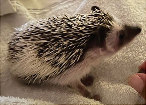
Fig. 1. African pygmy hedgehog Atelerix albiventris.
Dermatological examination revealed the presence of 3 similar lesions located exclusively on the abdomen: they were erythematous plaques, oval in shape, 2x1 cm in diameter, with well-defined, vesicular-pustular borders (Fig. 2). The patient complained of severe pruritus.
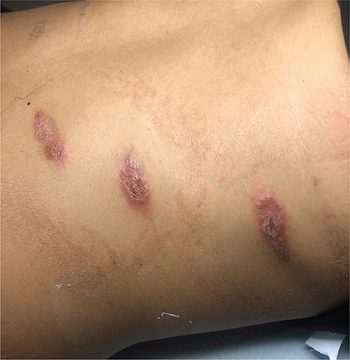
Fig. 2. Three similar, erythematous plaques, oval in shape, 2x1 cm in diameter, with well-defined, vesicular-pustular borders, located on the abdomen.
General physical examination did not reveal anything pathological.
What is your diagnosis?
Differential diagnosis 1: Multifocal impetigo
Differential diagnosis 2: Multifocal tinea corporis
Differential diagnosis 3: Superinfected eczema
Differential diagnosis 4: Inflammatory lesions caused by scratching
See next page for answer.
ANSWERS TO QUIZ
Multiple Inflammatory Lesions on the Abdomen: A Commentary
Diagnosis: Multifocal tinea corporis probably caused by Trichophyton erinacei transmitted by the African hedgehog Atelerix albiventris
Bacteriological examinations were negative. Direct microscopic examination, using 20% potassium hydroxide, was positive. Fungal cultures on Sabouraud dextrose agar (SDA) supplemented with chloramphenicol, SDA with chloramphenicol plus gentamicin, and SDA with chloramphenicol and cycloheximide were positive for Trichophyton erinacei. It was not possible to distinguish this species from other dermatophytes of the Trichophyton mentagrophytes species complex, such as Arthroderma benhamiae, because the sequence-based identification was not performed. However, it was possible to perform mycological examinations also on the patient’s hedgehog: a positivity for T. erinacei was detected in scales and prickles. The patient was successfully treated with terbinafine cream (2 applications/day for 3 weeks) and oral terbinafine (250 mg/day for 6 weeks).
T. erinacei was first described in 1960 by Marples and Smith (1). It was named in the past T. mentagrophytes var. erinacei and T. proliferans. T. erinacei is a zoophilic dermatophyte: 2 species of hedgehogs are the most frequent source of infection in humans: the African pygmy hedgehog or four-toed hedgehog (Atelerix albiventris) and the West European hedgehog (Erinaceus europaeus). Rare cases of human infections are transmitted by the Northern white-breasted hedgehog (Erinaceus roumanicus) (2). Other sources of human infections are humans themselves (3–5), guinea pigs, mice (4), soil (6), and even Asian elephants (7).
Tinea manuum is the most frequent manifestation of T. erinacei infection (8). Other not rare manifestations are tinea capitis (6, 9) and tinea barbae (5, 10): both can appear as a kerion (5, 6, 10). As with other human mycoses caused by zoophilic dermatophytes, clinical manifestations of T. erinacei infections are characterized by inflammatory lesions, especially pustular lesions. Tinea corporis is rare. To our knowledge, multifocal tinea corporis caused by T. erinacei has never been reported. Furthermore, some infections occurred on the face, arms, back, thighs, knees, legs, and toes. One case of proximal subungual onychomycosis has also been described. According to data in the literature, oral terbinafine is the therapy of choice (6): cases of resistance to fluconazole and itraconazole have been not infrequently reported.
REFERENCES
- Marples MJ, Smith JM. The hedgehog as a source of human ringworm. Nature 1960; 188: 867–868. https://doi.org/10.1038/188867b0
- Čmoková A, Kolařík M, Guillot J, Risco-Castillo V, Cabañes FJ, Nenoff P, et al. Host-driven subspeciation in the hedgehog fungus, Trichophyton erinacei, an emerging cause of human dermatophytosis. Persoonia 2022; 48: 203–218. https://doi.org/10.3767/persoonia.2022.48.06
- Rush-Munro FM, Smith JM. Further observations on Trichophyton erinacei and T. proliferans. Sabouraudia 1971; 9: 61–64. https://doi.org/10.1080/00362177185190151
- Padhye AA, Ajello L. The taxonomic status of the hedgehog fungus Trichophyton erinacei. Sabouraudia 1977; 15: 103–114. https://doi.org/10.1080/00362177785190181
- Sidwell RU, Chan I, Francis N, Bunker CB. Trichophyton erinacei kerion barbae from a hedgehog with direct osculatory transfer to another person. Clin Exp Dermatol 2014; 39: 38–40. https://doi.org/10.1111/ced.12197
- Romano C, Gianni C, Papini M. Tinea capitis in infants less than 1 year of age. Pediatr Dermatol 2001; 18: 465–468. https://doi.org/10.1046/j.1525-1470.2001.1861997.x
- Borges-Costa J, Martins MdL. Trichophyton erinacei skin infection after recreational exposure to an elephant in Southeast Asia. Pathog Glob Health 2014; 108: 58–59. https://doi.org/10.1179/2047773213Y.0000000117
- Gregory MW, English MP. Arthroderma benhamiae infection in the Central African hedgehog Erinaceus albiventris, and a report of a human case. Mycopathologia 1975; 55: 143–147. https://doi.org/10.1007/BF00491498
- Roger H, D’Incan M, Cambon M, Ferrier MC, Meyer M, Souteyrand P. Teigne inflammatoire à Trichophyton erinacei chez un enfant de trois ans. Ann Dermatol Venereol 1991; 118: 839–840.
- Simpson JR. Tinea barbae caused by Trichophyton erinacei. Br J Dermatol 1974; 90: 697–698. https://doi.org/10.1111/j.1365-2133.1974.tb06701.x