ORIGINAL REPORT
Targeting Complex Cutaneous Viral Infections in Search of Inborn Errors of Immunity
Ana Paula DO CARMO1, Amanda R. BATLLE1, Nyla T. Melo Lobao FRAGNAN2, Carlos A. S. MACHADO3, Rosemeire N. CONSTANTINO-SILVA2, Sarah E. HENRICKSON4-6 and Anete S. GRUMACH2
1Faculdade de Medicina, Centro Universitario FMABC, Santo Andre, SP, 2Discipline of Immunology, Faculdade de Medicina, Centro Universitario FMABC, Santo Andre, SP, 3Discipline of Dermatology, Faculdade de Medicina, Centro Universitario FMABC, Santo Andre, SP, Brazil, 4Department of Pediatrics, Perelman School of Medicine, University of Pennsylvania, 5Division of Allergy Immunology, Department of Pediatrics, The Children’s Hospital of Philadelphia, and 6Department of Microbiology, University of Pennsylvania, Philadelphia, PA, USA
Inborn errors of immunity are rare diseases and 50–80% present with dermatological manifestations. This study evaluated difficult-to-treat cutaneous human papillomavirus infections and their associations with immunological defects. Patients were recruited from the Dermatological Outpatient Clinic over 2 years. Patients reporting persistent common warts and/or a combination of molluscum contagiosum or more than 2 flat warts, with a clinical assessment of severe or persistent skin infection, met the clinical severity criteria for inclusion. Resistance to several therapies was also considered. A total of 632 patient records were analysed to clinically characterize the warts, laboratory data, treatments used and their responses, comorbidities, and family history. Among these, 459 cases were initially excluded from further evaluation. A questionnaire was provided by phone to 173 patients, among whom 47 patients were selected for an in-person consultation. Of these, 6 met the criteria for further evaluation. Immunological tests revealed neutropenia, low levels of immunoglobulin isotypes (IgA, IgM, and IgG), and reduced frequency of lymphocyte subsets. Family history, flat warts, and associated recurrent viral infections suggested the need for further immunological evaluation. Criteria are proposed for identifying patients with cutaneous warts that warrant additional evaluation for potential inborn errors of immunity.
SIGNIFICANCE
This study was stimulated by 2 main factors. Dermatological manifestations are closely linked to immune defects and family history has relevance to background identification of inborn errors of immunity. In the last 20 years, a high number of inborn errors of immunity was described for unique susceptibility to infectious agents. We aimed to evaluate patients with persistent warts selected from patients referred to dermatological surgery due to presumed unresponsive treatments. Evaluation of these patients led to the development of warning signs of inborn errors of immunity amongst these patients.
Key words: Papillomaviridae; cutaneous immunity; warts; HPV; inborn errors of immunity.
Citation: Acta Derm Venereol 2025; 105: adv41318. DOI: https://doi.org/10.2340/actadv.v105.41318.
Copyright: © 2025 The Author(s). Published by MJS Publishing, on behalf of the Society for Publication of Acta Dermato-Venereologica. This is an Open Access article distributed under the terms of the Creative Commons Attribution-NonCommercial 4.0 International License (https://creativecommons.org/licenses/by-nc/4.0/).
Submitted: Aug 10, 2024. Accepted after revision: Apr 25, 2025. Published: Jun 9, 2025.
Corr: Anete S. Grumach, MD, PhD, Clinical Immunology, Faculdade de Medicina, Centro Universitario Saude ABC, Av Lauro Gomes 2000, Santo Andre, Sao Paulo 09060-870, Brazil. E-mail: asgrumach@gmail.com
Competing interests and funding: The authors have no conflicts of interest to declare.
APPC received a scholarship from the Institutional Program for Scientific Initiation from the National Council of Research (PIBIC-CNPq). SEH received a grant from the Clinical Immunology Society, and ASG received a scholarship for research productivity from the National Council of Scientific and Technological Development (CNPq).
INTRODUCTION
Inborn errors of immunity (IEIs) are rare diseases with a prevalence of approximately 1:1,200 individuals (1). Among them, 50–80% have dermatological manifestations that may be associated with infectious or non-infectious disorders (2, 3). In addition, cutaneous symptoms precede the diagnosis of IEI in one-third of cases (4). In patients with concern for IEI, human papillomavirus (HPV) infections with characteristics that deviate from the usual pattern of evolution or that are resistant to treatment may suggest an IEI (5). HPV causes different skin lesions depending on the type of virus, its location, morphology, and genome. While most IEIs predispose individuals to a wide spectrum of viral infections, certain diseases enhance patient vulnerability to specific types of viruses (3, 6–8).
Host defence against HPV is provided by a system of factors, including functioning cellular immunity, T cells, natural killer (NK) cells, and innate immune cells. Hypofunctional T or NK cells are associated with an enhanced susceptibility to HPV (8). While many T cell disorders are associated with increased susceptibility to cutaneous warts, cutaneous warts are a cardinal feature of a subset of IEIs, such as the “warts, hypogammaglobulinemia, immunodeficiency, myelokathexis” (WHIM) syndrome. This syndrome is caused by heterozygous variants in the C-X-C chemokine receptor type 4 (CXCR4), resulting in CXCR4 gain-of-function; mutations in the epidermodysplasia verruciformis genes (EVER1 and EVER2); a dedicator of cytokinesis 8 (DOCK8) deficiency (autosomal recessive variants in DOCK8, causing combined immunodeficiency with elevated immunoglobulin E [IgE] levels) and a GATA-2 deficiency (7, 9–12).
Most patients with viral/common warts (66%) can expect spontaneous remission within 2 years, with the number of lesions not affecting prognosis (13, 14). However, no consensus currently exists regarding the definition of severe or persistent warts (15).
Therefore, the present study aimed to characterize patients with cutaneous HPV infections according to their clinical characteristics and responses to treatment. Furthermore, we aimed to propose practical criteria for dermatological evaluations that could indicate underlying immunological defects.
METHODS
This study had 2 phases; namely, an observational phase and an interventional phase. The medical records of patients who were referred to the Surgery Center of the Dermatology Outpatient Clinic for resection of cutaneous warts were reviewed and the following clinical metadata were collected: medical record number; patient name; sex; date of birth; age; wart characteristics (type, number, location, extent, and evolution); comorbidities (associated with immunosuppression and others); treatments used; lesion classification based on the treatment response (limited, resistant, and/or persistent); and family history, including consanguinity. Patients with predisposing diseases, such as oncological processes and/or immunosuppressive therapy, were excluded. No age limit was imposed for patient enrolment.
The first phase used retrospective data from patient files. The clinical manifestations used to select patients for the second phase were based on warning signs such as family history, consanguinity, persistent viral infections, and/or association with no response to treatment according to Relan and Lehman (16), confirmed by Cagdas et al. (17). After the first analysis, patients were contacted by telephone and a standard questionnaire was administered to identify patients with symptoms suggestive of increased risk of IEI for presential evaluation at the Immunology Clinic. The initial in-person consultations were followed by immunological screening for immunoglobulin (IgG, IgA, IgM, and IgE) measurement (via nephelometry); antibody response (according to age and immunization history); immunophenotyping of T lymphocytes and subpopulations (e.g., CD4 and CD8), B lymphocytes, and NK cells (via flow cytometry); CH50 (complement system haemolytic assay); and HIV serology. For patients with a high suspicion of immune defects based on clinical metadata and immunological evaluation, whole-exome sequencing (WES) was performed.
This study was approved by the Research Ethics Committee of the Faculdade de Medicina, Centro Universitario FMABC (number 52536915.5.0000.0082), and consent forms were signed by the patients and/or their responsible healthcare proxies.
RESULTS
Initially, 632 eligible patients were identified over the 2-year study period. A total of 141 patients were excluded: 114 due to non-verrucous lesions during the surgical evaluation, 5 due to immunosuppression, and 22 due to incomplete data. Of the 491 patients included in the present study, most were female (64.7%) with an average age of 54.3 years (range, 6–95 years). Regarding comorbidities, 11% of the patients had onychocryptosis/onychomycosis and seborrheic keratosis, and 8.8% had actinic keratosis. Hypertension (14.9%) and diabetes mellitus (6.3%) were common findings. The most frequent wart types were viral or common cutaneous (72%) and filiform (15.9%) warts, with the majority affecting the face (37.6%) and digits (20.6%). The most common initial therapeutic approaches were radiofrequency ablation (48.8%) and shaving (42.1%). Resistance to initial treatment (lack of regression of the treated lesions) was reported in the medical records of 118 of 491 (24%) patients, and 50 of the 118 (42.4%) patients underwent more than 5 cycles of treatment with different techniques (Table SI). An additional 318 of the 491 patients were excluded because subsequent medical appointments revealed that they were cured of their cutaneous infection or had a clinical condition compatible with standard HPV infection (no lesions after the initial treatment or were not considered refractory to resolution with fewer than 5 treatments). The remaining 173 patients were contacted by telephone; among these, 126 were excluded (109 who were cured or presented with classic, uncomplicated HPV infection, 15 receiving immunosuppression drugs, 1 due to incomplete data, and 1 due to death). Therefore, 47 patients were invited to undergo evaluation in the immunology clinic. After clinical evaluation, 6 patients (HIV-negative) underwent immunological screening and subsequent genetic analysis by WES (Fig. 1). A total of 6 patients showed variations in the initial immunological evaluation, including both humoral and cellular alterations. Besides humoral changes, high and low levels of different immunoglobulins and low neutrophil counts were observed. Cellular immune dysregulation was also identified, including alterations in T lymphocyte, B lymphocyte, and NK cell counts. However, none of the patients met the criteria for severe combined immunodeficiency (SCID) or hypomorphic SCID according to Shearer et al. (18). Pathogenic variants or variants of unknown significance were identified in 4 of the 6 (67%) patients analysed using WES.

Fig. 1. Patients with warts selected for immunologic evaluation.
Patient 1 was a woman referred for a nasal lesion previously confirmed as nodular and micronodular basal cell carcinoma of the skin in the nasal region. During the follow-up, she developed multiple flat warts that persisted even after several treatments. Physical examination revealed flat yellow-brown warts on the breasts, arms, forearms, and back (> 20). Although no consanguinity was identified, several family members reported having flat warts. Immunological evaluation resulted in IgG levels lower than the third percentile (p3) and IgM levels higher than the p97 for the patient’s age. No other relevant findings were reported. WES variants in myeloid differentiation primary response 88 (MYD88) and interferon regulatory factor 3 (IRF3).
Patient 2 was a man born to consanguineous parents. Among 11 siblings, 3 were stillbirths, a 7-year-old boy died of a fever of undetermined origin, and 1 sister had recurrent abortions and died of an unknown cause. The patient presented with vulgar and flat warts affecting the genitals, experienced recurrent tonsillitis up to 18 years of age, and experienced difficulty in healing the lesions. The immunological evaluation revealed decreased IgG and IgM levels and low TCD8+ and NK cell counts. WES revealed no variants.
Patient 3 was a man who reported several infections since infancy, with no unusual complications. Onychomycosis and tinea pedis were diagnosed on the patient’s 50th birthday. The family history was significant: the first and second siblings had died at 1 and 3 years of age, respectively, of unknown causes; the fifth sibling had contracted tetanus at 16 years of age and the sixth sibling had Kaposi’s sarcoma and was HIV-negative. No consanguinity was observed. The results of the immunological evaluation were unremarkable except for high TCD4+ cell counts. A pathogenic heterozygous variant of RAG1 was observed in the patient and his brother with Kaposi’s sarcoma. Testing for tumour necrosis factor receptor superfamily, member 4 (OX40) mutations revealed no variants.
Patient 4 was a woman who presented with extensive, resistant, and recurrent condylomata acuminata and warts on the vulva at 22 years of age. A vulvar biopsy showed high-grade vulvar squamous intraepithelial neoplasia (VIN II) associated with cytoarchitectural changes, suggestive of an HPV infection. One year later, flat viral warts appeared in diffuse spots on the patient’s body (front, neck, upper limbs, lower limbs, back, and pubis), as well as a single café-au-lait spot on the abdomen. Skin biopsy confirmed the presence of viral warts and fibroepithelial polyps. The patient had previously experienced 3 episodes of pneumonia. Her grandmother had undergone a hysterectomy for HPV infection. WES revealed compound heterozygous variants in JAK3 that were classified as pathogenic (Chr19:17955112del, c.115del, p.Gln39Serfs*108) or VUS (Chr19:17945804C > T, c.2056G > A, p.Asp686Asn). The pathogenic variant was inherited maternally.
Patient 5, the daughter of first-cousin parents, first developed multiple treatment-resistant bilateral plantar warts at 15 years of age. The patient also demonstrated vulgar warts in the dorsal and inframammary regions, molluscum on the chin, and basal cell carcinoma in the epicanthus. Biopsies of several warts performed 5 years later revealed atypia in the basal layer. Additional biopsies performed at 27 years of age revealed basal cell carcinoma, CPB, and squamous cell carcinoma in situ, suggestive of Bowen’s disease. Immunological consultations revealed the genealogy, and 2 other siblings had the same complaints. Examination showed neutropenia, reduced isohemagglutinins, lymphopenia affecting TCD4+ and B cells, and a low NK cell count. WES identified a VUS in JAK1.
Patient 6, the daughter of an inbred marriage, had resistant warts in the soles of her feet and her dorsal region since 15 years of age and presented with molluscum on the chin. The patient had also been treated for basal cell carcinoma of the epicanthus. Some episodes of axillary and inguinal furunculosis were also reported. Multiple warts were a common finding in several family members (Tables I–II, Fig. 2).
| Case | Sex | Age | Wart type | Comorbidities | Familial history |
| 1 | F | 66 | Flat | Fever blister, diabetes, arterial hypertension, varicose vein, obesity, hepatic steatosis | Maternal relatives with multiple warts |
| 2 | M | 51 | Vulgar and flat | Hepatitis B, recurrent tonsillitis | Inbreeding |
| 3 | M | 57 | Vulgar; herpes zoster; eosinophilic urticaria | Asthma; infected varicella; rubella; bilateral inguinal herniorrhaphy; autoimmune urticaria; tinea pedis; onychomycosis | Brother: Kaposi’s sarcoma |
| 4 | F | 33 | Viral and flat; acuminated condyloma; café-au-lait spots | Cervical cancer II; fibroepithelial polyp; viral warts; repeated sinusitis; pneumonia | Grandmother: hysterectomy because of HPV Inbreeding by maternal grandmother |
| 5 | F | 45 | Vulgar and flat; reddish and scaling-of plaques (perivascular dermatitis) | D. Bowen, basal-cell carcinoma, squamous cell carcinoma; pyogenic granuloma | Inbreeding† |
| 6 | F | 57 | Vulgar resistant; flat; molluscum contagiosum | Basal-cell carcinoma, arthrosis, dyslipidaemia, rhinitis, furunculosis, gastritis | Uncles with repeating warts; mother with warts |
| †Genealogy. F: female; M: male; HPV: human papillomavirus. | |||||
| Case | Immunological screening† | Other relevant results | Lymphocyte immunophenotyping (cells/mm³) | WES | |||||||
| IgG (mg/dL) | IgM (mg/dL) | IgA (mg/dL) | IgE (IU/ml) | CD3+ | CD4+ | CD8+ | CD19+ | NK cells | |||
| 1 | 734 (< P3) | 329 (> P97) | 241 (P75) | 99.3 | IgG Ab positive for CMV, EBV, and rubella. HIV-negative | 1,251 (P50) | 927 (P50) | 321 (P10) | 356 (P50) | 194 (P10) | MYD88 – likely pathogenic. IRF8 – VUS |
| 2 | 716 (< P3) | 32.8 (< P3) | 99.9 (P10) | 9 | Anti-HBsAg-positive, HIV and EB-negative | 1,418 (P50) | 1,032 (P90) | 166 (< P10) | 300 (P50) | 52 (< P10) | No variants to report |
| 3 | 1,398 (P97) | 127 (P50) | 296 (P75) | – | HIV-negative; IgG anti-herpes-positive | 2,668 (> P90) | 1,878 (> P90) | 691 (P90) | 495 (P90) | 196 (P50) | Pathogenic heterozygous variant in RAG1 c.1420C > T, p.(Arg474Cys) |
| 4 | 1,104 (P50) | 211 (> P97) | 237 (P75) | 20.5 | HIV, IgG anti-rubella, and anti-HBs-negative; CMV and EBV IgG-positive; Anti-B 1:16 | 1,014 (P10) | 633 (P50) | 364 (P50) | – | 15.6% (P90) | ‡JAK3 – Pathogenic (SCID) JAK3 – VUS MMP26 – VUS |
| 5 | 1,570 (> P97) | 154 (P75) | 197 (P50) | 25.5 | HIV-negative; rubella, anti-HBs, EBV, and CMV-positive; neutrophils 930 Anti-B 1:8 |
829 (< P10) | 451 (< P10) | 317 (P10-50) | 43 (< P10) | 93 (< P10) | JAK1 – VUS (Immunodeficiency and cancer) |
| 6 | 1,113 (P75) | 64 (< P3) | 175 (P50) | 32.6 | HIV and anti-HBs-negative; rubella, CMV, and EBV-positive; anti-A 1:4 anti-B 1:2 |
1,224 (P50) | 840 (P50) | 396 (P50) | 361 (P50) | 205 (P50) | JAK3 (two mutations) – VUS |
| †Immunological screening: blood count, immunoglobulins (mg/dL), isoagglutinin (anti-A and anti-B), vaccine response. ‡Patient 4 – Family exome result: Mother: JAK3 – pathogenic (SCID, AR) and RAG1 – pathogenic (Omenn syndrome, AR). Brother: RAG1 – pathogenic (Omenn syndrome, AR). IgG: immunoglobulin G; Ab: antibody; CMV: cytomegalovirus; EBV: Epstein–Barr virus; HIV: human immunodeficiency virus; HBsAg: hepatitis B surface antigen; JAK3: Janus kinase 3; SCID: severe combined immunodeficiency disease; AR: autosomal recessive; RAG1: recombination activating gene 1; MYD88: myeloid differentiation primary response 88; IRF8: interferon regulatory factor 8; VUS: variant of uncertain significance; MMP26: matrix metallopeptidase 26. Bold numbers represent results out of normal range. | |||||||||||
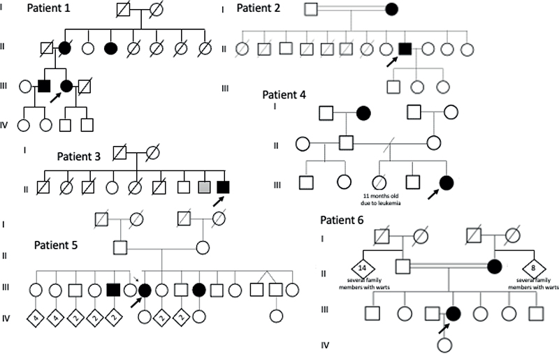
Fig. 2. Genealogy of the 6 patients selected for whole-exome sequencing. *The probands are pointed by arrows and the numbers of the genealogy correspond to patients in table 2
DISCUSSION
Cutaneous warts are a common dermatological and immunological complaint in primary care. The immunological investigation of patients with refractory skin viral infections is not routine in all dermatology practices, although it is increasingly recognized as a concerning clinical sign, which may indicate the presence of an IEI. In-depth studies of patients with specific patterns of susceptibility to certain infectious agents, including HPV, have revealed some IEIs including those associated with increased susceptibility to mycobacteria and HPV infections associated with monogenic defects (5). Several gene variants cause IEIs associated with HPV infection, such as epidermodysplasia verruciformis and DOCK8, ATM, and GATA2 deficiencies. Recurrent or resistant warts may also be symptoms associated with other infections or manifestations (19). The present study aimed to evaluate a large population seen by dermatologists to identify signs suggesting the need for further immunologic testing for underlying IEIs.
We assumed that patients treated for viral cutaneous warts and referred to the surgery centre had conditions more resistant to treatment than the rest of the affected population. Most patients received more than 1 type of treatment, primarily radiofrequency, shaving, cryotherapy, or chemical acid application. We excluded 482 patients from this cohort after the first screening of their medical records based on established clinical criteria (13–15). The present study identified family history and flat warts as characteristics to be further investigated. Notably, 2 families had consanguinity, and in all 6 patients a suspicious history of multiple warts and carcinomas influenced the decision to proceed with immunological tests (20, 21).
Cellular immunity provided by T and NK cells is the main host defence mechanism against HPV infection. These patients may have a functional deficit of CD4+ T cells and a tendency to reverse the Th1 to the Th2 response, depending on the severity of the infection (22, 23). A CD8+ cell deficit is observed in patients with cytological findings compatible with HPV lesions at all disease stages (23). Patient 2 had low TCD8+ and NK cell counts; however, WES revealed no pathogenic variants, although the parents were consanguineous. In contrast, patient 5 had lymphopenia, with increased TCD4+, B, and NK cell counts and neutropenia, which was associated with several skin carcinomas. The JAK1 variant in this patient is associated with 2 types of cutaneous carcinoma (squamous and basal cell) (24). The occurrence of HPV flat warts in patients treated with selective JAK1 inhibitor could suggest the relevance of our finding. JAK-1 inhibitors contribute to impairments in interferon-mediated antiviral responses, which can increase susceptibility to viral infections, and a patient under therapy with upadacitinib for atopic dermatitis was reported with flat warts relatively resistant to therapy (25).
RAG1 variants are associated with several phenotypes, hampering the interpretation of their effects (26, 27). Patient 3’s family history was associated with premature death and a higher occurrence of carcinomas. Considering that the patient’s brother was affected by Kaposi sarcoma and was negative for HIV, we searched for OX40 deficiency, as previously described (28). However, only the same RAG1 variant was found. The relevance of this variant was not considered in this patient due to the normal immunological profile, in the setting of infections and malignancy.
Patient 4 reported recurrent infections and carcinoma; however, the results of the basic immunological evaluation did not reveal any relevant abnormalities. A combined heterozygous mutation in JAK3 was identified, 1 of which was pathogenic; however, it was insufficient to determine its role in the patient’s manifestations. The association between JAK3 deficiency and HPV infection has been reported (29).
Hypo-IgM has recently been included in the classification of IEIs (30). While increased malignancy risk has been reported, 2 definitions for this condition have been used in the literature (31, 32), including IgM values in adults < 30 mg/dL independent of age (33) and levels below 2 standard deviations from normal (34). Thus, Patient 5 could be considered to have selective IgM deficiency, which may be related to the increased susceptibility to neoplasia.
A study applying targeted panel sequencing and WES to a large number of patients highly suspected of having IEIs (n = 878) identified disease-causing variants in 56% of the probands (35). Consanguineous families have a higher likelihood of monogenic IEIs (36), consistent with our observations in 3 of the 6 patients with consanguinity included in the present study. Furthermore, 2 (patients 4 and 5) showed variants.
This study has some limitations. First, HPV types could not be tested because of the recall of patients who had previously undergone the procedure. Second, specific functional assays to assess the impact of a VUS on protein function could not be performed; however, further tests will permit the description of the pathogenicity of these variants, although some cases (patients 4 and 5) showed appropriate correlations with clinical immune findings.
The results of the present analysis of a large, consecutive group of patients previously seen by dermatologists and who underwent a series of therapies suggest an optimized profile of patients with warts at greater risk for IEIs; namely: (i) a family history of recurrent warts, immunological defects, and consanguinity; (ii) the presence of flat warts; and (iii) the presence of additional viral cutaneous infections such as molluscum contagiosum and acuminated condyloma.
ACKNOWLEDGEMENTS
The authors would like to thank Alessandra Tolentino, who initiated the patient evaluations.
Editage (www.editage.com.br) is acknowledged for English-language editing.
REFERENCES
- Rubin Z, Pappalardo A, Schwartz A, Antoon JW. Prevalence and outcomes of primary immunodeficiency in hospitalized children in the United States. J Allergy Clin Immunol Pract 2018; 6: 1705–1710.e1. https://doi.org/10.1016/j.jaip.2017.12.002
- Berron-Ruiz A, Berron-Perez R, Ruiz-Maldonado R. Cutaneous markers of primary immunodeficiency diseases in children. Pediatr Dermatol 2000; 17: 91–96. https://doi.org/10.1046/j.1525-1470.2000.01721.x
- de Wit J, Brada RJK, van Veldhuizen J, Dalm VASH, Pasmans SGMA. Skin disorders are prominent features in primary immunodeficiency diseases: a systematic overview of current data. Allergy 2019; 74: 464–482. https://doi.org/10.1111/all.13681
- Moin A, Farhoudi A, Moin M, Pourpak Z, Bazargan N. Cutaneous manifestations of primary immunodeficiency diseases in children. Iran J Allergy Asthma Immunol 2006; 5: 121–126.
- Béziat V. Human genetic dissection of papillomavirus-driven diseases: new insight into their pathogenesis. Hum Genet 2020; 139: 919–939. https://doi.org/10.1007/s00439-020-02183-x
- Al-Herz W, Essa S. Spectrum of viral infections among primary immunodeficient children: report from a national registry. Front Immunol 2019; 10: 1231. https://doi.org/10.3389/fimmu.2019.01231
- Ruffner MA, Sullivan KE, Henrickson SE. Recurrent and sustained viral infections in primary immunodeficiencies. Front Immunol 2017; 8: 665. https://doi.org/10.3389/fimmu.2017.00665
- Dropulic LK, Cohen JI. Severe viral infections and primary immunodeficiencies. Clin Infect Dis 2011; 53: 897–909. https://doi.org/10.1093/cid/cir610
- Walter JE, Ballow M. The WHIM syndrome is no longer a whim. J Allergy Clin Immunol Pract 2019; 7: 1578–1579. https://doi.org/10.1016/j.jaip.2019.03.001
- de Jong SJ, Imahorn E, Itin P, Uitto J, Orth G, Jouanguy E, et al. Epidermodysplasia verruciformis: inborn errors of immunity to human beta-papillomaviruses. Front Microbiol 2018; 9: 1222. https://doi.org/10.3389/fmicb.2018.01222
- Albert MH, Freeman AF. Wiskott–Aldrich syndrome (WAS) and dedicator of cytokinesis 8- (DOCK8) deficiency. Front Pediatr 2019; 7: 451. https://doi.org/10.3389/fped.2019.00451
- Marcenaro E, Notarangelo LD, Orange JS, Vivier E. Editorial: NK cell subsets in health and disease: new developments. Front Immunol 2017; 8: 1363. https://doi.org/10.3389/fimmu.2017.01363
- Leto M, Santos Júnior GFD, Porro AM, Tomimori J. Human papillomavirus infection: etiopathogenesis, molecular biology and clinical manifestations. An Bras Dermatol 2011; 86: 306–317. https://doi.org/10.1590/S0365-05962011000200014
- Sterling JC, Handfield-Jones S, Hudson PM, British Association of Dermatologists. Guidelines for the management of cutaneous warts. Br J Dermatol 2001; 144: 4–11. https://doi.org/10.1046/j.1365-2133.2001.04066.x
- Leiding JW, Holland SM. Warts and all: human papillomavirus in primary immunodeficiencies. J Allergy Clin Immunol 2012; 130: 1030–1048. https://doi.org/10.1016/j.jaci.2012.07.049
- Relan M, Lehman HK. Common dermatologic manifestations of primary immune deficiencies. Curr Allergy Asthma Rep 2014; 14: 480. https://doi.org/10.1007/s11882-014-0480-2
- Cagdas D, Ayasun R, Gulseren D, Sanal O, Tezcan I. Cutaneous findings in inborn errors of immunity: an immunologist’s perspective. J Allergy Clin Immunol Pract 2023; 11: 3030–3039. https://doi.org/10.1016/j.jaip.2023.06.037
- Shearer WT, Dunn E, Notarangelo LD, Dvorak CC, Puck JM, Logan BR, et al. Establishing diagnostic criteria for severe combined immunodeficiency disease (SCID), leaky SCID, and Omenn syndrome: the Primary Immune Deficiency Treatment Consortium experience. J Allergy Clin Immunol 2014; 133: 1092–1098. https://doi.org/10.1016/j.jaci.2013.09.044
- Henrickson SE. Susceptibility to papillomavirus. In: Sullivan K, Stiehm RE. Stiehm’s immune deficiencies: inborn errors of immunity. 2nd ed. Amsterdam: Elsevier; 2020: p. 885–993. https://doi.org/10.1016/B978-0-12-816768-7.00040-5
- Subbarayan A, Colarusso G, Hughes SM, Gennery AR, Slatter M, Cant AJ, et al. Clinical features that identify children with primary immunodeficiency diseases. Pediatrics 2011; 127: 810–816. https://doi.org/10.1542/peds.2010-3680
- Eldeniz FC, Gul Y, Yorulmaz A, Guner SN, Keles S, Reisli I. Evaluation of the 10 warning signs in primary and secondary immunodeficient patients. Front Immunol 2022; 13: 900055. https://doi.org/10.3389/fimmu.2022.900055
- Sharma A, Rajappa M, Saxena A, Sharma M. Cytokine profile in Indian women with cervical intraepithelial neoplasia and cancer cervix. Int J Gynecol Cancer 2007; 17: 879–885. https://doi.org/10.1136/ijgc-00009577-200707000-00019
- Lee BN, Follen M, Shen DY, Malpica A, Adler-Storthz K, Shearer WT, et al. Depressed type 1 cytokine synthesis by superantigen-activated CD4+ T cells of women with human papillomavirus-related high-grade squamous intraepithelial lesions. Clin Diagn Lab Immunol 2004; 11: 239–244. https://doi.org/10.1128/CDLI.11.2.239-244.2004
- Trilla-Fuertes L, Gámez-Pozo A, Maurel J, Garcia-Carbonero R, Capdevila J, G-Pastrián L, et al. Description of the genetic variants identified in a cohort of patients diagnosed with localized anal squamous cell carcinoma and treated with panitumumab. Sci Rep 2021; 11: 7402. https://doi.org/10.1038/s41598-021-86966-w
- Seale E, Gavigan G. Exacerbation of human papillomavirus infection with initiation of upadacitinib for atopic dermatitis. JAAD Case Rep 2023; 36: 60–62. https://doi.org/10.1016/j.jdcr.2023.04.019
- Kumánovics A, Lee YN, Close DW, Coonrod EM, Ujhazi B, Chen K, et al. Estimated disease incidence of RAG1/2 mutations: a case report and querying the Exome Aggregation Consortium. J Allergy Clin Immunol 2017; 139: 690–692.e3. https://doi.org/10.1016/j.jaci.2016.07.027
- Delmonte OM, Villa A, Notarangelo LD. Immune dysregulation in patients with RAG deficiency and other forms of combined immune deficiency. Blood 2020; 135: 610–619. https://doi.org/10.1182/blood.2019000923
- Byun M, Ma CS, Akçay A, Pedergnana V, Palendira U, Myoung J, et al. Inherited human OX40 deficiency underlying classic Kaposi sarcoma of childhood. J Exp Med 2013; 210: 1743–1759. https://doi.org/10.1084/jem.20130592
- Laffort C, Le Deist F, Favre M, Caillat-Zucman S, Radford-Weiss I, Debré M, et al. Severe cutaneous papillomavirus disease after haemopoietic stem-cell transplantation in patients with severe combined immune deficiency caused by common γc cytokine receptor subunit or JAK-3 deficiency. Lancet 2004; 363: 2051–2054. https://doi.org/10.1016/S0140-6736(04)16457-X
- Tangye SG, Al-Herz W, Bousfiha A, Cunningham-Rundles C, Franco JL, Holland SM, et al. Human inborn errors of immunity: 2022 update on the classification from the International Union of Immunological Societies Expert Committee. J Clin Immunol 2022; 42: 1473–1507. https://doi.org/10.1007/s10875-022-01289-3
- Caka C, Cimen O, Kahyaoğlu P, Tezcan İ, Cagdas D. Selective IgM deficiency: follow-up and outcome. Pediatr Allergy Immunol 2021; 32: 1327–1334. https://doi.org/10.1111/pai.13497
- Batista CHR, Smanio MCM, Poltronieri PB, Resende LL, Kokron CM, Barros MT, et al. Selective IgM deficiency: evaluation of 75 patients according to different diagnostic criteria. Immunol Res 2024; 73: 15–22. https://doi.org/10.1007/s12026-024-09568-4
- Janssen LMA, Macken T, Creemers MCW, Pruijt JFM, Eijk JJJ, de Vries E. Truly selective primary IgM deficiency is probably very rare. Clin Exp Immunol 2018; 191: 203–211. https://doi.org/10.1111/cei.13065
- Campochiaro C, Atay S, Clark KEN, Ong V, Denton CP. Autoimmunity and immunodeficiency at the crossroad: autoimmune disorders as the presenting feature of selective IgM deficiency. BMJ Case Rep 2019; 12: e223180. https://doi.org/10.1136/bcr-2017-223180
- Platt CD, Zaman F, Bainter W, Stafstrom K, Almutairi A, Reigle M, et al; International Consortium for Immunodeficiencies. Efficacy and economics of targeted panel versus whole-exome sequencing in 878 patients with suspected primary immunodeficiency. J Allergy Clin Immunol 2021; 147: 723–726. https://doi.org/10.1016/j.jaci.2020.08.022
- Simon AJ, Golan AC, Lev A, Stauber T, Barel O, Somekh I, et al. Whole exome sequencing (WES) approach for diagnosing primary immunodeficiencies (PIDs) in a highly consanguineous community. Clin Immunol 2020; 214: 108376. https://doi.org/10.1016/j.clim.2020.108376