QUIZ SECTION
Haemorrhagic Bullous Wound Changes After a Knee Joint Replacement: A Quiz
Lina WEISS1, Matiar MADANCHI1 and Beda MÜHLEISEN1,2
1Department of Dermatology, University Hospital Basel, Burgfelderstrasse 101, CH-4055 Basel, and 2Department of Dermatology, Dermatopathology Training Center, University Hospital Basel, Basel, Switzerland. E-mail: lina.weiss@usb.ch
Citation: Acta Derm Venereol 2024; 104: adv41347. DOI: https://doi.org/10.2340/actadv.v104.41347.
Copyright: © 2024 The Author(s). Published by MJS Publishing, on behalf of the Society for Publication of Acta Dermato-Venereologica. This is an Open Access article distributed under the terms of the Creative Commons Attribution-NonCommercial 4.0 International License (https://creativecommons.org/licenses/by-nc/4.0/).
Published: Oct 24, 2024
A 76-year-old woman who was referred with suspicion of knee joint replacement-associated infection. She had undergone elective joint replacement at a specialized orthopaedic centre. Three days after surgery, infection markers in the blood remained significantly elevated, accompanied by subfebrile temperatures. No systemic source of infection was found. The patient reported increasing pain in the surgical area, accompanied by bullous wound changes. Surgical revision was performed 6 days after the initial operation. Brown, non-fetid liquid was evacuated, along with some haematoma, and a change of inlay was performed, alongside microbial sampling. Despite continued antibiotic therapy, the patient’s condition continuously deteriorated, prompting transfer to the University Hospital of Basel. On admission, the patient was sub-febrile with elevated infection markers in the blood. Extensive haemorrhagic bullae were present around the surgical suture (Fig. 1). Vascular assessment excluded peripheral arterial occlusive disease. Subsequently, a significant deterioration in the patient’s general condition and the wound was observed. Although initial microbial samples showed no growth of microbes, additional microbial sampling and wound edge biopsy were performed. A histologic examination showed a heavy deep perivascular and interstitial neutrophilic inflammation with admixed histiocytes and few lymphocytes (Fig. 2), with negative microbial stainings (PAS, Gram, Ziehl Neelsen) (Fig. 3).
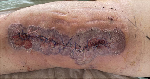
Fig. 1. Clinical image of the affected leg upon initial presentation.
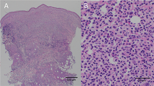
Fig. 2. Skin histology from the margin of the wound depicting a deep heavy predominantly neutrophilic infiltrate with admixed histiocytes and some lymphocytes. HE staining: (A) 40x magnification and (B) 400x magnification.
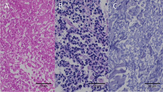
Fig. 3. Skin histology from the margin of the wound with negative microbial stainings for (A) bacteria (Gram staining, 400x magnification), (B) fungal elements (PAS staining, 400x), and (C) acid-fast bacilli (Ziehl Neelsen staining, 400x).
What is your diagnosis?
Differential diagnosis 1: Necrotizing fasciitis
Differential diagnosis 2: Cutaneous tuberculosis
Differential diagnosis 3: Bullous pyoderma gangrenosum
Differential diagnosis 4: Bullous contact dermatitis
See next page for answer.
ANSWERS TO QUIZ
Haemorrhagic Bullous Wound Changes After a Knee Joint Replacement: A Commentary
Diagnosis: Bullous pyoderma gangrenosum
Antibiotic therapy was changed to imipenem and clindamycin intravenously and concomitant high-dose systemic steroid therapy was initiated. Under this treatment regimen, the patient’s clinical state improved rapidly. All microbial cultures and eubacterial polymerase chain reaction (PCR) remained negative.
Five days after escalating antibiotic therapy it was discontinued, while sustaining systemic high-dose steroid therapy. The patient’s condition remained stable, and wound conditions improved. Subsequently, surgical reconstruction of the affected knee joint and tissue defect was performed over several months. As part of a continuous steroid reduction regimen for systemic immunosuppression, tumour necrosis factor (TNF)-alpha inhibitors were introduced. The patient tolerated all subsequent surgical interventions well. Immunosuppressive therapy was eventually able to be stopped after 8 months.
Pyoderma gangrenosum (PG) is a rare dermatological condition with most therapy recommendations based on clinical experience and case reports/series. It is mostly a clinical diagnosis supported by histological findings of deep dermal neutrophilic infiltrate (1, 2). Atypical (bullous) pyoderma gangrenosum (APG) is an even rarer sub-variant (1–3). Often initially misdiagnosed as an infectious process, it presents with bullous and ulcerative skin changes, sometimes with signs of necrosis (4–6). Diagnosis is made by excluding other disease entities. Generally, surgical interventions in this condition pose a risk of recurrence, leading to the pathergy phenomenon. PG has been known to manifest for the first time after surgical interventions, particularly in breast and orthopaedic surgery (5–8). In this context diagnosis is often delayed by mimicking postoperative wound infections, posing a peculiar challenge in postoperative care and management (5, 7–10).
Atypical pyoderma gangrenosum should be considered when anti-infective therapy yields insufficient improvement and dermatological evaluation should be sought. Perioperative management can be challenging and requires interdisciplinary cooperation. Often long-term systemic immunosuppression is necessary to achieve remission.
ACKNOWLEDGEMENT
Patient informed written consent was obtained regarding the anonymous publication of their medical information and images.
REFERENCES
- Powell FC, Collins S. Pyoderma gangrenosum. Clin Dermatol 2000; 18: 283–293. https://doi.org/10.1016/S0738-081X(99)00119-4
- Bennett ML, Jackson JM, Jorizzo JL, Fleischer ABJ, White WL, Callen JP. Pyoderma gangrenosum: a comparison of typical and atypical forms with an emphasis on time to remission. Case review of 86 patients from 2 institutions. Medicine (Baltimore) 2000; 79: 37–46. https://doi.org/10.1097/00005792-200001000-00004
- Callen JP, Jackson JM. Pyoderma gangrenosum: an update. Rheum Dis Clin North Am 2007; 33: 787–802, vi. https://doi.org/10.1016/j.rdc.2007.07.016
- Koester G, Tarnower A, Levisohn D, Burgdorf W. Bullous pyoderma gangrenosum. J Am Acad Dermatol 1993; 29: 875–878. https://doi.org/10.1016/0190-9622(93)70261-Q
- Tolkachjov SN, Fahy AS, Cerci FB, Wetter DA, Cha SS, Camilleri MJ. Postoperative pyoderma gangrenosum: a clinical review of published cases. Mayo Clin Proc 2016; 91: 1267–1279. https://doi.org/10.1016/j.mayocp.2016.05.001
- Ruebhausen MR, Mendenhall SD, Neumeister MW, Berry NN. Postsurgical pyoderma gangrenosum following carpal tunnel release: a rare disease following a common surgery. Eplasty 2017; 17: e10.
- Gulyas K, Kimble FW. Atypical pyoderma gangrenosum after breast reduction. Aesthetic Plast Surg 2003; 27: 328–331. https://doi.org/10.1007/s00266-003-3017-y
- Ebrad S, Severyns M, Benzakour A, Roze B, Derancourt C, Odri G-A, et al. Pyoderma gangrenosum after orthopaedic or traumatologic surgery: a systematic revue of the literature. Int Orthop 2018; 42: 239–245. https://doi.org/10.1007/s00264-017-3672-2
- Ramamurthi A, Adamson KA, Yang KJ, Sanger J, Ling-LeBlanc JP, Wilson B, et al. Management of postsurgical pyoderma gangrenosum following deep inferior epigastric perforator flap breast reconstruction: a role for a dermal regeneration template. Wounds Compend Clin Res Pract 2021; 33: E67–74. https://doi.org/10.25270/wnds/2021.e6774
- To D, Wong A, Montessori V. Atypical pyoderma gangrenosum mimicking an infectious process. Case Rep Infect Dis 2014; 2014: 589632. https://doi.org/10.1155/2014/589632