Chronic Upper Eyelid Oedema in a Young Boy: A Quiz
Jurand Domański1, Piotr K. Krajewski2, Wojciech Baran2 and Jacek C. Szepietowski2
1Students Research Group of Experimental Dermatology, Department of Dermatology, Venereology and Allergology and 2Department of Dermatology, Venereology and Allergology, Wroclaw Medical University, Chalubinskiego 1, PL-50-368 Wroclaw, Poland. E-mail: jacek.szepietowski@umed.wroc.pl
An 8-year-old boy was admitted to the dermatology department for diagnosis of eyelid swelling with accompanying lacrimal gland inflammation that had been present for over 18 months. Since the beginning of his symptoms the patient had been repeatedly hospitalized and diagnosed in numerous hospital wards. Previous laboratory tests showed strongly positive anti-nuclear antibodies, but no perinuclear anti-neutrophil cytoplasmic antibodies, cytoplasmic anti-neutrophil cytoplasmic antibodies, rheumatoid factor or IgG4 antibodies. Epstein-Barr virus, HIV and Toxocara canis infection were excluded. Schirmer’s test was negative. Magnetic resonance imaging of the orbits and salivary glands revealed enlargement of the left submandibular and parotid glands, enlargement of both lacrimal glands, enlargement of submandibular lymph nodes and massive inflammatory changes of the paranasal sinuses.
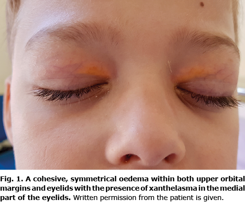
On admission to our ward the patient presented with non-painful, non-itchy cohesive oedema of both upper eyelids with the presence of xanthelasma (Fig. 1).

Laboratory analyses excluded dermatomyositis. A subtle presence of anti-Sjögren’s-syndrome-related antigen A autoantibodies and a lack of anti-Sjögren’s-syndrome-related antigen B autoantibodies were detected in the extractable nuclear antigens profile. Significantly positive centromere protein B, eosinophilia and negative antigliadin antibodies, anti-tissue transglutaminase antibodies, anti-endomysial antibodies were found. One month later, laboratory diagnostics revealed elevated proinflammatory protein serum amyloid A and serum IgG4. Histopathological analysis of a biopsy from the labial gland showed IgG4 deposits. Ultrasonography imaging revealed enlarged submandibular and parotid glands with very numerous enlarged lymph nodes in the area of these glands. An irregular hypoechogenic area was found in the left submandibular salivary gland. Positron emission tomography examination demonstrated increased metabolism of 18f-fluorodeoxyglucose tracer in the salivary glands, lacrimal glands and lymph nodes of the diaphragm, stomach and peritoneum. During gastroscopy mucosal sections were taken, which revealed chronic inflammation (mononuclear infiltration) and infiltration of IgG4-positive cells.
What is your diagnosis? See next page for answer.
Chronic Upper Eyelid Oedema in a Young Boy: A Commentary
Acta Derm Venereol 2021; 101: adv00615.
Diagnosis: Mikulicz’s disease
The disease was described in 1888 by Jan Mikulicz- Radecki, the “father of surgery”, head of the Surgery Department at Jagiellonian University, Kraków 1882–1887, at the University of Königsberg 1887–1890 (formerly Krolewicz, Poland, currently Kaliningrad, Russia) and from 1890 to 1905 at the University of Breslau (currently Wrocław) (1). Due to the clinical and histological similarity, Mikulicz’s disease (MD) has been identified in the past as a subtype of Sjögren’s syndrome (SS). However, the discovery and systematization in the 21st century of IgG4-related diseases (IgG4-RD) has given grounds for the independent categorization of MD (2, 3). IgG4-RD covers a group of multi-organ immune-mediated conditions whose clinical features often mimic a variety of malignant, infectious, and inflammatory disorders (4).
Helpful tools for differentiating IgG4-RD from other diseases are the guidelines developed in 2019 by American College of Rheumatology (ACR) and European League Against Rheumatism (EULAR) (5). The authors of these documents caution that the criteria are not intended for use in clinical practice as a basis for establishing a diagnosis of IgG4-RD. Therefore, IgG4-RD can be diagnosed even in patients who fail to fulfil the ACR/EULAR classification criteria (5). MD is often confused with other conditions of the IgG4-RD group (Küttner’s tumour) or other diseases, such as lymphoma, SS, MALT or sarcoidosis (4, 6). The characteristic features of IgG4 group diseases are inflammatory infiltration, fibrosis and pseudotumour formation in single or multiple organs, including the pancreas, bile ducts, lungs, aorta, retroperitoneal space, lymph nodes, lacrimal glands, oculomotor muscles and orbit (7). The head and neck is the one of the most common sites of lesion localization in IgG4-RD (7, 8).
There are no established main or secondary criteria for diagnosis of MD. Basically, the differentiation of MD is based on: (i) general features of IgG4-RD diseases, as well as (ii) a predilection for some more specific symptoms:
- typical localization and character of lesions (symmetrical involvement of the submandibular and parotid glands and the lacrimal gland. The enlarged glands are not painful and (often) function normally. The swellings are persistent and persist for more than 3 months) (2);
- increased serum IgG4 concentration (this criterion is not necessarily required to be fulfilled in all IgG-RD) (5);
- characteristic changes in histopathological examination (pathognomonic symptoms for IgG4+ diseases: the abundant infiltration of IgG4-positive cells, storiform fibrosis).
Several papers have reported associations of rhinosinusitis with MD (9–11). The literature describes many other symptoms more or less typical for MD. The most important of them, in relation to the case reported here (along with the differentiation with SS), are summarized in Table SI.
It has been found that MD is the most common manifestation of IgG4-RD in childhood (12). Patients with MD respond well to glucocorticosteroids treatment; however, too rapid dose reduction leads to recurrence of disease symptoms. Thus, the recommended period of dose reduction is between 3 and 6 months (13). It is of utmost importance to note that the clinical picture of MD becomes clearer with the duration of the disease; hence symptomatology at an early stage of the disease may be incomplete. This causes additional difficulties in establishing the correct diagnosis (13). In the current case, SS was suspected on several occasions. Undoubtedly, the initial progress of the disease was deceptive; however, prospective analysis confirmed the diagnosis of MD.
The authors have no conflicts of interest to declare
REFERENCES
- Sikorska D, Grzybowski A. [Jan Mikulicz-Radecki - his impact on the development of rheumatology]. Forum Reumatol 2017; 3: 1–4 (in Polish).
- Himi T, Takano K, Yamamoto M, Naishiro Y, Takahashi H. A novel concept of Mikulicz’s disease as IgG4-related disease. Auris Nasus Larynx 2012; 39: 9–17.
- Manger B, Schett G. Jan Mikulicz-Radecki (1850–1905): return of the surgeon. Ann Rheum Dis 2021; 80: 8–10.
- Kamisawa T, Zen Y, Pillai S, Stone JH. IgG4-related disease. Lancet 2015; 385: 1460–1471.
- Wallace ZS, Naden RP, Chari S, Choi H, Della-Torre E, Dicaire JF, et al. The 2019 American College of Rheumatology/European League Against Rheumatism Classification Criteria for IgG4-Related Disease. Arthritis Rheumatol 2020; 72: 7–19.
- Goto H, Takahira M, Azumi A, Japanese Study Group for Ig GROD. Diagnostic criteria for IgG4-related ophthalmic disease. Jpn J Ophthalmol 2015; 59: 1–7.
- Sebastian A, Donizy P, Wiland P. IgG4-Related Disease and the Spectrum of Mimics in Rheumatology. Chronic Autoimmune Epithelitis - Sjogren’s Syndrome and Other Autoimmune Diseases of the Exocrine Glands. 2019.
- Gallo A, Martellucci S, Fusconi M, Pagliuca G, Greco A, De Virgilio A, et al. Sialendoscopic management of autoimmune sialadenitis: a review of literature. Acta Otorhinolaryngol Ital 2017; 37: 148–154.
- Piao Y, Wang C, Yu W, Mao M, Yue C, Liu H, et al. Concomitant occurrence of Mikulicz’s disease and immunoglobulin G4-related chronic rhinosinusitis: a clinicopathological study of 12 cases. Histopathology 2016; 68: 502–512.
- Moteki H, Yasuo M, Hamano H, Uehara T, Usami S. IgG4-related chronic rhinosinusitis: a new clinical entity of nasal disease. Acta Otolaryngol 2011; 131: 518–526.
- Aboulenain S, Miquel TP, Maya JJ. Immunoglobulin G4 (IgG4)-related sialadenitis and dacryoadenitis with chronic rhinosinusitis. Cureus 2020; 12: e9756.
- Lu H, Teng F, Zhang P, Fei Y, Peng L, Zhou J, et al. Differences in clinical characteristics of IgG4-related disease across age groups: a prospective study of 737 patients. Rheumatology (Oxford) 2021; 60: 2635–2646.
- Kamiński B. [Mikulicz’s disease and Sjögren’s syndrome as the main autoimmune disorders involving salivary glands]. Medical Studies/Studia Medyczne. 2020; 36: 211–218 (in Polish).