SHORT COMMUNICATION
Brentuximab Vedotin-induced Tumour Lysis Syndrome in Mycosis Fungoides: A Case Report
Berenika OLSZEWSKA1,2*, Anna ZARYCZAŃSKA1,2, Roman J. NOWICKI1,2 and Małgorzata SOKOŁOWSKA-WOJDYŁO1,2
1Department of Dermatology, Venereology and Allergology, Faculty of Medicine, Medical University of Gdansk, Mariana Smoluchowskiego 17, PL-80-214, Gdańsk, and 2Department of Dermatology, Venereology and Allergology, University Clinical Centre, Gdańsk, Poland.
*E-mail: berenika.olszewska@gumed.edu.pl
Citation: Acta Derm Venereol 2024; 104: adv41982. DOI https://doi.org/10.2340/actadv.v104.41982.
Copyright: © 2024 The Author(s). Published by MJS Publishing, on behalf of the Society for Publication of Acta Dermato-Venereologica. This is an Open Access article distributed under the terms of the Creative Commons Attribution-NonCommercial 4.0 International License (https://creativecommons.org/licenses/by-nc/4.0/).
Submitted: Sep 18, 2024. Accepted after revision: Oct 15, 2024. Published: Nov 5, 2024
INTRODUCTION
Tumour lysis syndrome (TLS) is a life-threatening condition characterized by metabolic disturbances that occur spontaneously or following the initiation of cancer therapy. The metabolic abnormalities typical of TLS manifests as hyperkalaemia, hyperphosphatemia, hypocalcaemia, and hyperuricemia, potentially leading to severe renal impairment, cardiac arrhythmia, seizure, and possibly multi-organ failure and death (1, 2). TLS most frequently occurs in patients with haematologic malignancies and solid tumours undergoing treatment with chemotherapeutic agents or novel targeted therapies, including brentuximab vedotin (BV), a CD30-directed antibody-drug conjugate (ADC). BV is approved for the treatment of classical Hodgkin’s lymphoma and other CD30-expressing lymphomas, including primary cutaneous T-cell lymphomas (pCTCLs). It has proved to be generally well tolerated, with the most commonly reported adverse events being peripheral sensory/motor neuropathy, followed by neutropenia. BV treatment may also result in life-threatening adverse events (AEs), including TLS. However, TLS occurrence in pCTCLs following treatment with a BV has not been reported yet. Here, we present the first case of TLS induced by BV in a patient with mycosis fungoides (MF).
CASE PRESENTATION
A 40-year-old man was diagnosed with MF stage IB (T3N0M0B0) in 2010. After 5 years of effective treatment with phototherapy (narrow band UVB, PUVA-bath, PUVA), localized radiotherapy, and acitretin, he experienced rapid aggravation of skin lesions, exhibiting erythroderma, and a high blood tumour burden of Sézary cells (absolute counts of atypical Sezary cells 3.3 × 10⁹/L), meeting haematologic criteria for B2 blood involvement. Despite multiple treatment methods, including bexarotene, interferon alpha-2a, and pegylated interferon, erythroderma persisted (Fig. 1), and the patient experienced progression in 2020, leading to lymphadenopathy (N3), visceral involvement (spleen and liver), and CD30+ expression confirmed in skin lesions.

Fig. 1. Persistent erythroderma.
The histopathological examination ruled out large-cell transformation of MF. Cytoreductive treatment with gemcitabine was initiated to reduce the lymphoma burden before BV implementation. Prior to BV initiation, his baseline laboratory values were: WBC 19.58 × 10⁹/L, neutrocytes 13.79 × 10⁹/L, haemoglobin 11 g/dL, serum creatinine 0.96 mg/dL, lactate dehydrogenase 2801 U/L, CRP 206 mg/L, ALT 102 U/l, AST 112 U/l, bilirubin 4.1 mg/dL, and normal range of electrolytes. The day after treatment initiation, he presented in poor general condition, with lymphadenopathy, yellowish skin discoloration, and increased inflammatory markers. Subsequent laboratory results showed WBC 12.49 × 10⁹/L, neutrocytes 8.7 × 10⁹/L, haemoglobin 9.4 g/dL, lactate dehydrogenase 8665 U/L, CRP 372 mg/L, bilirubin 7.11 mg/dL, and abnormal TLS-related serum values (Fig. 2). The abnormalities in uric acid, phosphorus, calcium, and creatinine, along with acute renal failure, were consistent with TLS according to Cairo and Bishop criteria (2). He was treated with intravenous fluids, rasburicase, calcium carbonate, allopurinol, broad-spectrum antibiotics (piperacillin/tazobactam, vancomycin, meropenem), tramadol, morphine, furosemide, haemodialysis due to anuria, and filgrastim followed by transfusions for pancytopenia. Despite extensive treatment, renal and hepatic parameters worsened, pancytopenia persisted, and the patient’s general condition deteriorated. The patient died of multiorgan failure on day 12 of hospitalization.
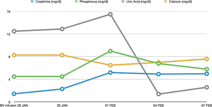
Fig. 2. Patient’s selected serum laboratory values after BV infusion, meeting the Cairo and Bishop criteria for TLS (2).
DISCUSSION
The incidence of TLS varies across different types of malignancies and therapeutic methods.
TLS is most common in high-grade NHL and acute leukaemia, whereas pCTCLs are classified as TLS low-risk diseases, with a defined risk below 1% (2).
While TLS is most frequently associated with cytotoxic therapy, it has also been observed following the use of steroids, methotrexate, monoclonal antibodies, tyrosine kinase inhibitors, and chimeric antigen receptor (CAR) T cells (3, 4). Moreover, novel, highly effective therapies may increase the risk of TLS in malignancies previously considered low risk, such as solid tumours (3).
The incidence of TLS following BV administration in pCTCLs remains unreported; thus, our case is the first to describe TLS occurring after BV treatment in a patient with MF. Pro et al. (5) reported TLS in 1 of 58 patients (1.7%) in a phase II trial of single-agent brentuximab for systemic anaplastic large-cell lymphoma. TLS in pCTCL is rare, with reported cases including spontaneous TLS in “SS preceded by MF” (6) and steroid-induced TLS in MF. (7). Despite lack of reported cases of TLS in pCTCL after BV treatment, it must be emphasized that patients with rapidly proliferating tumour and high tumour burden are at greater risk of developing TLS in pCTCLs (8). The presented case draws attention to modern and targeted anti-cancer therapies, which, due to their high efficacy, carry an increased risk of inducing TLS. Early risk stratification for TLS is crucial prior to the initiation of cancer treatment, with key predictors including renal dysfunction, hyponatremia, metastatic or large tumour burden, male sex, splenomegaly, and elevated creatinine, uric acid, and lactate dehydrogenase levels (9). Given the high mortality rate associated with TLS, ranging from 7% to 51%, (10, 11), it is essential to identify high-risk patients and implement appropriate prophylaxis to prevent TLS development.
ACKNOWLEDGEMENT
Conflict of interest disclosures: MSW has been a consultant and received compensation for lectures and the preparation of informational materials for doctors and patients from Takeda, Swixx, Celgene, and Recordati. BO, AZ, RJN have no conflicts of interest to declare.
REFERENCES
- Adeyinka A, Bashir K. Tumor lysis syndrome. 2022 Oct 31. In: StatPearls (Internet). Treasure Island (FL): StatPearls Publishing; 2024.
- Cairo MS, Coiffier B, Reiter A, Younes A; TLS Expert Panel. Recommendations for the evaluation of risk and prophylaxis of tumour lysis syndrome (TLS) in adults and children with malignant diseases: an expert TLS panel consensus. Br J Haematol 2010; 149: 578–586. https://doi.org/10.1111/j.1365-2141.2010.08143.x
- Sury K. Update on the prevention and treatment of tumor lysis syndrome. J Onco Nephrol 2019; 3: 19–30. https://doi.org/10.1177/2399369319837212
- Howard SC, Trifilio S, Gregory TK, Baxter N, McBride A. Tumor lysis syndrome in the era of novel and targeted agents in patients with hematologic malignancies: a systematic review. Ann Hematol 2016; 95: 563–573. https://doi.org/10.1007/s00277-015-2585-7
- Pro B, Advani R, Brice P, Bartlett NL, Rosenblatt JD, Illidge T et al. Brentuximab vedotin (SGN-35) in patients with relapsed or refractory systemic anaplastic large-cell lymphoma: results of a phase II study. J Clin Oncol 2012; 30: 2190–2196. https://doi.org/10.1200/JCO.2011.38.0402
- Abdelmouttalib A, Elghtaibi FZ, Sialiti S, Senouci, K. Sézary syndrome preceded by mycosis fungoides and complicated by tumor lysis syndrome and macrophage activation syndrome. Our Dermatol Online 2022; 13: 114–115. https://doi.org/10.7241/ourd.20221.32
- Kopterides P, Lignos M, Mavrou I, Armaganidis A. Steroid-induced tumor lysis syndrome in a patient with mycosis fungoides treated for presumed pneumocystis carinii pneumonia. Am J Hematol 2005; 80: 309. https://doi.org/10.1002/ajh.20444
- Brentuximab vedotin (Adcteris). European Medicines Agency (EMA) [accessed October 16, 2016]. Available from: https://www.ema.europa.eu
- Wilson FP, Berns JS. Tumor lysis syndrome: new challenges and recent advances. Adv Chronic Kidney Dis 2014; 21: 18–26. https://doi.org/10.1053/j.ackd.2013.07.001
- Stephanos K, Picard L. Pediatric oncologic emergencies. Emerg Med Clin North Am 2018; 36: 527–535. https://doi.org/10.1016/j.emc.2018.04.007
- Darmon M, Guichard I, Vincent F, Schlemmer B, Azoulay E. Prognostic significance of acute renal injury in acute tumor lysis syndrome. Leuk Lymphoma. 2010; 51: 221–227. https://doi.org/10.3109/10428190903456959