QUIZ SECTION
Small Papule on the Eyelid of an Early Adolescent Male: A Quiz
Nneka EDE1 and Brett KEELING2,3
1Dell Medical School at the University of Texas at Austin, Austin, 2Division of Dermatology, Dell Medical School at the University of Texas at Austin, Austin, and 3Clinical Pathology Associates, Austin, TX, USA. E-mail: nnekaede@utexas.edu
Citation: Acta Derm Venereol 2025; 105: adv42066. DOI: https://doi.org/10.2340/actadv.v105.42066.
Copyright: 2025 © The Author(s). Published by MJS Publishing, on behalf of the Society for Publication of Acta Dermato-Venereologica. This is an Open Access article distributed under the terms of the Creative Commons Attribution-NonCommercial 4.0 International License (https://creativecommons.org/licenses/by-nc/4.0/).
Published: Feb 18, 2025.
A 12-year-old male with no pertinent past medical history presented to clinic with an asymptomatic solitary growth on the left upper eyelid. His mother stated the growth had been present for 4–5 months. There was no history of trauma or any relevant family history. The growth had not changed significantly since it first appeared. The physical exam revealed a firm yellowish-white papule on the medial aspect of the left upper eyelid (Fig. 1). A biopsy of the lesion was obtained (Fig. 2). Haematoxylin and eosin-stained sections revealed dermal deposits of calcium with overlying reactive epithelium. Only a sparse chronic dermal inflammatory infiltrate was present. Following the biopsy, there was no reoccurrence.
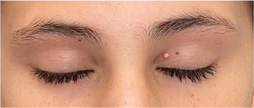
Fig. 1. Clinical appearance. Smooth yellowish-white firm papule on the medial crease of the left upper eyelid.
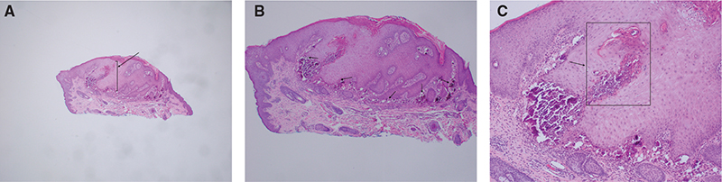
Fig. 2. H&E stained sections. The lesion at different magnifications demonstrates pseudoepitheliomatous epidermal hyperplasia (black arrow, A) overlying multifocal calcium deposits (black arrows, B) with evidence of early trans-epidermal elimination (black arrow, C) and chronic dermal histiocytic inflammation. (A) haematoxylin-eosin stain 20x; (B) haematoxylin-eosin stain 40x; (C) haematoxylin-eosin stain 200x.
What is your diagnosis?
Differential diagnosis 1: Molluscum contagiosum
Differential diagnosis 2: Pilomatricoma
Differential diagnosis 3: Epidermal inclusion cyst
Differential diagnosis 4: Subepidermal calcified nodule
Differential diagnosis 5: Chalazion (style)
See next page for answer.
ANSWERS TO QUIZ
Small Papule on the Eyelid of an Early Adolescent Male: A Commentary
Diagnosis: Subepidermal calcified nodule
This is a case of a subepidermal calcified nodule (SCN) of the eyelid in an early adolescent. SCN is a form of calcinosis cutis that typically present on the face of children. SCN presents as a small, firm, solitary papule or nodule with a verrucous surface. It can present at birth or develop throughout childhood and predominates in males in a 2:1 ratio (1). It is commonly located in the head and neck region and can frequently involve the eyelid (2, 3). Furthermore, SCN tends to appear lighter than the surrounding skin in patients with darker complexions (4). Affected individuals typically have normal serum levels of calcium and phosphate, and there is usually no history of trauma to the area of the lesions, suggesting a spontaneous dystrophic origin (2, 3).
Overarchingly, calcinosis cutis represents a unique family of conditions characterized by abnormal deposition of calcium within the skin. These conditions can be identified by the appearance of singular or multiple white or yellowish nodules or plaques on the skin, with the number of nodules depending on the extent of calcium deposition. The presence of these calcified deposits in the skin often leads to discomfort and can significantly impact the quality of life due to pain, potential infection, and their cosmetic appearance.
The classification of calcinosis is based on the underlying aetiology. Dystrophic calcinosis, the most common type, occurs in areas of the skin that have suffered damage or are affected by a disease process and is often associated with connective tissue diseases, such as scleroderma or dermatomyositis (5). Metastatic calcinosis results from systemic conditions, such as hyperparathyroidism, renal failure, or certain malignancies, that have led to elevated calcium or phosphate levels in the blood and resulted in calcium deposits within the skin (5). Iatrogenic calcinosis follows medical procedures or treatments, particularly when calcium or phosphate-containing substances are introduced into the skin (5). Lastly, idiopathic calcinosis occurs without any identifiable systemic disease or skin injury, making its diagnosis particularly challenging. Subepidermal calcified nodule (SCN) is an uncommon subtype of idiopathic calcinosis cutis also referred to as a SCN calcification or Winer nodular calcinosis, as it was first described by Winer in 1952 as sweat gland hamartoma (4–6). It is primarily observed in children and manifests as single or multiple painless, yellow-white nodules with no known systemic association (4). Currently, there are only a few reported cases in the literature.
The pathogenesis of SCN remains largely unknown, but there are many proposed mechanisms. The first proposed mechanism suggests the aggregation of calcified granules within the stroma eventually leads to a mass (2, 3). The second postulates that a calcified lesion forms first and then is subsequently reabsorbed leading to the creation of calcified granules (2, 3). Other mechanisms include deposition of calcium and phosphate from degranulated mast cells and increased levels of gamma carboxyglutamic acid within the soft tissue (2, 3). The histopathology of SCN reveals focal spherical calcium deposits beneath the epidermis. The epidermis is often characterized as hyperkeratotic, with papillomatosis and overlying parakeratosis with dermal basophilic spherules on H&E staining (3, 7). The deposits stain with a Von Kossa stain and histiocytes enveloping the calcium deposits may be confirmed with CD68 immunoperoxidase staining (3, 7). The calcium deposits work their way through the epidermis as a process of self-resolution (7).
The differential diagnosis for SCN based on clinical examination includes juvenile xanthogranuloma, epidermal inclusion cysts, verruca vulgaris, cutaneous horns, molluscum contagiosum, and, less likely, malignant tumours (2, 3). The histologic differential diagnosis for SCN includes disorders that have calcification or some calcification mimicking component such as gout (1). Utilizing both clinical and histopathological features of SCN is key to making an accurate diagnosis.
Treatment for SCN involves surgical excision and histopathologic evaluation to provide both cosmetic and symptomatic relief (3). Surgery is considered the definitive approach because the lesion is benign and because there is a lack of effective alternative treatments. Intralesional corticosteroid injection may be used for other forms of calcinosis cutis; however, it has limited efficacy in SCN due to the absence of a responsive stroma and inflammatory cell population (2, 3). Furthermore, the spheroidal calcium deposits characteristic of SCN are expected to be refractory to pharmacologic lysis (2, 7).
In summary, SCN of the eyelid is a rare condition that manifests as solitary, painless, yellowish-white nodules with papillomatous features. The condition’s presentation in the first two decades of life, its benign nature, and the absence of systemic abnormalities or history of trauma all help to point to the condition’s uniqueness among other calcinosis cutis subtypes. Clinicians should be aware of SCN and SCN’s potential to mimic other eyelid lesions to ensure an accurate diagnosis and appropriate patient care.
REFERENCES
- Bagci B, Karakas C, Gokden M. Subepidermal calcified nodule in a child with neurofibromatosis type 1. Cureus 2022; 14: e23261. https://doi.org/10.7759/cureus.23261
- Juzych LA, Nordby CA. Subepidermal calcified nodule. Pediatr Dermatol 2001; 18: 238–240. https://doi.org/10.1046/j.1525-1470.2001.018003238.x
- Kim HJ, Johnson PB, Kropinak M, et al. Subepidermal calcified nodules of the eyelid. Ophthal Plast Reconstr Surg 2009; 25: 489–490. https://doi.org/10.1097/IOP.0b013e3181b80b52
- Shields CL, Marous MR, Casey MG, et al. Subepidermal calcified nodule in the periocular region: a report of 6 cases. Ophthalmology 2016; 123: 671–673. https://doi.org/10.1016/j.ophtha.2015.10.015
- Reiter N, El-Shabrawi L, Leinweber B, Berghold A, Aberer E. Calcinosis cutis: part I. Diagnostic pathway. J Am Acad Dermatol 2011; 65: 1–12; quiz 13–14. https://doi.org/10.1016/j.jaad.2010.08.038
- Winer LH. Solitary congenital nodular calcification of the skin. AMA Arch Derm Syphilol 1952; 66: 204–211. https://doi.org/10.1001/archderm.1952.01530270062008
- Nguyen J, Jakobiec FA, Hanna E, Fay A. Subepidermal calcified nodule of the eyelid. Ophthal Plast Reconstr Surg 2008; 24: 494–495. https://doi.org/10.1097/IOP.0b013e31818be82c