QUIZ SECTION
Persistent Alopecic Plaque on the Scalp in a Woman with Breast Cancer: A Quiz
Yujie WEN and Yang WANG*
Department of Dermatology and Venereology, Peking University First Hospital; Beijing Key Laboratory of Molecular Diagnosis on Dermatoses; National Clinical Research Center for Skin and Immune Diseases, 8 Xishiku Street, Xicheng District, Beijing, PR China. *E-mail: yangwang_dr@bjmu.edu.cn
Citation: Acta Derm Venereol 2025; 105: adv42120. DOI: https://doi.org/10.2340/actadv.v105.42120.
Copyright: © 2025 The Author(s). Published by MJS Publishing, on behalf of the Society for Publication of Acta Dermato-Venereologica. This is an Open Access article distributed under the terms of the Creative Commons Attribution-NonCommercial 4.0 International License (https://creativecommons.org/licenses/by-nc/4.0/).
Published: Jan 8, 2025.
A 44-year-old woman presented to the dermatology department with an alopecic plaque on the scalp, which had persisted for 7 years and became ulcerated for 2 months (Fig. 1A). She has a 10-year history of hormone receptor-positive, HER2-positive breast cancer, for which she underwent surgery followed by adjuvant treatment with tamoxifen. When she first noticed focal hair loss on the scalp 7 years ago, multi-metastatic breast cancers were found in her supraclavicular lymph nodes, lung, and sacrum. She was treated with trastuzumab in combination with paclitaxel and capecitabine. The metastatic lesions responded well to chemotherapy and trastuzumab, while the hair loss persisted and was diagnosed with alopecia areata. All imaging studies did not reveal any evidence of metastasis during the past 7 years, despite an unexplained elevated serum CA15-3 level (147U/ml, normal range 0–25U/mL).
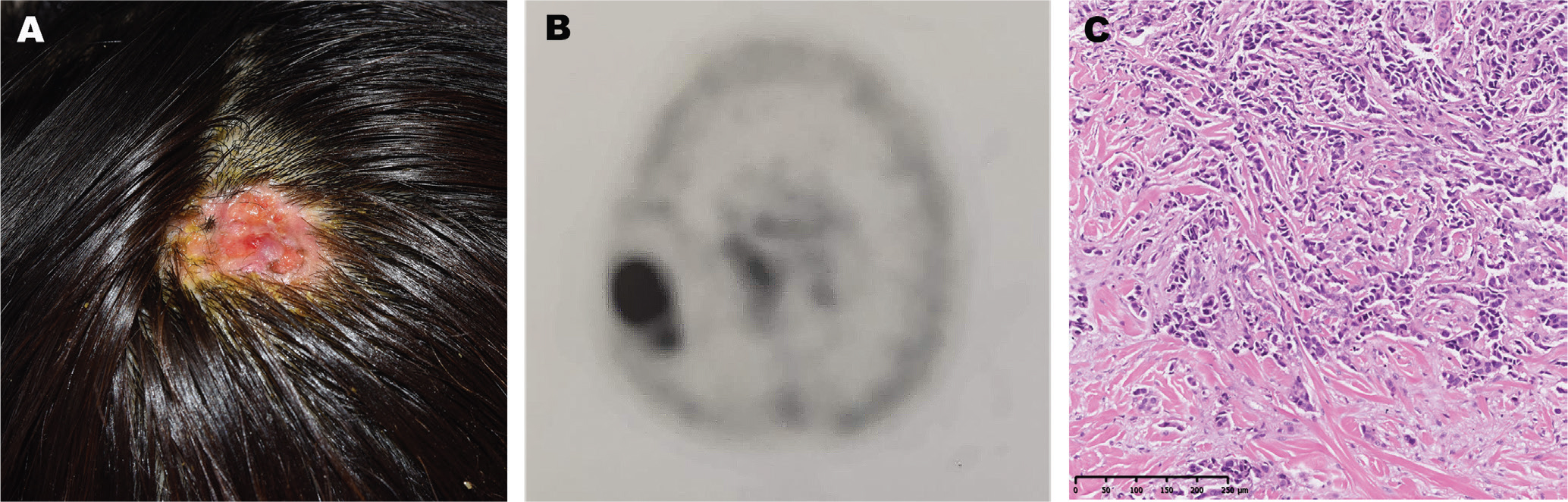
Fig. 1. (A) The ulcerated alopecic plaque on the right parietal area of the scalp. (B) A positron emission tomography-computed tomography scan shows increased glucose uptake of the skin lesion. (C) Skin biopsy specimen reveals atypical epithelioid cells lying between the collagen bundles in the dermis (haematoxylin–eosin, original magnification ×100).
She discontinued trastuzumab 2 months prior to this visit, and the alopecic plaque on the scalp became thickened and ulcerated. A positron emission tomography-computed tomography (PET-CT) scan showed increased glucose uptake (standardized uptake value [SUV], 10.0) of the skin lesion on the right parietal area (Fig. 1B). A skin biopsy specimen was obtained from the ulcerated lesion (Fig. 1C).
What is your diagnosis?
Differential diagnosis 1: Squamous cell carcinoma
Differential diagnosis 2: Alopecia areata
Differential diagnosis 3: Alopecia neoplastica associated with metastatic breast cancer
Differential diagnosis 4: Basal cell carcinoma
See next page for answer.
ANSWERS TO QUIZ
Persistent Alopecic Plaque on the Scalp in a Woman with Breast Cancer: A Commentary
Diagnosis: Alopecia neoplastica associated with metastatic breast cancer
A skin biopsy revealed clusters of large, atypical epithelioid cells lying between the thick collagen bundles in the dermis. The tumour cells were immunopositive for CK7, hormone receptors, and HER2. Pilosebaceous units were absent in this area. A diagnosis of metastatic breast cancer was made. She received 6 cycles of treatment with vinorelbine tartrate, pyrotinib maleate, and trastuzumab. The ulcerative plaque gradually regressed with an alopecic scar. However, an abnormal serum CA15-3 remained (113.4U/mL, normal range 0–25 U/mL). A skin biopsy on the scar confirmed the persistence of metastatic breast cancer. The alopecic area on the scalp was then surgically removed. After the excision, a PET-CT scan showed no evidence of abnormal glucose uptake, and the level of serum CA15-3 decreased. No recurrence was observed during follow-ups.
Cutaneous metastasis accounts for about 2% of all skin cancers, and the incidence is increasing due to improved survival of patients with cancer (1). The most common tumour that metastasizes to the skin is breast cancer, with an incidence of 24% (2). It has been reported that the scalp accounted for 4% of body-site cutaneous metastases (3). However, the scalp involvement is uncommon in breast cancer. The incidence of scalp metastases caused by breast cancer is about 1.2% (4). Scalp involvement has been regarded as a sign of progression or widespread metastatic disease. A metastatic lesion on the scalp tends to present as single or multiple asymptomatic, well-defined alopecic plaques, often red-pink in colour (5). Histological examination usually reveals infiltration of neoplastic cells in the dermis or subcutaneous tissue with loss of pilosebaceous units. Of note, there is often dense dermal fibroplasia in the metastatic scalp tissue (3). The dense dermal fibrosis on the scalp may be responsible for the inadequate response to systemic therapy and therefore serve as a reservoir for dormant metastatic breast cancer. However, this hypothesis remains speculative at present, and further research is needed to explore how this factor might influence the behaviour of metastatic breast cancer.
The scalp metastasis of breast cancer can resemble various inflammatory or malignant diseases causing alopecia. It is easy for this to be misdiagnosed as alopecia areata (6), especially in the dormant state, as in our case. The presence of erythema and ulceration may help to differentiate alopecia neoplastica from alopecia areata (7). Alopecia neoplastica may be secondary to a series of malignancies, including basal cell carcinoma, squamous cell carcinoma, malignant melanoma, extramammary Paget’s disease, angiosarcoma, cutaneous T-cell lymphoma, as well as metastatic breast cancers (3). The clinical history and histology patterns helped to coin the diagnosis. Systemic chemotherapeutics targeting the primary tumour appear to be the standard treatment for metastatic breast cancers, but skin metastasis, especially lesions on the scalp, is frequently refractory to systemic therapy (8). In this scenario, as in our case, surgical removal of the metastatic lesions contributes to a high response rate and low recurrence rather than using systemic therapy alone, especially for isolated lesions, which are refractory to systemic treatments (1).
Alopecia neoplastica may present as the initial sign of an undiagnosed malignancy. Clinicians should pay attention to any persistent alopecic plaque occurring in a patient with a history of breast cancer, especially if accompanied by erythema or ulceration. The histological feature of dense dermal fibrosis may partially explain the resistance to systemic therapy in some patients, which also indicates the requirement for alternative treatments. Recognizing the varied presentations of cutaneous metastasis is pivotal to early intervention for rapidly progressive cancers.
REFERENCES
- Strickley JD, Jenson AB, Jung JY. Cutaneous metastasis. Hematol Oncol Clin North Am 2019; 33: 173–197. https://doi.org/10.1016/j.hoc.2018.08.008
- Krathen RA, Orengo IF, Rosen T. Cutaneous metastasis: a meta-analysis of data. South Med J 2003; 96: 164–167. https://doi.org/10.1097/01.SMJ.0000053676.73249.E5
- Suchonwanit P, McMichael AJ. Alopecia in association with malignancy: a review. Am J Clin Dermatol 2018; 19: 853–865. https://doi.org/10.1007/s40257-018-0378-1
- Lemieux J, Amireault C, Provencher L, Maunsell E. Incidence of scalp metastases in breast cancer: a retrospective cohort study in women who were offered scalp cooling. Breast Cancer Res Treat 2009; 118: 547–552. https://doi.org/10.1007/s10549-009-0342-0
- Rolz-Cruz G, Kim CC. Tumor invasion of the skin. Dermatol Clin 2008; 26: 89–102, viii. https://doi.org/10.1016/j.det.2007.08.004
- Lin WL, Lin WC, Jung SM, Yang CH, Hong HS. Breast cancer metastasized to the scalp mimicking alopecia areata: alopecia neoplastica. Breast J 2007; 13: 94–95. https://doi.org/10.1111/j.1524-4741.2006.00372.x
- Conner KB, Cohen PR. Cutaneous metastasis of breast carcinoma presenting as alopecia neoplastica. South Med J 2009; 102: 385–389. https://doi.org/10.1097/SMJ.0b013e31819b0c26
- Waldman RA, Finch J, Grant-Kels JM, Stevenson C, Whitaker-Worth D. Skin diseases of the breast and nipple: benign and malignant tumors. J Am Acad Dermatol 2019; 80: 1467–1481. https://doi.org/10.1016/j.jaad.2018.08.066