SHORT COMMUNICATION
Palbociclib-induced Toxic Epidermal Necrolysis: A Case Report with Review of All Cases Secondary to Cyclin-dependent Kinase 4/6 Inhibitors
Aurore DEVOLDERE1, Clélia VANHAECKE1, Pauline SOIBINET2, Brahim AZZOUZ3 and Manuelle VIGUIER4
1Dermatology Department, Reims University Hospital, Reims, 2Medical Oncology Department, Godinot Institute, Reims, 3Regional Centre For Pharmacovigilance, Reims University Hospital, Reims, and 4Reims Champagne Ardenne University, Dermatology Department, Reims University Hospital, IRMAIC UR7509, Reims, France. E-mail: adevoldere@chu-reims.fr
Citation: Acta Derm Venereol 2025; 105: adv42291. DOI: https://doi.org/10.2340/actadv.v105.42291.
Copyright: © 2025 The Author(s). Published by MJS Publishing, on behalf of the Society for Publication of Acta Dermato-Venereologica. This is an Open Access article distributed under the terms of the Creative Commons Attribution-NonCommercial 4.0 International License (https://creativecommons.org/licenses/by-nc/4.0/).
Submitted: Oct 25, 2024. Accepted after revision: Dec 18, 2024. Published: Jan 14, 2025.
Competing interests and funding: The authors have no conflicts of interest to declare.
INTRODUCTION
The cyclin-dependent kinase 4/6 inhibitors, combined with anti-hormonal drugs, are first-line drugs recently used in oncology for treating metastatic hormone-dependent breast cancer. Here we describe a case of toxic epidermal necrolysis (TEN) induced by palbociclib in a patient with metastatic breast cancer. Improvement in the skin healing process was spectacular with the tumour necrosis factor inhibitor etanercept, but the patient died 72 h later from respiratory distress possibly secondary to tumour progression. A review of the literature identified 7 cases of severe muco-cutaneous reactions secondary to CDK 4/6 inhibitor treatment, with several clinical features different from TEN associated with antibiotic, antigout, and antiepileptic agents. Induced subacute lupus erythematosus represents the main differential diagnosis, leading to significant therapeutic challenges.
CASE REPORT
A woman in her 70s received treatment for inoperable brain, pulmonary, and pleural metastatic breast cancer with hormonal therapy (anastrozole) for 10 weeks, with the addition, 3 weeks later, of a cyclin-dependent kinase (CDK) 4/6 inhibitor (palbociclib). Seven weeks after the initiation of palbociclib, exanthema developed on the arms and extended to the rest of the body including the face, without fever. Palbociclib was stopped 5 days after the exanthema onset. Six days after the discontinuation of palbociclib, the woman was referred because of extensive skin eruption with fever. Physical examination showed diffuse exanthema with bullae and positive Nikolsky’s sign, with skin detachment close to 60% of the body surface area (Fig. 1). Oropharyngeal erosions and cheilitis were present. Laboratory tests revealed pancytopenia (haemoglobin level 10.3 g/dL, neutrophil count 0.6 G/L, platelet count 64 G/L), slightly increased C-reactive protein level (7.3 mg/L), normal liver and renal function, negative serology for Mycoplasma pneumoniae, and negative results for antinuclear, anti-double stranded DNA, antidesmoglein 1/3, anti-BP180, and anti-BP230 antibodies. A skin biopsy revealed dermis without epidermis, haemorrhagic suffusions, and no specific dermal lymphocytic infiltrate. Direct cutaneous immunofluorescence was negative. No other medication than anastrozole and palbociclib was taken in the last 3 months.
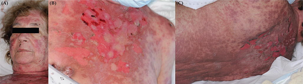
Fig. 1. (A) Facial erythema, cheilitis, erosions, and detachment of the neckline at admission. (B) Erythematobullous eruption on the trunk at admission. (C) Back skin detachment with positive Nikoslky’s sign at admission.
We concluded that this represented toxic epidermal necrolysis (TEN) due to palbociclib. The Severity-of-Illness Score for Toxic Epidermal Necrolysis (SCORTEN) was 3. After 72 h with supportive care, because of the extended skin detachment and persistence of widespread inflammatory erythema, etanercept was administered via a single injection of 50 mg. Remarkable skin improvement was observed within 48 h, with the disappearance of erythema, cessation of the extended detachment, and evidence of re-epidermization. Nevertheless, 3 days after etanercept treatment, the patient died of respiratory distress, potentially due to brain herniation or progression of lung metastases by a rebound effect after targeted therapy discontinuation.
DISCUSSION
Here we report a case of TEN induced by palbociclib, a CDK 4/6 inhibitor. CDKs are a large family of serine threonine kinases that play an important role in regulating cell-cycle progression (1). Three CDK 4/6 inhibitors are currently approved: palbociclib, abemaciclib, and ribociclib. They are used as first-line treatment for metastatic hormone-dependent breast cancer, combined with anti-hormonal drugs. These new treatments are associated with significant improvement in overall survival and progression-free survival (1). Frequent adverse effects are cytopenia, diarrhoea, nausea, and vomiting (2).
The skin toxic effects include mostly alopecia, dryness, or pruritus. To date, serious skin drug reactions are uncommon (3). We identified 7 cases (4–6), including ours, of severe bullous or erosive muco-cutaneous reactions induced by a CDK 4/6 inhibitor (Table I). The patients were all women with metastatic breast cancer, from 50 to 77 years old, who received ribociclib (4 patients), palbociclib (2 patients), or both (1 patient). Delay after drug introduction ranged from 15 days to 4 months, with a median and mean time of 28 and 39.5 days, respectively. All patients had a painful rash and mucosal damage (oral, anal, or ocular). One had acute renal failure (7), one had dyspnoea (8) and one had mild liver dysfunction with no symptoms reported (8). After discontinuation of the drug in all patients, methylprednisolone, ciclosporin, and/or etanercept were given in 4 of the 7 patients, in addition to supportive care. Except for our patient, no patient died. Although the authors classified these cases as erythema multiforme, Stevens–Johnson syndrome (SJS), or TEN, some of the cases are puzzling, including 2 related to the use of palbociclib. In our opinion, the differential diagnosis of induced subacute lupus erythematosus should have been discussed and ruled out. Indeed, induced lupus erythematosus is well linked to the use of CDK 4/6 inhibitors (3, 9). In one case classified as SJS, exanthema was described as annular erosions on the face and trunk associated with mucocutaneous damage with interface dermatitis and dermal lymphocytic infiltrates on skin biopsy (10). In another case, both the delay of exanthema onset (4 months) and the pathological findings, showing vacuolar degeneration of the dermo–epidermal junction, apoptotic keratinocytes, and dense perivascular infiltration of mononuclear cells within an oedematous papillary dermis, were also potentially consistent with lupus erythematosus (7). In both cases, a search for antinuclear antibodies was lacking.
| Age, years | Culprit drug | Time from introduction | Cutaneous signs | Mucosal damage | Fever (°C) | Pathological findings | TEN treatment | Outcome | Ref. |
| 63 | Palbociclib | 3 weeks | Pink-to-violaceous eroded plaques (face, trunk, legs) | Ocular symptoms, buccal erosions, cheilitis | Unk | Vacuolar interface dermatitis, focal full-thickness epidermal necrosis, mild superficial perivascular lymphocytic infiltrate with pigment incontinence and dermal oedema Negative DIF results |
Cyclosporine 5 mg/kg/day x 1 week then 2.5 mg/kg x 2 weeks | Rash transformed into psoriasiform dermatitis (Koebner phenomenon) | (10) |
| 77 | Ribociclib | 4 months | Painful symmetric erythematous plaques (limbs, chest, back), detachment > 20% BSA | Gluteal and perianal necrohaemorragic erosions, mouth pain, dysphagia | > 38.8 | Vacuolar degeneration of the DEJ with scattered apoptotic keratinocytes No DIF mentioned |
Etanercept 40 mg once + cyclosporine 4 mg/kg/day | Full re-epithelization with residual hypopigmentation | (7) |
| 70 | Ribociclib | 4 days after second course | Diffuse painful rash (4 extremities, torso, perineum), detachment 10% BSA | Oral mucosa spared Perineum and rectum damage |
Unk | Described as compatible with SJS diagnosis. No DIF mentioned | None | Eventual improvement | (4) |
| 46 | Ribociclib | 20 days | Painful rash (trunk, neck, extremities) | Oral mucosa (ulcers) | > 38 | Unk | Methylprednisolone 2 mg/kg/day for 5 days | Resolution after 15 days | (8) |
| 67 | Ribociclib | 15 days | Painful erythematous maculopapular rash (face, arms, trunk), haemorrhagic blisters, detachment 30% BSA | Dry eyes, buccal erosions | Unk | Destruction of the epithelium by forming blisters and abscesses No DIF mentioned |
None | Gradual improvement in the next few weeks | (5) |
| 50 | Ribociclib then palbociclib | 1 month after ribociclib and then 1 month after palbociclib | Erythematous, pink-to-violaceous, annular papules with dusky centres and bullae on hands | Aphthous lesions (hard palate) at introduction of palbociclib | Unk | Lichenoid skin reaction: epidermal and subepidermal vesiculation, apoptotic keratinocytes in all levels of the epidermis and moderate lymphohistiocytic infiltration in the upper dermis No DIF mentioned |
None | Rash gradually abated after discontinuation of ribociclib, relapse when introduction of palbociclib 1 month later, lesions subsided within the next month | (6) |
| 71 | Palbociclib | 7 weeks | Exanthema with bullae and detachment > 60% BSA | Oropharyngeal erosions, cheilitis | > 38.6 | Dermis without epidermis, haemorrhagic suffusions, no specific dermal lymphocytic infiltrate Negative DIF results |
Etanercept 50 mg once | Re-epidermization Death (respiratory distress) |
Our case |
| BSA: body surface area; DEJ: dermo–epidermal junction; DIF: direct immunofluorescence; EM: erythema multiforme; REF: Reference; SJS: Stevens–Johnson Syndrome; TEN: toxic epidermal necrolysis; Unk: unknown. | |||||||||
Our patient fulfils the criteria for authentic TEN (11), with lupus erythematosus ruled out, and could therefore be considered as the first certified report of TEN induced by palbociclib. The 29-h half-life of palbociclib leads to drug elimination close to 6 days after interruption, which potentially explains the initial aggravation of symptoms in our patient. The only other drug recently introduced was anastrozole, 10 weeks before the beginning of the symptoms. Nevertheless, cutaneous side effects are uncommon with anastrozole and other aromatase inhibitors, and no case of TEN or generalized erythema multiforme has been reported so far (12). Importantly, most of the previous cases of TEN associated with CDK 4/6 inhibitors had atypical presentations, with different features as compared with TEN associated with antibiotics, antigout, and antiepileptic agents. The time between drug initiation and the first symptoms seems longer than in classical TEN, as illustrated in our case (7 weeks). Although fever is 1 of the diagnostic criteria for TEN/SJS (11), in 4 of 7 cases febrile eruption was not reported. Moreover, whereas classical TEN shows sudden deterioration of general condition with high fever and multiple locations of mucosal damage, TEN related to CDK 4/6 inhibitors can be more insidious at the beginning, with mucosal involvement subtler or absent. The remarkable efficacy of etanercept in our case is also an additional argument favouring TEN, with underlying T cell activation mechanism rather than lupus or direct skin toxicity due to CDK4/6.
Medical consensus on TEN treatment agrees with the need to stop the imputable drug as soon as possible and supportive care is essential (13). There are currently no guidelines on the use of immunosuppressive agents. The use of etanercept, as in our patient, was reported in an open-label randomized controlled trial of comparison with corticosteroids in 91 patients: etanercept produced a non-statistically significant lower mortality rate, a mortality rate lower than the rate predicted with the SCORTEN score, and lower time required for complete skin healing and fewer adverse effects, notably gastrointestinal bleeding, than corticosteroids (14).
Prescribers of CDK 4/6 inhibitors should be aware of the risk of TEN, its uncommon clinical presentation and its life-threatening potential. With erosive or blistering exanthema in patients receiving CDK 4/6 inhibitors, direct cutaneous immunofluorescence and serological autoimmune assessment is needed to exclude lupus disease because its treatment relies on systemic corticosteroids. Although drug re-challenge can be debated in induced lupus erythematosus, it is excluded in TEN, with major consequences on the survival of patients.
REFERENCES
- Spring LM, Wander SA, Andre F, Moy B, Turner NC, Bardia A. Cyclin-dependent kinase 4 and 6 inhibitors for hormone receptor-positive breast cancer: past, present, and future. Lancet 2020; 395: 817–827. https://doi.org/10.1016/S0140-6736(20)30165-3
- Wekking D, Lambertini M, Dessì M, Denaro N, Bardanzellu F, Garrone O, et al. CDK4/6 inhibitors in the treatment of metastatic breast cancer: focus on toxicity and safety. Semin Oncol 2023; 50: 131–139. https://doi.org/10.1053/j.seminoncol.2024.01.002
- Sibaud V, Sollena P. Dermatologic toxicities to inhibitors of cyclin-dependent kinases CDK 4 and 6: an updated review for clinical practice. Ann Dermatol Vénéréologie 2023; 150: 208–212. https://doi.org/10.1016/j.annder.2022.11.013
- Widmer S, Grossman M. Chemotherapy patient with Stevens–Johnson syndrome presents to the emergency department: a case report. Am J Emerg Med 2018; 36: 1325.e3–1325.e4. https://doi.org/10.1016/j.ajem.2018.04.019
- Kowalczyk A, Radecka B. Stevens–Johnson syndrome in breast cancer patient treated with ribociclib. Oncol Clin Pract 2023; 19: 59–62. https://doi.org/10.5603/OCP.2022.0004
- Vrana E, Mylona S, Bobos M, Kontovinis L, Papazisis K. Ribociclib and palbociclib-induced erythema multiforme: a case report. Oxf Med Case Rep 2022; 2022: omac116. https://doi.org/10.1093/omcr/omac116
- López-Gómez V, Yarza R, Muñoz-González H, Revilla E, Enrech S, González-Valle O, et al. Ribociclib-related Stevens–Johnson syndrome: oncologic awareness, case report, and literature review. J Breast Cancer 2019; 22: 661–666. https://doi.org/10.4048/jbc.2019.22.e44
- Bozkaya Y, Ay B. Toxic epidermal necrolysis associated with ribociclib in a patient with metastatic breast cancer. Breast J 2020; 26: 773–774. https://doi.org/10.1111/tbj.13570
- Raschi E, Fusaroli M, La Placa M, Ardizzoni A, Zamagni C, Poluzzi E, et al. Skin toxicities with cyclin-dependent kinase 4/6 inhibitors in breast cancer: signals from disproportionality analysis of the FDA adverse event reporting system. Am J Clin Dermatol 2022; 23: 247–255. https://doi.org/10.1007/s40257-021-00645-0
- Karagounis T, Vallurupalli M, Nathan N, Nazarian R, Vedak P, Spring L, et al. Stevens–Johnson syndrome-like eruption from palbociclib in a patient with metastatic breast cancer. JAAD Case Rep 2018; 4: 452–454. https://doi.org/10.1016/j.jdcr.2017.12.015
- Ingen-Housz-Oro S, Duong TA, Bensaid B, Bellon N, De Prost N, Dévy L, et al. Epidermal necrolysis: French national diagnosis and care protocol (PNDS; protocole national de diagnostic et de soins). Orphanet J Rare Dis 2018; 13: 56. https://doi.org/10.1186/s13023-018-0793-7
- Kim YJ, Cohen PR. Anastrozole-induced dermatitis: report of a woman with an anastrozole-associated dermatosis and a review of aromatase inhibitor-related cutaneous adverse events. Dermatol Ther 2020; 10: 221–229. https://doi.org/10.1007/s13555-020-00353-3
- Kridin K, Brüggen MC, Chua SL, Bygum A, Walsh S, Nägeli MC, et al. Assessment of treatment approaches and outcomes in Stevens–Johnson syndrome and toxic epidermal necrolysis: insights from a Pan-European multicenter study. JAMA Dermatol 2021; 157: 1182–1190. https://doi.org/10.1001/jamadermatol.2021.3154
- Wang CW, Yang LY, Chen CB, Ho HC, Hung SI, Yang CH, et al. Randomized, controlled trial of TNF-α antagonist in CTL-mediated severe cutaneous adverse reactions. J Clin Invest 2018; 128: 985–996. https://doi.org/10.1172/JCI93349