RESEARCH LETTER
Seroconversion of QuantiFERON-TB Gold Test due to Mycobacterium marinum Infection in a Patient with Psoriasis Treated with Adalimumab
Grigorios THEODOSIOU1,2*, Annarita ANTELMI3 and Åke SVENSSON1,2
1Department of Dermatology, Skåne University Hospital, Malmö, 2Department of Clinical Sciences, Faculty of Medicine, Lund University, Malmö, and 3Department of Occupational and Environmental Dermatology, Skåne University Hospital, Lund University, Malmö, Sweden. *E-mail: gregtheodosiou@yahoo.com; grigorios.theodosiou@skane.se; grigorios.theodosiou@med.lu.se
Citation: Acta Derm Venereol 2024; 104: adv42336. DOI: https://doi.org/10.2340/actadv.v104.42336.
Copyright: © 2024 The Author(s). Published by MJS Publishing, on behalf of the Society for Publication of Acta Dermato-Venereologica. This is an Open Access article distributed under the terms of the Creative Commons Attribution 4.0 International License (https://creativecommons.org/licenses/by/4.0/).
Submitted: Oct 24, 2024. Accepted after revision: Nov 5, 2024. Published: Nov 25, 2024
To the Editor,
Mycobacterium marinum (M. marinum) is a free-living mycobacterium species first isolated by Aronson in saltwater fish in 1926 (1). M. marinum causes disease in many fish species and infection in human following contact with infected fishes or contaminated water. M. marinum infections are rare with an estimated incidence between 0.04 and 0.27 cases per 100,000 persons annually (2). The first report of human disease caused by M. marinum was reported in 1951 in those using a contaminated pool in Örebro, Sweden (3).
A 44-year-old male of Caucasian origin was referred to the outpatient clinic of the Department of Dermatology in Malmö. He had a 20-year medical history of severe psoriasis and he had been treated for 2 years with adalimumab after insufficient results with methotrexate therapy. Before administration of adalimumab a QuantiFERON-TB Gold test was negative, and a chest X-ray did not reveal any radiographic evidence of active or prior Mycobacterium tuberculosis infection (MTB). The patient experienced good clinical response; however, 16 months later he developed asymptomatic erythematous scaly nodular skin lesions in an irregular, roughly linear distribution localized on his left hand and underarm (Fig. 1A–C). No systemic symptoms were mentioned. The physical examination was otherwise unremarkable. The patient mentioned performing daily cleaning of a freshwater aquarium at home, often without gloves.

Fig. 1. (A–C) Multiple erythematous papules and nodules on the patient’s left hand and underarm. (D, E) Regression of the lesions after the discontinuation of adalimumab and 8 weeks’ treatment with clarithromycin and ethambutol.
A skin biopsy of a nodule was taken and the histopathological examination showed necrotizing and granulomatous inflammation (Fig. 2). Furthermore, a skin biopsy culture from one of the skin nodules was positive for M. marinum. The QuantiFERON-TB Gold test was found to be positive and chest tomography was performed with normal findings. The diagnosis of fish-tank granuloma was made and the therapy with adalimumab was discontinued. Antibiotic treatment with ethambutol (1,200 mg/day) and clarithromycin (500 mg twice daily) was initiated. The nodular lesions went into regression (Fig. 1D, E) within 4 months; thereafter the antibiotics were discontinued. Adalimumab was reintroduced without any reactivation of the skin infection.
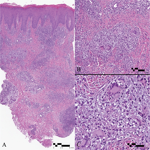
Fig. 2. (A) Granulomatous inflammation with numerous Langhans-type giant cells (H&E x10). (B) H&E x 40 (C) H&E x 100.
M. marinum is a non-tuberculous mycobacterium, whose natural habitat is fresh or saltwater, particularly enclosures of water that are not often replenished, e.g. swimming pools and aquarium tanks. Human infection may be caused by direct injury by fish or occur after cutaneous trauma and exposure to contaminated water (2–4), manifests after an incubation time of 2–3 weeks, and is usually limited to the skin and soft tissues in immunocompetent patients. Slow-growing erythematous nodules, typically localized on the hands, elbows, or knees, and sometimes painful and ulcerated, can spread following the distribution of the lymphatic system (2–4). Disseminated cutaneous involvement has been reported in immunosuppressed individuals and in a child (4, 5).
Differential diagnosis includes sporotrichosis and leishmaniasis as well as other atypical mycobacterial infections. The diagnosis of M. marinum is often delayed due to insidious clinical presentation. A history of exposure to aquatic environments or marine animals in combination with the occurrence of single or multiple granulomatous skin nodules should raise suspicion of M. marinum infection, confirmed by performing a nonmotile acid-fast bacillus (AFB) culture of the tissue (2).
Tumour necrosis factor alpha (TNF-α) inhibitors are commonly used in the treatment of psoriasis and other inflammatory dermatoses. Adalimumab is a fully human IgG1 antibody to TNF-α. One important concern with the use of TNF-α inhibitors has been the risk of reactivation of latent tuberculosis as well as other opportunistic infections, especially mycobacterial infections. Several cases of M. marinum infections have been reported in patients treated with various TNF-α inhibitors (6). It is therefore essential that before initiating TNF-α inhibitors, patients should be screened for latent TB infection with QuantiFERON Gold test or purified protein derivate (PPD) test.
The QuantiFERON-TB Gold test is an interferon-gamma (IFN-γ) release assay and considered a modern alternative to the tuberculin skin test (TST, PPD, or Mantoux). The antigens used in QuantiFERON-TB Gold test are ESAT-6 and CFP-10, both present in M. tuberculosis but not in most non-tuberculosis mycobacteria, except for M. marinum, M. kansasii, and M. szulgai (7). In a study by Kobashi et al., the QuantiFERON-TB test was positive in 58% of patients with M. marinum infections (8).
The present case highlights the need for dermatologists to maintain a high degree of suspicion for atypical mycobacterial infections in patients treated with TNF-α inhibitors. Furthermore, M. marinum infection can lead to seroconversion of the QuantiFERON Gold test and this may be used in clinical praxis as a quick diagnostic tool.
REFERENCES
- Aaronson JD. Spontaneous tuberculosis in salt water fish. J Infect Dis 1926; 39: 315–320. https://doi.org/10.1093/infdis/39.4.315
- Harris DM, Keating MR. Mycobacterium marinum: current recommended pharmacologic therapy. J Hand Surg Am 2009; 34: 1734–1735. https://doi.org/10.1016/j.jhsa.2009.06.027
- Norden A, Linell FA. A new type of pathogenic mycobacterium. Nature 1951; 168: 826. https://doi.org/10.1038/168826a0
- Meybeck A, Tetart M, Baclet V, Alcaraz I, Blondiaux N, Peytavin G, et al. A disseminated Mycobacterium marinum infection in a renal transplant HIV-infected patient successfully treated with a bedaquiline-containing antimycobacterial treatment: a case report. Int J Infect Dis 2021; 107: 176–178. https://doi.org/10.1016/j.ijid.2021.04.054
- Briand C, Gaudart A, Demonchy D, Hubiche T. Subacute multifocal abscesses related to a Mycobacterium marinum infection in a child. J Pediatr 2021; 230: 255–256. https://doi.org/10.1016/j.jpeds.2020.11.066
- Timoney I, Lynch M, Timoney L, Feeney E, Kirby B. Mycobacterium marinum infection contracted from seaweed wrap in a psoriasis patient undergoing treatment with adalimumab. Dermatol Online J 2017; 23: 13030. https://doi.org/10.5070/D3238036011
- Pai M, Riley LW, Colford JM. Interferon-gamma assays in the immunodiagnosis of tuberculosis: a systematic review. Lancet Infect Dis 2004; 4: 761–776. https://doi.org/10.1016/S1473-3099(04)01206-X
- Kobashi Y, Mouri K, Yagi S, Obase Y, Miyashita N, Okimoto N, et al. Clinical evaluation of the QuantiFERON-TB Gold test in patients with non-tuberculous mycobacterial disease. Int J Tuberc Lung Dis 2009; 13: 1422–1426.