QUIZ SECTION
Linear Hyperpigmented and Depressed Patches Following Blaschko’s Lines: A Quiz
Lin WANG1,2, Yiyun WU3, Hongkai GAO4 and Cheng TAN, MD2*
1Yancheng TCM Hospital Affiliated to Nanjing University of Chinese Medicine, Yancheng, Jiangsu, China, 2Department of Dermatology and 3Department of Ultrasound, Affiliated Hospital of Nanjing University of Chinese Medicine, 155 Hanzhong Road, Nanjing 210029, China, and 4Wuxi TCM Hospital Affiliated to Nanjing University of Chinese Medicine, Wuxi, Jiangsu, China. *E-mail: tancheng@yeah.net
Citation: Acta Derm Venereol 2025; 105: adv42345. DOI: https://doi.org/10.2340/actadv.v105.42345.
Copyright: © 2025 The Author(s). Published by MJS Publishing, on behalf of the Society for Publication of Acta Dermato-Venereologica. This is an Open Access article distributed under the terms of the Creative Commons Attribution-NonCommercial 4.0 International License (https://creativecommons.org/licenses/by-nc/4.0/).
Published: Jan 24, 2025.
Competing interests and funding: The authors have no conflicts of interest to declare.
A 24-year-old male developed brown atrophic patches in a band-like arrangement on his right arm. There was no evidence of prior inflammation. Physical examination revealed multiple brown atrophic patches extending from the right axilla to the forearm, arranged in a band-like pattern (Fig. 1A). Palpation showed no induration or sclerosis. Partial leukonychia was noted on the fingernails (Fig. 1B). A skin biopsy of the atrophic lesion demonstrated hyperpigmentation of the basal layer, normal collagen fibres in the mid-dermis, and a sparse perivascular lymphocytic infiltrate (Fig. 2A). Ultrasound elastography shows that the stiffness of the tissue exhibits no significant difference and may even be slightly lower than that of normal tissue, which aligns with the pathological findings (Fig. 2B). High-frequency ultrasound findings showed a thinner dermis (–28.1%), and a slightly thicker subcutaneous layer (+ 5.4%) compared with normal skin (Fig. 2C, D).

Fig. 1. Clinical presentation. (A) Hyperpigmented depressed patches along Blaschko’s lines on the right arm. (B) Partial leukonychia with distal reddishness.
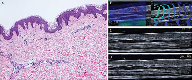
Fig. 2. Histopathological and ultrasound findings. (A) Basilar layer hyperpigmentation with normal collagen fibres and sparse perivascular infiltration of lymphocytes in the dermis. (B) Elastography measurements showed skin stiffness of 13.5 kPa on the affected side and 14.6 kPa on the normal side. (C and D) Ultrasonic images of the lesion indicated that the thickness of the dermis and subcutaneous tissue on the affected side was 1.28 mm and 2.71 mm, respectively, compared with 1.78 mm and 2.57 mm on the normal side (×100, HE stain).
What is your diagnosis?
Differential diagnosis 1: Idiopathic atrophoderma of Pasini and Pierini
Differential diagnosis 2: Lichen striatus
Differential diagnosis 3: Linear morphea
Differential diagnosis 4: Linear atrophoderma of Moulin
See next page for answer
ANSWERS TO QUIZ
Linear Hyperpigmented and Depressed Patches Following Blaschko’s Lines: A Commentary
Diagnosis: Linear Atrophoderma of Moulin
Histopathological examination showed hyperpigmentation of the basal layer, normal collagen fibres in the mid-dermis, and a sparse perivascular lymphocytic infiltrate (Fig. 2A). Skin ultrasound examination using a 50-mm linear probe operating at 24 MHz (Canon Aplio i900; https://eu.medical.canon/) indicated that the thickness of the dermis and subcutaneous tissue on the affected side was 1.28 mm and 2.71 mm, respectively, compared with 1.78 mm and 2.57 mm on the normal side (Fig. 2C and D). Elastography measurements showed skin stiffness of 13.5 kPa on the affected side and 14.6 kPa on the normal side (Fig. 2B). These findings are consistent with the linear atrophoderma of Moulin (LAM). After reassuring the patient of the benign nature of the condition, no treatment was recommended. The lesions remained stable at the 3-month follow-up visit.
LAM is defined by linear, hyperpigmented, depressed patches that follow Blaschko’s lines, typically without preceding inflammation or a sclerotic appearance. It primarily affects children and adolescents and tends to occur sporadically (1). LAM presents clinically as a unilateral band-like or linear dermatosis of varying size that follows Blaschko’s lines on the trunk and extremities. It is asymptomatic, with no systemic involvement or progression. LAM shows a diversity of manifestations, including an inflammatory type, bilateral involvement, telangiectatic lesions, and spotty lentiginosis within the lesions. Additionally, leukonychia, including partial leukonychia as observed in this patient, may appear on the fingernails (2). Our case fulfils the diagnostic criteria for LAM (3): (i) onset ranging from infancy to middle age; (ii) presence of hyperpigmented, mildly atrophic, unilateral lesions along Blaschko’s lines; (iii) absence of scleroderma; (iv) a stable, nonprogressive course without remission; and (v) histological findings of basal layer hyperpigmentation, with normal dermis and unaffected connective tissue and elastic fibres. LAM can be distinguished from idiopathic atrophoderma of Pasini and Pierini by its characteristic Blaschkoid distribution of lesions. Other pigmentary dermatoses that follow Blaschko’s lines, such as lichen striatus, linear morphea, naevoid hypermelanosis, and focal dermal hypoplasia, can be excluded based on the absence of sclerosis and inflammatory infiltrates. The underlying causes of LAM remain debated. Moulin et al. and earlier studies suggested that subcutaneous atrophy contributes to its clinical presentation (4). Recently, ultra-high-frequency ultrasound has been adopted as a non-invasive method to assess lesion thickness in LAM. New ultrasound evidence suggests that clinical atrophy is mainly due to a reduction in dermal tissue (5), with only a few severe cases involving the subcutaneous layer. In our patient, ultrasound elastography shows that the stiffness of the tissue exhibits no significant difference and may even be slightly lower than that of normal tissue, which aligns with the pathological findings. High-frequency ultrasound findings showed a thinner dermis (–28.1%) and a slightly thicker subcutaneous layer (+5.4%) than normal skin. In LAM, the Blaschkoid distribution may indicate mosaicism resulting from a postzygotic mutation during an early developmental stage. Other predisposing factors may include autoimmunity or connective tissue diseases (6). LAM develops gradually in the first few years without apparent inflammation, then becomes non-sclerotic and static. Given the absence of effective treatment options for LAM, reassuring patients of its benign nature and conducting regular follow-ups is appropriate.
ACKNOWLEDGEMENT
Funding sources: The study was financially supported in part by the 3rd Leading Talent Project for Chinese Medicine in Jiangsu Province.
REFERENCES
- Zhang LW, Li CH, Fu LX, Zhou PM, Meng HM, Shen X, et al. Linear atrophoderma of Moulin is due to the decreased dermal thickness. Skin Res Technol 2022; 28: 646–648. https://doi.org/10.1111/srt.13175
- Villani AP, Amini-Adle M, Wagschal D, Balme B, Thomas L. Linear atrophoderma of moulin: report of 4 cases and 20th anniversary case review. Dermatology 2013; 227: 5–9. https://doi.org/10.1159/000347110
- Tang C, Wang P. Atypical linear atrophoderma of Moulin complicated with elevated immunoglobulin M: a case report. Clin Cosmet Investig Dermatol 2023; 16: 193–196. https://doi.org/10.2147/CCID.S398441
- Norisugi O, Makino T, Hara H, Matsui K, Furuichi M, Shimizu T. Evaluation of skin atrophy associated with linear atrophoderma of Moulin by ultrasound imaging. J Am Acad Dermatol 2011; 65: 232–233. https://doi.org/10.1016/j.jaad.2009.12.005
- Li C, Zhang L, Zheng L, Lu Y, Liu Y. Linear atrophoderma of Moulin: dermal or subcutaneous atrophy? Australas J Dermatol 2020; 61: e365–e366. https://doi.org/10.1111/ajd.13255
- Tan SK, Tay YK. Linear atrophoderma of Moulin. JAAD Case Rep 2016; 2: 10–12. https://doi.org/10.1016/j.jdcr.2015.10.005