ORIGINAL REPORT
Epidemiology and Management of Actinic Keratosis in France: A General Population Survey (REAKT)
Brigitte DRÉNO1, Pierre LÉVY2, Gregory CAILLET3, Chantal TOUBOUL4, Jean-Michel JOUBERT5 and Jean Michel AMICI6
1Nantes University, INSERM, CNRS, Immunology and New Concepts in Immunotherapy, UMR 1302/EMR6001, Nantes, 2Paris-Dauphine University, PSL University, LEDA[LEGOS], Paris, 3Medical Affairs Department, Almirall SAS, Paris, 4Cerner Enviza France, Paris, 5Government Affairs Department, Almirall SAS, Paris, and 6Department of Dermatology, Saint-André Hospital, Bordeaux University Hospital, Bordeaux, France
The objective of this retrospective observational study was to estimate the prevalence of actinic keratosis (AK) in individuals aged ≥ 40 years in France, to describe the characteristics of affected patients, and to describe treatments. A representative panel of 20,000 households with ≥ 1 member aged ≥ 40 years were invited to participate. Participants who reported AK lesions diagnosed by a physician were eligible. The study questionnaire collected data on demographics, lesion characteristics, Fitzpatrick phototype, diagnosis, and treatments. In total, 15,246 questionnaires (78.5%) were returned and 639 responders were eligible. The adjusted prevalence of AK was 4.03% (95% CI: 3.73–4.35). Prevalence is probably underestimated due to data collection by self-report and low awareness of AK. 177 participants (27.7%) were aged < 65 years. AK was diagnosed by a dermatologist for 521 participants (81.6%). Some 200 participants (31.3%) had no lesions at the time of the survey and 243 (37.9%) had never been treated; 312 participants (78.6%) were prescribed physical treatment, principally cryotherapy; and 125 (31.5%) were prescribed topical treatment, principally 5-fluorouracil or imiquimod. In conclusion, improving diagnosis of AK in everyday clinical practice is important to ensure that all individuals with AK are treated optimally and encouraged to take sun protection measures to prevent progression to SCC.
SIGNIFICANCE
Actinic keratosis is a chronic skin disease caused by cumulative exposure to ultraviolet light presenting as rough keratotic lesions on sun-exposed areas. As no recent data exist on the burden of actinic keratosis in France, a questionnaire survey was performed in a representative panel of 20,000 households with at least member aged ≥ 40 years. Some 4.0% of participants declared that they had actinic keratosis lesions diagnosed by a physician; ~30% of these individuals had never been treated and ~50% reported no improvement since diagnosis. Improved patient awareness and physician involvement are essential to improve diagnosis and optimize patient management.
Key words: actinic keratosis; epidemiology; disease management; squamous cell carcinoma; surveys and questionnaires.
Citation: Acta Derm Venereol 2025; 105: adv42372. DOI: https://doi.org/10.2340/actadv.v105.42372.
Copyright: 2025 © The Author(s). Published by MJS Publishing, on behalf of the Society for Publication of Acta Dermato-Venereologica. This is an Open Access article distributed under the terms of the Creative Commons Attribution-NonCommercial 4.0 International License (https://creativecommons.org/licenses/by-nc/4.0/).
Submitted: Nov 12, 2024; Accepted after revision: Dec 2, 2024; Published: Jan 26, 2025.
Corr: Professor Brigitte Dréno, Nantes Université, INSERM, CNRS, Immunology and New Concepts in Immunotherapy, INCIT, UMR 1302/EMR6001, 22 boulevard Benoni Goullin, FR-44000 Nantes, France. E-mail: brigitte.dreno@atlanmed.fr
Competing interests and funding: BD has received consulting fees from Almirall SAS; PL has received honoraria for study development from Almirall SAS; GC is an employee of Almirall SAS, the sponsor of the study; CT is an employee of Cerner Enviza who carried out the study sponsored by Almirall SAS; J-MJ is an employee of Almirall SAS, the sponsor of the study; JMA has received consulting fees from Almirall SAS.
INTRODUCTION
Actinic keratosis is a chronic skin disease caused by sun exposure (1), presenting as rough keratotic lesions typically on exposed areas, like the face, scalp, or back of the hands (1). The principal risk factors include fair skin, cumulative sun exposure, old age, and male gender (2). Spontaneous resolution is rare and recurrence is common even following treatment (3). The principal health concern with AK lesions is the risk of progression to squamous cell carcinoma (SCC). Although only a minority of AK lesions evolve to SCC, it is believed that most SCCs arise from a pre-existing AK (4–7). The factors influencing progression from AK to SCC remain poorly understood.
The prevalence of AK is rising due to longer life expectancy and a history of frequent sun exposure. To limit the risk of developing SCC, targeted screening programmes are necessary, which in turn require epidemiological studies to collect local data on the epidemiology and current management of AK. However, such data in Europe are limited. The only prevalence data from France published in the last 25 years come from a nationwide cross-sectional survey of community dermatologists conducted in 2004 (8).
We have now undertaken a retrospective observational study in a representative sample of the French general population aged ≥ 40 years old (REAKT study). The study aimed to estimate the prevalence of AK, to describe the characteristics of affected participants, and to describe their care management.
MATERIALS AND METHODS
The REAKT study was conducted in a representative sample of the French general population aged ≥ 40 years from a household panel (METASKOPE). Data were collected using a paper questionnaire. The survey was implemented by Cerner Enviza (Paris, France) between 14 November and 19 December 2022. A Scientific Committee oversaw the design and implementation of the study, and the interpretation of the data.
Data source
The METASKOPE panel consists of a sample of 20,000 households participating on a voluntary basis. The panel is representative of the French population in terms of sex, age, gender, socioeconomic class, region of residence, and population size of the municipality of residence. Panellists are invited to complete questionnaires on a regular basis (around once a month) on a variety of topics, including health-related issues. The METASKOPE panel has been widely used in medical research to collect data on epidemiology and care provision in France over the last 30 years (9–15).
Participants
Households containing at least 1 member aged ≥ 40 were eligible. Each such household in the METASKOPE panel was invited to participate; a total of 19,426 invitations were sent by post. If more than 1 household member was aged ≥ 40 years, then each was invited to complete the questionnaire.
Data collection
METASKOPE panellists who agreed to participate completed the REAKT study questionnaire, which was sent in the same envelope as the invitation. This questionnaire (provided in Appendix S1) contained 25 questions, some of which had several sub-questions. The total time to complete the questionnaire was around 15 minutes. The majority took the form of yes/no closed questions or multiple-choice questions. The questionnaire opened with 2 screening questions. The first question was the following: “Have you ever had, or do you have at the moment, one or more actinic keratosis lesions? These are scabby skin lesions or lesions that are rough to the touch, also called sun keratosis.” Individuals who did not report such lesions stopped the questionnaire at this point. The second question asked whether their AK lesions had been diagnosed by a physician: “Have your lesions been diagnosed as actinic keratosis by a physician”. Those patients who replied that their lesions had been diagnosed by a physician were eligible for the study.
The study questionnaire collected information on demographics, characteristics of AK lesions, diagnosis, and treatments. Participants identified their Fitzpatrick phototype (16) from reference photographs and were provided with a checklist of typical lesion sites (face, scalp, back of hands, arms, ears, neck, legs of feet, or other). A list of approved treatments of AK in France was provided to participants to identify treatments that they had been offered (see Table SI for list and reimbursement status). They were invited to identify their most recent treatments (up to 3 choices possible) and these data were used to describe treatment sequences.
Statistical analysis
Three populations of interest were identified and analysed for this study. The full study population consisted of all participants returning a study questionnaire and was used for estimation of the self-reported prevalence of AK in the French general population, analysis of patient and disease characteristics, and description of the diagnostic pathway. The treated population consisted of all participants receiving at least 1 treatment and was used for analysis of types of treatments received. This population was also analysed by age group (< 65 years vs ≥ 65 years). The “treatment sequences documented” population consisted of all participants reporting information on treatment adherence, switches, and discontinuation for up to 3 of their most recent treatments.
The demographic structure of the sample (by age, gender, and region of residence), was compared with the structure of the French general population aged ≥ 40 years in 2023 (17). From this comparison, an adjusted prevalence of AK could be estimated by weighting using raking-adjusted statistics (18).
Data were controlled, validated, and analysed centrally using Dasie software( adn-soft, Paris, France; https://adn-soft.com/logiciels/) version 2.4.84 for descriptive and bivariate analyses.
RESULTS
Study population
A total of 15,246 individual questionnaires were returned (response rate of 78.5%). Of these responders, 936 (6.1%) declared current or previous AK lesions and 639 (5.5%) that these lesions had been diagnosed by a physician. These 639 participants made up the study population. Of these, 397 participants (62.1%) made up the treated population, of whom 364 (91.7%) provided information on up to 3 most recently used treatments (“treatment sequences documented” population). A flow diagram illustration the selection of participants is presented in Fig. 1.
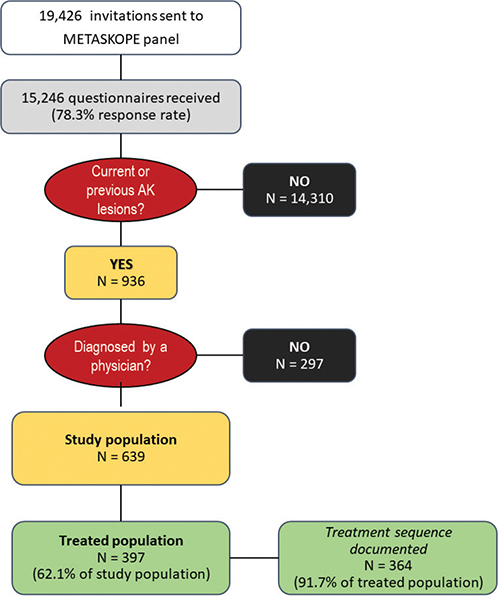
Fig. 1. Selection of participants. AK: actinic keratosis.
Prevalence
The adjusted prevalence of AK was 4.03% (95% CI: 3.73–4.35) in individuals aged ≥ 40 years old (5.92% [CI: 5.40–6.49] in individuals aged ≥ 65 years old and 2.38% [CI: 2.07–2.74] in individuals aged 40–65 years old). Adjusted prevalence was similar in men (4.31% [CI: 3.86–4.80]) and women (3.79% [CI: 3.39–4.23]), and increased with age (Fig. 2). The age- and gender-adjusted prevalence of AK also varied geographically (Fig. S1), being highest in coastal regions and displaying a North–South gradient. This distribution only poorly matched that of the participants’ phototype (Pearson correlation coefficient: 0.05; p = 0.43, Fig. S1).
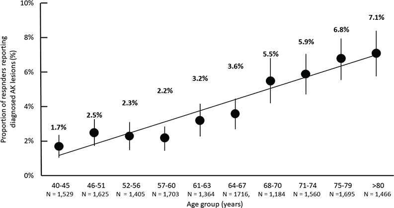
Fig. 2. Prevalence of self-reported actinic keratosis as a function of age.
Participant and disease characteristics
Participant and disease characteristics in the full study population are presented in Table I. The mean age of participants was 69.6 ± 10.6 years and women were slightly over-represented. Participants under the age of 65 constituted 27.7% of the sample (n = 177). Most participants (76.5%) were Phototype II or III. Moreover, 150 participants (20.3%) had a previous history of treatment for skin cancer.
| Factor | Full study population (n = 639) |
| Age, mean ± SD | 69.6 ± 10.6 |
| Median [range] | 70.25 [40–98] |
| Gender, n (%) | |
| Men | 296 (46.3%) |
| Women | 343 (53.7%) |
| Phototype, n (%) | |
| I | 90 (14.1%) |
| II | 276 (43.2%) |
| III | 213 (33.3%) |
| IV | 26 (4.1%) |
| V | None |
| VI | 1 (0.2%) |
| Not reported | 33 (5.2%) |
| Medical history, n (%) | |
| Treated for skin cancera | 130 (20.3%) |
| Received anticancer therapyb | 60 (9.4%) |
| Treated for another skin disorder | 44 (6.9%) |
| Undergone transplantation | 3 (0.5%) |
| At least 1 of the above | 208 (32.6%) |
| Not reported | 51 (8.0%) |
| Time since diagnosis, n (%) | |
| < 6 months | 95 (14.8%) |
| 6 months to 1 year | 59 (9.2%) |
| 1 to 3 years | 169 (26.5%) |
| > 3 years | 301 (47.1%) |
| Not reported | 15 (2.4%) |
| Disease activity, n (%) | |
| Current lesions | 432 (67.6%) |
| No current lesions | 200 (31.3%) |
| Not reported | 7 (1.1%) |
| Change since diagnosis, n (%) | |
| Worsened | 56 (8.8%) |
| No change | 220 (34.4%) |
| Improved | 81 (12.7%) |
| Completely resolved | 200 (31.3%) |
| Not reported | 82 (12.8%) |
| Number of current lesions, n (%) | n = 432 |
| Only 1 | 103 (23.8%) |
| 2–3 | 156 (36.1%) |
| 4–10 | 109 (25.2%) |
| 11–25 | 35 (8.1%) |
| > 25 | 15 (3.5%) |
| Not reported | 14 (3.2%) |
| aSpecified as “melanoma, squamous cell carcinoma or basocellular carcinoma”. bSpecified as “chemotherapy, radiotherapy or immunotherapy”. SD: standard deviation. |
|
The diagnosis of AK was made > 3 years prior to the survey for 301 participants (47.1%) and lesions had resolved completely by the time of the survey for 200 participants (31.3%). For the 439 participants with current lesions, 259 (60.0%) reported ≤ 3 current lesions. The most frequent lesion sites reported were the face, scalp, or lower extremities (Fig. S2).
Diagnosis
The diagnosis was made by a dermatologist for 521/639 participants in the full study population (81.6%) and by a GP for 111 participants (17.4%). Diagnosis by a dermatologist was made after referral by a GP in 104 of these cases (20.0%) (Table SII). The presence of AK lesions was the reason for consulting a dermatologist for 287 participants (55.1%), and was discovered during a consultation for another reason in 117 participants (22.5%). The median interval between lesion appearance and diagnosis was 17 months, although there was wide variation.
Treatment of actinic keratosis
Overall, 397 participants (62.1%) had received AK treatment at least once and these constituted the treatment population (Table II). Of these, 312 participants (78.6%) reported receiving physical treatment, principally cryotherapy; 125 (31.5%) reported using topical treatment, principally imiquimod or 5-fluorouracil. Dermatologists were responsible for prescription of 114/149 (76.5%) individual topical treatments and 382/417 (91.6%) individual physical treatments (Table III).
| Factor | Treatment population (n = 397) n (%) | Age 40–65 years (n = 101) n (%) | Age > 65 years (n = 296) n (%) | p-value (χ² test) |
| Topical treatments | 125 (31.5%) | 40 (39.6%) | 85 (28.7%) | 0.019 |
| Imiquimod | 49 (12.3%) | 15 (14.9%) | 34 (11.5%) | |
| 5-fluorouracil | 51 (12.9%) | 11 (10.9%) | 40 (13.5%) | |
| Sodium diclofenac | 22 (5.5%) | 13 (12.9%) | 9 (3.0%) | |
| Methyl aminolaevulinate | 14 (3.5%) | 3 (3.0%) | 4 (1.4%) | |
| Ingenol mebutate | 11 (2.8%) | 1 (1.0%) | 10 (3.4%) | |
| Nicotinamide | 7 (1.8%) | 3 (3.0%) | 4 (1.4%) | |
| Physical treatments | 312 (78.6%) | 66 (63.4%) | 246 (83.1%) | 0.3735 |
| Cryotherapy | 253 (63.7%) | 47 (46.5%) | 206 (69.6%) | |
| Photodynamic therapy | 22 (5.5%) | 6 (5.9%) | 16 (5.4%) | |
| Laser therapy | 38 (9.6%) | 11 (10.9%) | 27 (9.2%) | |
| Electrocoagulation | 37 (9.3%) | 9 (8.9%) | 28 (9.5%) | |
| Othera | 6 (1.5%) | 1 (1.0%) | 5 (1.69%) | |
| Treatment not identified | 28 (7.1%) | 6 (5.9%) | 22 (7.4%) | |
| aFusidic acid, ciclopirox, hyaluronic acid, salicylic acid/betamethasone or sunscreens. | ||||
| Factor | Topical treatments (n = 149) n (%) | Physical treatments (n = 417) n (%) | |
| Physician prescribing treatment | |||
| GP | 22 (14.8%) | 8 (1.9%) | |
| Dermatologist | 114 (76.5%) | 382 (91.6%) | |
| Othera | – | 3 (0.7%) | |
| Missing data | 13 (8.7%) | 24 (5.8%) | |
| Duration of treatment | Imiquimod (n = 52) | 5-fluorouracil (n = 55) | Not applicable |
| ≤ 2 weeks | 10 (19.2%) | 14 (25.5%) | |
| 2–3 weeks | 7 (13.5%) | 8 (14.5%) | |
| 3–4 weeks | 14 (26.9%) | 16 (29.1%) | |
| 1–2 months | 11 (21.2%) | 5 (9.1%) | |
| > 2 months | 5 (9.6%) | 5 (9.1%) | |
| Missing data | 5 (9.6%) | 7 (12.7%) | |
| Treatment stopped | Not applicable | ||
| Before disappearance of lesions | 22 (14.8%) | ||
| Reason for stopping | |||
| End of treatment course | 11 (50.0%) | ||
| Poor tolerability of treatment | 4 (18.2%) | ||
| Treatment ineffective | 2 (9.1%) | ||
| Treatment duration considered too long | 2 (9.1%) | ||
| Treatment too problematic/complicated | 2 (9.1%) | ||
| Treatment too time-consuming | – | ||
| Missing data | 1 (4.6%) | ||
| Management post-treatment | |||
| Prescription renewed | 24 (16.1%) | 25 (6.0%) | |
| Treatment changed | 17 (11.4%) | 16 (3.8%) | |
| Regular surveillance | 1 (0.7%) | 26 (6.2%) | |
| All treatment stopped | 48 (32.2%) | 105 (25.2%) | |
| No follow-up consultation | 26 (17.4%) | 101 (24.2%) | |
| Treatment ongoing | 21 (14.1%) | 67 (16.1%) | |
| Other | 2 (1.3%) | 5 (1.2%) | |
| Missing data | 10 (6.7%) | 72 (17.3%) | |
| Subsequent treatments | n = 69 | n = 133 | |
| With same treatment as initially | 16 (23.2%) | 81 (60.9%) | |
| With different treatment | 53 (76.8%) | 51 (38.4%) | |
| Missing data | 0 | 1 (0.7%) | |
| Reason for retreatment | N = 69 | N = 133 | |
| Due to lesion persistence | 17 (24.6%) | 13 (9.8%) | |
| Due to lesion recurrence | 29 (42.0%) | 56 (42.1%) | |
| To prevent lesion recurrence | 14 (20.3%) | 31 (23.3%) | |
| Other | 3 (4.3%) | 0 | |
| Missing data | 3 (8.7%) | 33 (24.8%) | |
| aENT physician in 1 case and surgeon in another, and not reported in the third. 2Dermatologist retired from practice, dermatologist decided not to pursue treatment, neglect, forgot treatment. |
|||
Participants aged ≤ 65 years used topical treatments more frequently than older patients (39.6% vs 28.7% respectively) whereas older patients used physical treatments more frequently (63.4% vs 83.1%; p = 0.019 for the interaction age group × treatment group; χ² test). The individual topical treatment used also varied between the age groups (Table II). For example, 32.5% of participants ≤ 65 years (n = 40) declared using sodium diclofenac, 27.5% 5-fluorouracil and 2.5% ingenol mebutate. In the older age group (n = 85), the proportions were 10.6%, 47.0%, and 11.8% respectively.
Of 397 treated patients, 370 provided more detailed information on up to 3 of their most recent treatments: 231 (62.4%) reported a single treatment, 76 (20.5%) 2 treatments, and 63 (17.0%) 3 treatments. The mean number of treatments was 2.23 ± 1.47. Seventy-one participants (19.5%) were under treatment at the time of the survey. When further treatment was given, the original treatment was renewed for 97 patients (46.9%). Switching between topical and physical treatments was less common than remaining in the same class (Fig. 3). Overall, 40 treated participants (11.0%) switched from physical treatment to topical treatment and 25 (6.9%) from topical to physical treatment. Within each class, changes to pattern of use between sequential treatments were limited (Table SIII). Electrocoagulation and photodynamic therapy were used more often later in the treatment sequence at the expense of cryotherapy (p = 0.0027; χ² test).
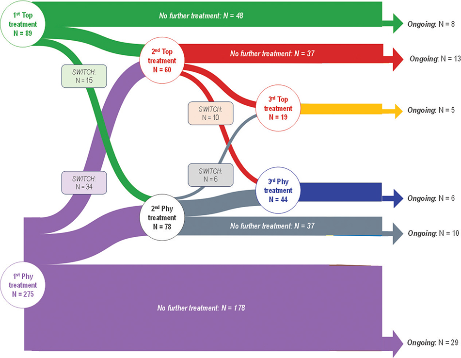
Fig. 3. Treatment sequences. Phy: physical treatments; Top: topical treatments. One patient did not provide information on the type of treatment.
For topical treatments, the treatment duration was between 1 and 4 weeks for the majority of patients (mean: 38.6 ± 60.9 days; median: 23.3 days) (Table III). Because the recommended treatment durations differ between treatments, treatment duration was analysed individually for imiquimod and 5-fluorouracil, for which sufficient treatments were available. For imiquimod (recommended duration: up to 4 weeks), 16 treatments (30.8%) were used for over 4 weeks. For 5-fluorouracil (recommended duration: 3–4 weeks), 10 treatments (18.2%) were used for over 4 weeks and 22 (40.0%) for less than 3 weeks.
The reason for stopping topical treatment was the end of the prescribed treatment course in most cases (50.0%); poor tolerability accounted for 4 of the early discontinuations (18.2%). After the end of the topical treatment course, all treatment was stopped in 48 cases (32.2%) and no follow-up consultation was made in 26 cases (17.5%). Treatment was stopped before complete lesion resolution for 22/149 (14.8%) of topical treatments (Table III). The principal reason for retreatment with a topical treatment was lesion recurrence (42.0%), followed by lesion persistence (24.6%) (Table III). For physical treatments, no further treatment was prescribed or there was no follow-up consultation in 49.4% of cases (Table III). The principal reason for retreatment following a physical treatment was lesion recurrence (42.1% of cases).
DISCUSSION
The REAKT survey provides the first update of the epidemiology of AK in France since 2008. The survey identified 639 participants aged ≥ 40 years with self-reported, physician-diagnosed AK. As expected, the majority of these cases (57.3%) had a fair skin phototype (Fitzgerald I or II). They frequently lived in coastal regions and in southern regions of France where sun exposure may be highest.
The overall prevalence was estimated from self-report by the participant to be 4.0% (5.9% in participants aged ≥ 65 years) corresponding to a potential 1.5 million individuals aged ≥ 40 years with AK in France. Awareness of AK in the general population in Europe is low (6–7%) (23, 24), leading many individuals to consider their lesions to be a “normal” feature of skin ageing and not to seek medical advice, and this may have led to an underestimate of the true prevalence. Studies where the diagnosis of AK was established by a dermatologist through systematic screening, such as in the earlier French study (8), have provided much higher estimates. For example, prevalence estimates were 15.4% in men and 5.9% in women aged ≥ 40 years in North-West England (19), 23% in people aged ≥ 60 years in South Wales (20), and, in particular, 49% in men and 28% in women aged ≥ 45 years in the Netherlands (21). On the other hand, in a large German general population study, prevalence was 4.1% in men and 6.8% in women aged ≥ 50 years, which is consistent with the present study. In our study, the prevalence of AK increased progressively with age. The finding that 28% of participants reporting AK were under 65 years of age, and that prevalence rose smoothly with age from a rate of 1.7% in participants aged 40–45 years, was a novel finding compared with studies performed in previous decades (8, 19, 20). Nonetheless, the more recent studies from the Netherlands (21) and Germany (22) have also both reported a significant proportion of patients under 60 years of age and with a progressive increase from the age of 40. Earlier appearance of AK may reflect higher cumulative sun exposure, perhaps due to people more frequently taking holidays in the sun in recent decades.
Previous studies have consistently demonstrated a higher prevalence of AK in men than in women (19, 21, 22). In our study, no significant difference between men (4.3%) and women (3.8%) was observed. A possible explanation is that our data were obtained by self-report and there may be gender differences in awareness of and concern about AK lesions, with women being more likely to have sought medical advice for their lesions compared with men.
Referral to a dermatologist appears to be the key step to more comprehensive management of AK in France, because dermatologists are responsible for diagnosis in > 80% of cases and treatment initiation in > 85% of cases. However, GPs also play an important role, diagnosing 17% of cases or referring to a dermatologist for diagnostic confirmation in 20% of cases.
Around half our cases were diagnosed with AK serendipitously during consultations for other reasons. Moreover, a relatively high proportion of participants (20%) had a history of skin cancer, suggesting that they were already followed by a dermatologist, which may have facilitated diagnosis. These findings indicate the importance of screening and diagnosis of AK so that it can be treated in a timely fashion.
Even though all participants had been diagnosed with AK by a physician, 37.9% never received treatment. For patients who received treatment, one-third received topical treatment and two-thirds physical therapy, principally cryotherapy. In a previous survey of AK management in France (25), cryotherapy was used in 92% of cases, and topical treatments, principally sodium diclofenac, were used exclusively as a complement to cryotherapy, and never alone. A later survey (2018) reported prescription of physical therapy alone in 53% of cases, topical treatments in only 20% and a combination of both in 27% (26). Taken together, this suggests that treatment of AK has evolved over the last 20 years with a shift towards topical treatments, either alone or in combination, following the introduction of a wider range of treatments in France (imiquimod and mebutate ingenol) over this period. This may also reflect growing awareness of the need to treat the skin surrounding the AK lesion itself to reduce the risk of recurrence and of development of SCC (field cancerization theory) (27). We also observed important differences in treatments prescribed between older and younger participants, with more older participants receiving cryotherapy and more younger patients receiving topical therapy. This may be due to dermatologists believing that older patients will be less adherent to treatment. It should be noted that the number of lesions in this study was rather low, with 60% of participants reporting no more than 3 lesions. This may have influenced the treatment patterns observed, as practice guidelines recommend treating isolated lesions preferentially with cryotherapy, and multiple lesions with topical therapies (alone or combined with physical treatment) (28, 29). We observed that sequential treatments were sometimes required, notably due to recurrence of lesions. Switching between topical and physical treatments was uncommon. We also noted that > 30% of imiquimod and 5-fluorouracil treatment courses lasted for < 2 weeks, which may have been insufficient to ensure complete elimination of lesions. Indeed, 50% of topical treatments were stopped for reasons other than reaching the end of the prescribed treatment course and 14.8% were stopped before lesions had disappeared. One quarter of retreatments were motivated by lesion persistence following the original treatment. For around one-half of treatments (49.6% of topical treatments and 49.4% of physical treatments), participants were either never retreated or did not consult again.
In around one-third of the study population of (200/639 participants), AK lesions had disappeared by the time of the survey. After initial treatment, around one-quarter of participants were never treated again, and in another quarter no follow-up consultation was attended. Given that AK lesions frequently recur, follow-up consultations are important for performing skin checks and to renew treatment if necessary. In addition, a third of participants declared never having received treatment for their lesions.
Strengths and limitations
The strengths of this study include the general population setting, the requirement for physician diagnosis of the lesions, the large number of individuals sampled, and the high response rate (> 75%). The principal limitation is that the information is based on self-report, and may not always be fully accurate. As awareness of AK in the general public in Europe has been reported to be very low, and only a minority of individuals seek a skin cancer check from a dermatologist (24), capture of all individuals in the METASKOPE panel with AK may not have been exhaustive. In addition, given that the AK diagnosis may date from several years prior to the survey, recall error for information on disease and treatment history may be significant.
Conclusion
Actinic keratosis is a chronic disease that requires long-term photoprotection and topical or physical treatment as soon as the lesions are diagnosed, to reduce the risk of field cancerization. The REAKT study estimates the self-reported prevalence of AK in individuals aged ≥ 40 in France to be 4%. This relatively low estimate may indicate limited awareness of AK in the general population and measures to increase awareness of the disease are needed. We observed that over one-quarter of participants reporting AK lesions were aged < 65 years. This novel finding should encourage early management in order to reduce the risk of progression to SCC, and thus reduce the burden of long-term management. Currently, dermatologists are principally responsible for diagnosis and treatment of AK; however, given that access to dermatologists in France may be complicated, GPs could play a larger role in screening for AK in at-risk patients and prescribing topical treatments where necessary. We also observed increased use of topical treatments, particularly in younger patients, compared with previous studies, which is a positive trend towards early treatment of the affected photoexposed area. Nonetheless, treatment duration was frequently short and potentially insufficient, illustrating the need for effective and well-tolerated topical treatments that can potentially be used over the long term. Finally, many participants were never retreated or never consulted again, indicating that follow-up of patients with AK after treatment should be improved.
ACKNOWLEDGEMENTS
This study was funded by Almirall SAS.
IRB approval status: The survey was conducted in accordance with the ESOMAR International Code on Market and Social Practice, the EphMRA Code of Conduct, relevant current French and European legislation, and Good Epidemiological Practice guidelines. Analyses performed using the METASKOPE panel have been approved by the Commission Nationale de l’Informatique et des Libertés (CNIL), the French national data protection authority. Ethical committee approval is not required for this form of survey in France.
REFERENCES
- de Oliveira ECV, da Motta VRV, Pantoja PC, Ilha CSO, Magalhães RF, Galadari H, et al. Actinic keratosis: review for clinical practice. Int J Dermatol 2019; 58: 400–407. https://doi.org/10.1111/ijd.14147
- Rosen T, Lebwohl MG. Prevalence and awareness of actinic keratosis: barriers and opportunities. J Am Acad Dermatol 2013; 68: S2–9. https://doi.org/10.1016/j.jaad.2012.09.052
- Werner RN, Sammain A, Erdmann R, Hartmann V, Stockfleth E, Nast A. The natural history of actinic keratosis: a systematic review. Br J Dermatol 2013; 169: 502–518. https://doi.org/10.1111/bjd.12420
- Reinehr CPH, Bakos RM. Actinic keratoses: review of clinical, dermoscopic, and therapeutic aspects. An Bras Dermatol 2019; 94: 637–657. https://doi.org/10.1016/j.abd.2019.10.004
- Goldenberg G, Perl M. Actinic keratosis: update on field therapy. J Clin Aesthet Dermatol 2014; 7: 28–31.
- Feldman SR, Fleischer AB, Jr. Progression of actinic keratosis to squamous cell carcinoma revisited: clinical and treatment implications. Cutis 2011; 87: 201–207.
- Fernandez Figueras MT. From actinic keratosis to squamous cell carcinoma: pathophysiology revisited. J Eur Acad Dermatol Venereol 2017; 31: 5–7. https://doi.org/10.1111/jdv.14151
- Bernard P, Dupuy A, Sasco A, Brun P, Duru G, Nicoloyannis N, et al. Basal cell carcinomas and actinic keratoses seen in dermatological practice in France: a cross-sectional survey. Dermatology 2008; 216: 194–199. https://doi.org/10.1159/000112925
- Reach G, Pellan M, Crine A, Touboul C, Ciocca A, Djoudi Y. Holistic psychosocial determinants of adherence to medication in people with type 2 diabetes. Diabetes Metab 2018; 44: 500–507. https://doi.org/10.1016/j.diabet.2018.06.001
- Briot K, Grange L, Cortet B, Feron JM, Chauvin P, Coulomb A, et al. Real-world care for individuals aged over fifty with fractures in France: evidence for a wide care gap – The EPIFRACT Study. Joint Bone Spine 2020; 87: 467–473. https://doi.org/10.1016/j.jbspin.2020.04.007
- Mosnier-Pudar H, Hochberg G, Eschwege E, Halimi S, Virally ML, Guillausseau PJ, et al. How patients’ attitudes and opinions influence self-care behaviours in type 2 diabetes. Insights from the French DIABASIS Survey. Diabetes Metab 2010; 36: 476–483. https://doi.org/10.1016/j.diabet.2010.08.004
- Cortet B, Chauvin P, Feron JM, Grange L, Coulomb A, Launois R, et al. Fragility fractures in France: epidemiology, characteristics and quality of life (the EPIFRACT study). Arch Osteoporos 2020; 15: 46. https://doi.org/10.1007/s11657-019-0674-2
- Charles MA, Basdevant A, Eschwege E. [Prevalence of obesity in adults in France: the situation in 2000 established from the OBEPI Study]. Ann Endocrinol (Paris) 2002; 63: 154–158.
- Charles MA, Eschwege E, Basdevant A. Monitoring the obesity epidemic in France: the Obepi surveys 1997–2006. Obesity (Silver Spring) 2008; 16: 2182–2186. https://doi.org/10.1038/oby.2008.285
- Eschwege E, Basdevant A, Crine A, Moisan C, Charles MA. Type 2 diabetes mellitus in France in 2012: results from the ObEpi survey. Diabetes Metab 2015; 41: 55–61. https://doi.org/10.1016/j.diabet.2014.11.007
- Fitzpatrick TB. The validity and practicality of sun-reactive skin types I through VI. Arch Dermatol 1988; 124: 869–871. https://doi.org/10.1001/archderm.1988.01670060015008
- Institut National de la Statistique et des Études Économiques. Bilan démographique 2017. 2018.
- Deville J-C, Sarndal C-E, Sautory O. Generalized raking procedures in survey sampling. J Am Stat Assoc 1993; 88: 1013–1020. https://doi.org/10.1080/01621459.1993.10476369
- Memon AA, Tomenson JA, Bothwell J, Friedmann PS. Prevalence of solar damage and actinic keratosis in a Merseyside population. Br J Dermatol 2000; 142: 1154–1159. https://doi.org/10.1046/j.1365-2133.2000.03541.x
- Harvey I, Frankel S, Marks R, Shalom D, Nolan-Farrell M. Non-melanoma skin cancer and solar keratoses. I. Methods and descriptive results of the South Wales Skin Cancer Study. Br J Cancer 1996; 74: 1302–1307. https://doi.org/10.1038/bjc.1996.534
- Flohil SC, van der Leest RJ, Dowlatshahi EA, Hofman A, de Vries E, Nijsten T. Prevalence of actinic keratosis and its risk factors in the general population: the Rotterdam Study. J Invest Dermatol 2013; 133: 1971–1978. https://doi.org/10.1038/jid.2013.134
- Schaefer I, Augustin M, Spehr C, Reusch M, Kornek T. Prevalence and risk factors of actinic keratoses in Germany – analysis of multisource data. J Eur Acad Dermatol Venereol 2014; 28: 309–313. https://doi.org/10.1111/jdv.12102
- Halpern AC, Kopp LJ. Awareness, knowledge and attitudes to non-melanoma skin cancer and actinic keratosis among the general public. Int J Dermatol 2005; 44: 107–111. https://doi.org/10.1111/j.1365-4632.2005.02090.x
- MacKie RM. Awareness, knowledge and attitudes to basal cell carcinoma and actinic keratoses among the general public within Europe. J Eur Acad Dermatol Venereol 2004; 18: 552–555. https://doi.org/10.1111/j.1468-3083.2004.00993.x
- Bernard P, Dupuy A, Brun P, Sasko A, Duru G, Nicoloyannis N, et al. Évaluation médico-économique de la prise en charge des carcinomes basocellulaires superficiels et des kératoses actiniques multiples par les dermatologues français. Ann Dermatol Venereol 2007; 134: 527–533. https://doi.org/10.1016/S0151-9638(07)89263-5
- Savary J, Tine MC, Weber AC, Dorey J. Management and clinical practice of multiple face and scalp actinic keratosis in France. J Mark Access Health Policy 2019; 7: 1605787. https://doi.org/10.1080/20016689.2019.1605787
- Aggarwal I, Puyana C, Chandan N, Jetter N, Tsoukas M. Field cancerization therapies for the management of actinic keratosis: an updated review. Am J Clin Dermatol 2024; 25: 391–405. https://doi.org/10.1007/s40257-023-00839-8
- Dréno B, Amici JM, Basset-Seguin N, Cribier B, Claudel JP, Richard MA. Management of actinic keratosis: a practical report and treatment algorithm from AKTeam™ expert clinicians. J Eur Acad Dermatol Venereol 2014; 28: 1141–1149. https://doi.org/10.1111/jdv.12434
- Werner RN, Stockfleth E, Connolly SM, Correia O, Erdmann R, Foley P, et al. Evidence- and consensus-based (S3) Guidelines for the Treatment of Actinic Keratosis – International League of Dermatological Societies in cooperation with the European Dermatology Forum – Short version. J Eur Acad Dermatol Venereol 2015; 29: 2069–2079. https://doi.org/10.1111/jdv.13180