QUIZ SECTION
Yellow Nodule on the Right Nipple and Multiple Yellowish Plaques on the Face: A Quiz
Yue-Tong QIAN, Jia-Wei LIU and Dong-Lai MA*
Department of Dermatology, State Key Laboratory of Complex Severe and Rare Diseases, Peking Union Medical College Hospital, Chinese Academy of Medical Sciences and Peking Union Medical College, National Clinical Research Center for Dermatologic and Immunologic Diseases, Beijing 100730, China. *E-mail: mdonglai@sohu.com
Citation: Acta Derm Venereol 2025; 105: adv42478. DOI: https://doi.org/10.2340/actadv.v105.42478.
Copyright: © 2025 The Author(s). Published by MJS Publishing, on behalf of the Society for Publication of Acta Dermato-Venereologica. This is an Open Access article distributed under the terms of the Creative Commons Attribution-NonCommercial 4.0 International License (https://creativecommons.org/licenses/by-nc/4.0/).
Published: Jan 3, 2025
A 53-year-old woman developed multiple asymptomatic yellow nodular lesions and plaques on her face (Fig. 1A) that had been gradually increasing in size over the past 15 years. The lesions initially appeared as papules on the nasal tip and later spread, coalescing into plaques. Additionally, she found a yellow nodule on her right nipple (Fig. 1B). She had been treated with topical tacrolimus and zinc oxide, but there was no improvement. She had a history of benign thyroid nodules. The results of laboratory investigations, including a complete blood cell count, serum chemistry panel, liver and kidney function tests, lipid profile, autoimmunity testing, and immunoglobulins, were all negative or within normal ranges. Chest computed tomography results were generally normal. There were no obvious abnormalities in the bone marrow smear.
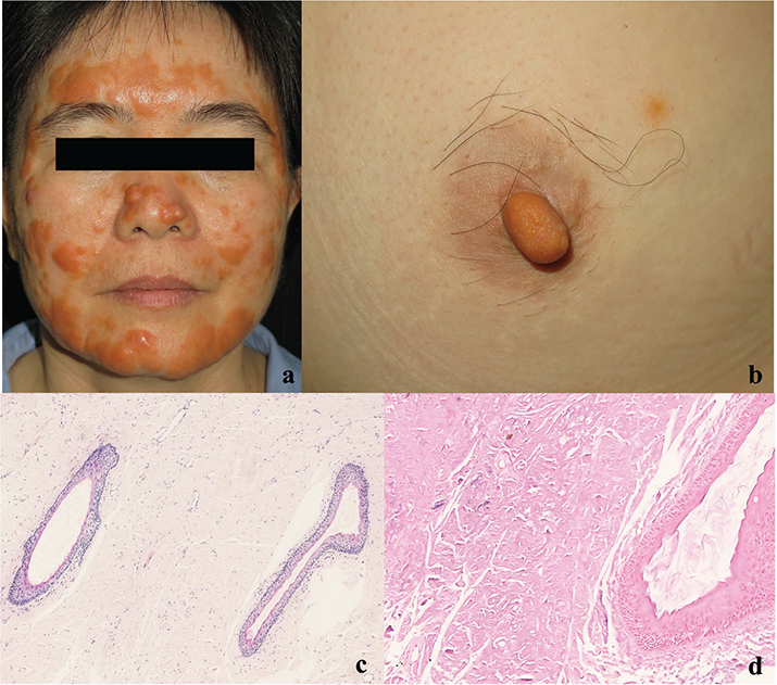
Fig. 1. (A) Multiple yellowish nodules coalesce into plaques on the face. (B) Solitary, yellow nodule on the right nipple. (C) Haematoxylin-eosin staining of the lesion revealed massive nodular homogeneous, amorphous, and slightly basophilic deposits throughout the dermis as well as scattered perivascular lymphocytes and plasma cells (original magnification×100). (D) Verhoeff-van Gieson stain revealed decrease and loss of elastic fibres in the dermis (original magnification×200).
What is your diagnosis?
Differential diagnosis 1: Adult-onset colloid milium
Differential diagnosis 2: Nodular colloid degeneration
Differential diagnosis 3: Nodular amyloidosis
Differential diagnosis 4: Xanthoma
See next page for answer.
ANSWERS TO QUIZ
Yellow Nodule on the Right Nipple and Multiple Yellowish Plaques on the Face: A Commentary
Diagnosis: Nodular colloid degeneration
A 4-mm punch biopsy specimen was obtained from the nasal tip. Histopathological examination revealed extensive nodular homogeneous, amorphous, and slightly basophilic deposits throughout the dermis, along with scattered perivascular lymphocytes and plasma cells (Fig. 1C). Periodic acid Schiff staining, Alcian blue staining, and Congo red staining were all negative. Verhoeff-van Gieson staining revealed a decrease and loss of elastic fibres in the dermis (Fig. 1D). Based on these findings, a diagnosis of nodular colloid degeneration (NCD) was made. Topical 0.025% retinoic acid for 6 months showed no improvement.
Skin colloid degeneration is a distinct clinicopathological entity characterized by colloid material deposits in the dermis, of which 5 clinical variants are recognized: adult-onset colloid milium (ACM), juvenile colloid milium (JCM), pigmented, NCD or paracolloid, and acral keratosis with eosinophilic dermal deposits (1). The most common type is ACM, primarily affecting middle-aged individuals on sun-exposed skin, whereas JCM starts before puberty. Both ACM and JCM lesions present with dome-shaped yellowish translucent papules and contain gelatinous material.
The aetiology of skin colloid degeneration remains unknown. Degenerated collagen, serum proteins, fibroblast secretions, and cytokeratin fragments have been considered. Broken and reduced elastic fibres in Verhoeff-van Gieson stain and electron microscopy examinations suggests that they might be degenerate elastic fibres (1). As the disease most commonly occurs in sun-exposed areas, long-term sun exposure has been postulated as an aetiologic factor of nodular colloid degeneration, but involvement of non-sun-exposed areas has been described (2, 3).
NCD or paracolloid is clinically manifested by an asymptomatic single large nodule or multiple soft to rubbery papules, plaques, or nodules. The lesions most commonly occur on face, ears, neck, and dorsal hands. Foreskin involvement has been reported as an exceptional case (3). NCD is a relatively rare condition and misdiagnoses are highly probable. The unusual plane xanthoma-like appearance of the case sets it apart from other entities of skin colloid degeneration, which supports the view that NCD may present with more diverse clinical manifestations than previously known (2, 4). Our current case is the first report of NCD occurring on the nipple. Skin diseases of the nipple and areola complex (NAC) are numerous and sometimes difficult to diagnose. A retrospective study revealed that common diseases of NAC include eczema, Paget’s disease, adenoma of the nipple, seborrheic keratosis, cutaneous metastasis of breast cancer, and soft fibroma (5, 6). This case suggests that NCD should be considered in the presence of yellow nipple nodules. The cause for the nipple involvement is unclear, and further studies may be required to elucidate the pathogenesis.
Histologically, there are various amounts of amorphous eosinophilic homogeneous material expanding the papillary and reticular dermis. Many irregular clefts can be observed in the deposits of amorphous colloid-like material, with only occasional pyknotic nuclear debris and scattered nuclei of fibroblasts in the dermis. Amyloid stains, including Congo red, crystal violet, methyl violet, and thioflavin-T, were negative.
The main differential diagnosis includes ACM, nodular amyloidosis, and planar xanthomata. ACM most commonly presents as dome-shaped yellowish translucent papules on sun-exposed areas. In contrast to NCD, in which deposits are found throughout the dermis, ACM represents an extreme degree of actinic damage centred on the upper dermal elastic fibres and the eosinophilic amorphous, clefted deposits are typically separated from the epidermis by a grenz zone containing normal collagen. Nodular amyloidosis has a predilection for females, presenting with solitary or multiple pink-brown nodules with waxy appearance. Histologically, the deposits of amyloid are present in both the papillary and reticular dermis and may involve the subcutaneous fat. Plasma cells are characteristically seen around blood vessels and at the margin of amyloid deposits. The deposits of nodular amyloidosis usually demonstrate positivity for amyloid stains. Planar xanthomata are typically soft yellow macules or plaques. Diffuse or generalized plane xanthomatosis may be associated with systemic disorder. The characteristic lipid-laden foam cells within the superficial dermis can be distinguished from NCD histopathologically.
Currently, there is no effective treatment for this condition. The most preventive and available treatment is sun protection. Treatment modalities such as surgical excision, cryotherapy, diathermy, erbium or CO2 laser resurfacing, and retinoic acid have shown varying outcomes in different cases (1, 2, 7).
REFERENCES
- Rozas-Muñoz E, Venegas-Rebolledo O, Madariaga JA. Multiple yellowish plaques in a cobblestone pattern on the tongue. JAMA Dermatol 2022; 158: 1440–1441. https://doi.org/10.1001/jamadermatol.2022.4751
- Ghanadan A, Kamyab-Hesari K, Daneshpajouh M, Balighi K, Normohammadpour P. Nodular colloid degeneration of the skin: report of three cases with review and update. Indian Dermatol Online J 2014; 5: S36–S39. https://doi.org/10.4103/2229-5178.144527
- Preston PW, Orpin SD, Muc R, Zaki I. Penile colloid degeneration. Clin Exp Dermatol 2006; 31: 674–676. https://doi.org/10.1111/j.1365-2230.2006.02196.x
- Chu H, Kim HJ, Lee MG. Xanthoma-like multiple yellowish nodular colloid degeneration on the face and scalp. J Eur Acad Dermatol Venereol 2017; 31: e195–e196. https://doi.org/10.1111/jdv.13937
- Wu C, Jia QN, Fang K, Zeng YP. Skin diseases of the nipple and areola complex: a case series study from China. Front Med (Lausanne) 2023; 10: 1136482. https://doi.org/10.3389/fmed.2023.1136482
- Cinotti E, Galluccio D, Tognetti L, Habougit C, Manganoni AM, Venturini M, et al. Nipple and areola lesions: review of dermoscopy and reflectance confocal microscopy features. J Eur Acad Dermatol Venereol 2019; 33: 1837–1846. https://doi.org/10.1111/jdv.15727
- Paradisi A, Lugli AP, Palmisano G, Annessi G, Capponi A, De Simone C, et al. Nodular colloid degeneration on the face: clinical, histopathological and imaging findings. J Eur Acad Dermatol Venereol 2024; 38: e943–e945. https://doi.org/10.1111/jdv.20002