QUIZ SECTION
Multiple Erythematous Annular Scaly Plaques: A Quiz
Masahiro OKA
Department of Dermatology, Kita-Harima Medical Center, 926-250 Ichiba-cho, Ono City, Hyogo 675-1392, Japan. E-mail: masahiro_oka@kitahari-mc.jp
Citation: Acta Derm Venereol 2025; 105: adv42771. DOI: https://doi.org/10.2340/actadv.v105.42771.
Copyright: 2025 © The Author(s). Published by MJS Publishing, on behalf of the Society for Publication of Acta Dermato-Venereologica. This is an Open Access article distributed under the terms of the Creative Commons Attribution-NonCommercial 4.0 International License (https://creativecommons.org/licenses/by-nc/4.0/).
Published: Mar 3, 2025.
A 79-year-old Japanese woman presented with 2-year history of multiple asymptomatic skin lesions on the extremities. Dermatological examination revealed multiple, sharply demarcated, erythematous annular scaly plaques with elevated margins and central atrophy that were widely scattered, predominantly on the extensor surface of the upper and lower extremities (Fig. 1). She had been diagnosed with uveitis of the left eye 6 months previously. Two skin lesion biopsy specimens were examined. In the specimen obtained from a plaque on the right upper arm, hyperkeratosis, parakeratosis, irregularly elongated rete ridges, and pseudoepitheliomatous hyperplasia were observed in the epidermis in both cut surfaces derived from 1 cut (Fig. 2A, B). Aggregates of inflammatory cells were present in the horny layer (Fig. 2C) and epidermis (Fig. 2D) on 1 of the 2 cut surfaces. Noncaseating epithelioid granulomas containing multinucleated giant cells were scattered from the upper dermis to the lower dermis (Fig. 2A, E). Superficial granulomas were in partial contact with the epidermis (Fig. 2A, B). Inflammatory cells in the horny layer and epidermis were positive for CD68 (Fig. 2F, G). Similar histological and immunohistochemical findings were observed in a specimen obtained from a plaque on the right lower leg (Fig. 2H, I).
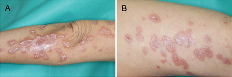
Fig. 1. Clinical findings. Multiple erythematous annular scaly plaques with elevated margins and central atrophy on (A) extensor surface of right upper extremity and (B) left thigh.
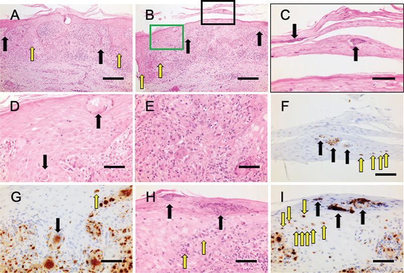
Fig. 2. Histological and immunohistochemical findings of specimens from the right upper arm (A–G) and right lower leg (H, I). (A, B) Hyperkeratosis, parakeratosis, irregularly elongated rete ridges (black arrows), and pseudoepitheliomatous hyperplasia (yellow arrows) in the epidermis in both cut surfaces derived from 1 cut (haematoxylin and eosin, original magnification ×40). (C) Magnified image of the inset marked with black line in (B). Aggregations of inflammatory cells in the horny layer (black arrows) (haematoxylin and eosin, original magnification ×400). (D) Magnified image of the inset marked with green line in (B). Aggregations of inflammatory cells in the epidermis (black arrows) (haematoxylin and eosin, original magnification ×400). (E) Non-caseating epithelioid granulomas containing multinucleated giant cells in the dermis in the cut surface of (B) (haematoxylin and eosin, original magnification ×400). (F) Positive staining with CD68 in aggregated (black arrows) and scattered (yellow arrows) inflammatory cells in the horny layer in the cut surface of (B) (CD68, original magnification ×400), which indicates transepidermal elimination of sarcoidal granulomas (TESG). (G) Positive staining with CD68 in aggregated inflammatory cells in the epidermis (black arrow) and in an inflammatory cell in the epidermis (yellow arrow) in the cut surface of (B) (CD68, original magnification ×400), indicating TESG. Many inflammatory cells in the dermis are also positive for CD68. (H) Aggregations of inflammatory cells in the horny layer (black arrows) and the epidermis (yellow arrows) (haematoxylin and eosin, original magnification ×400). (I) Positive staining with CD68 in aggregated inflammatory cells in the horny layer (black arrows) and scattered inflammatory cells in the epidermis (yellow arrows) (CD68, original magnification ×400), which indicates TESG. Bars indicates 250 μm in (A and B) and 100 μm in (C, D, E, F, G, H, and I), respectively.
What is your diagnosis?
Differential diagnosis 1: Necrobiosis lipoidica
Differential diagnosis 2: Annular elastolytic giant cell granuloma
Differential diagnosis 3: Psoriasis vulgaris
Differential diagnosis 4: Psoriasiform sarcoidosis
See next page for answer.
ANSWERS TO QUIZ
Multiple Erythematous Annular Scaly Plaques: A Commentary
Diagnosis: Psoriasiform sarcoidosis
Granulomatous skin lesions are categorized into 6 types according to cellular constituents and associated changes: (i) tuberculoid, (ii) sarcoidal, (iii) necrobiotic, (iv) suppurative, (v) foreign body, and (vi) histoid-type granuloma (1). Because of the presence of dermal sarcoidal granulomas, the skin lesions in the present case were considered to be cutaneous sarcoidosis (skin manifestations of sarcoidosis). Cutaneous sarcoidosis is classified into specific lesions with histologically evident noncaseating granulomas and nonspecific lesions that develop as a result of a reactive process that does not form granulomas (2). Specific lesions are further divided into common forms such as papules, nodules, plaques, and macules, and uncommon forms such as ulcerative, ichthyosiform, and psoriasiform sarcoidosis. Nonspecific lesions included erythema nodosum.
Transepidermal elimination is a phenomenon characterized by the elimination or extrusion of altered substances from the dermis through the epidermis. This phenomenon has been observed in several diseases; however, transepidermal elimination of sarcoidal granulomas (TESG) is exceptionally rare in cutaneous sarcoidosis. In the present case, inflammatory cells in the horny layer and epidermis were positive for CD68, indicating TESG.
Based on the clinical, histological, and immunohistochemical findings, the skin lesions in the present case were diagnosed as psoriasiform sarcoidosis presenting TESG.
Screening for sarcoidal lesions in other organs was performed. Chest computed tomography (CT) scan revealed no lung (including the hilar and mediastinal lymph nodes) abnormalities. Neither echocardiography nor electrocardiography revealed any abnormal findings suggestive of sarcoidal lesions. A CT scan of the abdomen revealed no lymphadenopathies. Musculoskeletal ultrasonography revealed subdeltoid bursitis and tendon synovitis of the biceps longus head of the right shoulder. Positron emission tomography was not performed because the patient refused the examination. The results of routine laboratory examinations, including blood count, liver function, renal function, and C-reactive protein concentration, were all within normal limits. Serum angiotensin-converting enzyme and calcium levels were within normal limits. The patient was diagnosed with sarcoidosis with cutaneous (psoriasiform sarcoidosis), ocular (uveitis of the left eye), and articular (arthritis of the right shoulder) involvement. Treatment with topical betamethasone butyrate propionate was ineffective. Treatment with methotrexate was presented to the patient, but she refused treatment and was lost to follow-up.
Psoriasiform sarcoidosis is an established but rare form of sarcoidosis-specific skin lesion. In general, this form of cutaneous sarcoidosis exhibits erythematous scaly plaques mimicking psoriasis, and histopathologically shows dermal sarcoidal granulomas and psoriasiform epidermal changes in the overlying epidermis.
In addition to the rare clinical form of psoriasiform of sarcoidosis, the cutaneous lesions in the present case had another feature of TESG. We analysed 10 cases of cutaneous sarcoidosis histologically showing TESG (3–10), including the present case, as reported in the English literature. The mean patient age was 53.2 years (range, 19–80 years). Five cases occurred between the ages of 10 and 40 and the remaining 5 cases occurred in patients in their 70s and 80s. None of the patients was in their 50s or 60s. Thus, the age distribution of the 10 patients showed 2 peaks, with the first peak of incidence between the ages of 10 and 40 and the second peak of incidence in the over-70s. On the other hand, approximately 70% of total sarcoidosis cases occur between 25 and 40 years of age, with a second peak of incidence over 50 years of age. Thus, although the age distribution in patients with cutaneous sarcoidosis with TESG has 2 peaks, similar to total sarcoidosis, the second peak shifts towards older age. Women were more affected than men, with a female-to-male ratio of 7:3, which is consistent with the fact that sarcoidosis is more prevalent in women. All male patients were under 30. Cutaneous lesions were either papules or plaques. Papular lesions were located mainly on the face, whereas plaque lesions, especially relatively large lesions, were, rather, present on the extremities. Two cases including the present case were psoriasiform sarcoidosis. Histologically, all patients had noncaseating epithelioid granulomas in the dermis. The histological findings of TESG can be divided into 2 types. One type is that dermal granulomas are excreted in small nests of histiocytes or scattered histiocytes (histiocyte-evident type) (6–10), as observed in the present case. In the other type, dermal granulomas are excreted as relatively large necrobiotic connective tissue, keratotic material, or cell debris, which cannot be clearly recognized as granulomas, through the transepidermal canal or by partial destruction of the epidermis (histiocyte-non-evident type) (3–5). Interestingly, cutaneous lesions of the histiocyte-evident type are clinically present mainly as relatively large plaques, whereas those of non-evident type appear mainly as papules or small plaques less than 2 cm in diameter.
REFERENCES
- Chakrabarti S, Pal S, Biswas B, Bose K, Pal S, Pathak S. Clinico-pathological study of cutaneous granulomatous lesions: a 5 yr experience in a tertiary care hospital in India. Iran J Pathol 2016; 11: 54–60.
- Koneti J, Cherukuri SP, Gadde S, Kalluru R, Chikatimalla R, Dasaradhan T. Sarcoidosis and its dermatological manifestations: a narrative review. Cureus 2022; 14: e28053. https://doi.org/10.7759/cureus.28053
- Batres E, Klima M, Tschen J. Transepidermal elimination in cutaneous sarcoidosis. J Cutan Pathol 1982; 9: 50–54. https://doi.org/10.1111/j.1600-0560.1982.tb01041.x
- Goette DK, Odom RB. Transepidermal elimination of granulomas in cutaneous tuberculosis and sarcoidosis. J Am Acad Dermatol 1986; 14: 126–128. https://doi.org/10.1016/S0190-9622(86)70016-9
- Yoneda K, Kakurai M, Umemoto N, Nakai K, Kubota Y, Demitsu T. Sarcoidosis presenting transepidermal elimination. Eur J Dermatol 2011; 22: 286–287. https://doi.org/10.1684/ejd.2011.1373
- Yamamoto T, Motoki Y. Transepidermal elimination of granuloma in a patient with sarcoidosis and systemic sclerosis overlap. Eur J Dermatol 2012; 21: 616–617. https://doi.org/10.1684/ejd.2012.1659
- Ismail A, Beckum K, McKay K. Transepidermal elimination in sarcoidosis: a frequent finding. J Cutan Pathol 2014; 41: 22–27. https://doi.org/10.1111/cup.12253
- Yamamoto T, Nishibu A, Eishi Y. Transepidermal elimination of sarcoidal granuloma in the lesional skin of psoriasiform sarcoidosis. J Dermatol 2017; 44: 100–101. https://doi.org/10.1111/1346-8138.13440
- Yamamoto T, Chen KR. Perforating plaque-type pretibial sarcoidosis with granulomatous phlebitis. Am J Dermatopathol 2020; 42: 225–226. https://doi.org/10.1097/DAD.0000000000001522
- Honda A, Matsumura N, Yamamoto T. Transepidermal elimination in cutaneous sarcoid plaques on the face: a possible role of M2 type macrophage. Australas J Dermatol 2020; 51: 168–169. https://doi.org/10.1111/ajd.13195